

The DOI matches its Crossref record; 9 of the 9 references it cites were found in a registry or an index.
Plasma Proteomic Signature Predicts Myeloid Neoplasm Risk
Published version of a preprint
Abstract
Purpose: Clonal hematopoiesis (CH) is thought to be the origin of myeloid neoplasms (MN). Yet, our understanding of the mechanisms driving CH progression to MN and clinical risk prediction of MN remains limited. The human proteome reflects complex interactions between genetic and epigenetic regulation of biological systems. We hypothesized that the plasma proteome might predict MN risk and inform our understanding of the mechanisms promoting MN development. Experimental Design: We jointly characterized CH and plasma proteomic profiles of 46,237 individuals in the UK Biobank at baseline study entry. During 500,036 person-years of follow-up, 115 individuals developed MN. Cox proportional hazard regression was used to test for an association between plasma protein levels and MN risk. Results: We identified 115 proteins associated with MN risk, of which 30% (N = 34) were also associated with CH. These were enriched for known regulators of the innate and adaptive immune system. Plasma proteomics improved the prediction of MN risk (AUC = 0.85; P = 5×10⁻⁹) beyond clinical factors and CH (AUC = 0.80). In an independent group (N = 381,485), we used inherited polygenic risk scores (PRS) for plasma protein levels to validate the relevance of these proteins toMNdevelopment. PRS analyses suggest that most MN-associated proteins we identified are not directly causally linked toMN risk, but rather represent downstream markers of pathways regulating the progression of CH to MN. Conclusions: These data highlight the role of immune cell regulation in the progression of CH to MN and the promise of leveraging multi-omic characterization of CH to improveMN risk stratification. See related commentary by Bhalgat and Taylor, p. 3095
Overview
The claims and statistics of this paper haven’t been checked yet.
Run the check — free, no account needed. It reads the open-access text and checks the sources it cites.
Integrity signals
Registry
Cites
Statistics
Figures
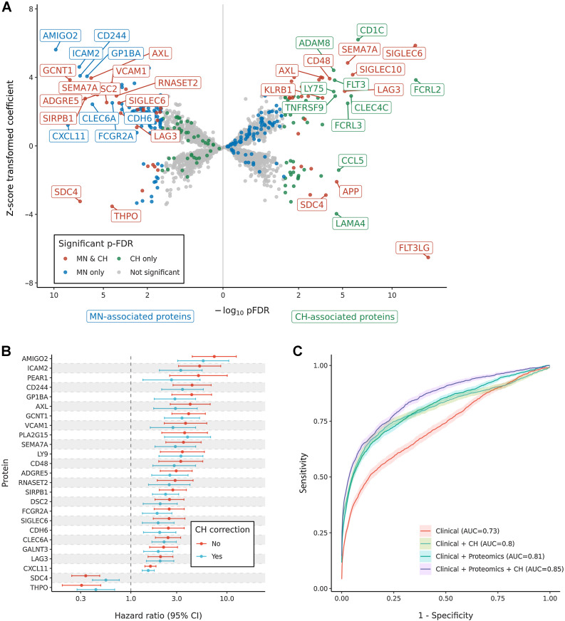
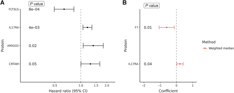
Verification
Claims the paper makes
The claims and statistics of this paper haven’t been checked yet.
Run the check — free, no account needed. It reads the open-access text and checks the sources it cites.
Claims other papers make about this one
Each is checked against this paper’s own text: does it say what the citing paper says it does?
In the citing paper
“Recent studies characterizing genomic53–55, proteomic56 and clinical predictors53 of hematologic cancer have enabled the development of disease-specific prognostic tools57,58.”
From Germline genetic variation impacts clonal hematopoiesis landscape and progression to malignancy (2025) · cites this paper as [56]
Self-check
Percentages, sums and reported differences re-computed from the paper’s own numbers.
The self-check needs the paper’s full text.
Citations
Cites
Sanderson, E., Glymour, M. M., Holmes, M. V., Kang, H., Morrison, J., Munafò, M. R., Palmer, T., Schooling, C. M., Wallace, C., Zhao, Q., et al. (2022) · Nature Reviews Methods Primers · doi:10.1038/s43586-021-00092-5 · Cited by 2,229
Plasma proteomic associations with genetics and health in the UK Biobank
Sun, B. B., Chiou, J., Traylor, M., Benner, C., Hsu, Y.-H., Richardson, T. G., Surendran, P., Mahajan, A., Robins, C., Vasquez-Grinnell, S. G., et al. (2023) · Nature · doi:10.1038/s41586-023-06592-6 · Cited by 1,880
The support of human genetic evidence for approved drug indications
Nelson, M. R., Tipney, H., Painter, J. L., Shen, J., Nicoletti, P., Shen, Y., Floratos, A., Sham, P. C., Li, M. J., Wang, J., et al. (2015) · Nature Genetics · doi:10.1038/ng.3314 · Cited by 1,666
Proteogenomic Characterization Reveals Therapeutic Vulnerabilities in Lung Adenocarcinoma
Gillette, M. A., Satpathy, S., Cao, S., Dhanasekaran, S. M., Vasaikar, S. V., Krug, K., Petralia, F., Li, Y., Liang, W.-W., Reva, B., et al. (2020) · Cell · doi:10.1016/j.cell.2020.06.013 · Cited by 820
Proteogenomic Analysis of Human Colon Cancer Reveals New Therapeutic Opportunities
Vasaikar, S., Huang, C., Wang, X., Petyuk, V. A., Savage, S. R., Wen, B., Dou, Y., Zhang, Y., Shi, Z., Arshad, O. A., et al. (2019) · Cell · doi:10.1016/j.cell.2019.03.030 · Cited by 799
Inherited causes of clonal haematopoiesis in 97,691 whole genomes
Bick, A. G., Weinstock, J. S., Nandakumar, S. K., Fulco, C. P., Bao, E. L., Zekavat, S. M., Szeto, M. D., Liao, X., Leventhal, M. J., Nasser, J., et al. (2020) · Nature · doi:10.1038/s41586-020-2819-2 · Cited by 733
Chronic infection drives Dnmt3a-loss-of-function clonal hematopoiesis via IFNγ signaling
Hormaechea-Agulla, D., Matatall, K. A., Le, D. T., Kain, B., Long, X., Kus, P., Jaksik, R., Challen, G. A., Kimmel, M., & King, K. Y. (2021) · Cell Stem Cell · doi:10.1016/j.stem.2021.03.002 · Cited by 329
Inflammatory Pathophysiology as a Contributor to Myeloproliferative Neoplasms
Fisher, D. A. C., Fowles, J. S., Zhou, A., & Oh, S. T. (2021) · Frontiers in Immunology · doi:10.3389/fimmu.2021.683401 · Cited by 115
Self-citationAlanazi, B., Munje, C. R., Rastogi, N., Williamson, A. J. K., Taylor, S., Hole, P. S., Hodges, M., Doyle, M., Baker, S., Gilkes, A. F., et al. (2020) · Leukemia · doi:10.1038/s41375-019-0596-4 · Cited by 64
Research works printed but not linked: 62
- Genovese, 2014, Clonal hematopoiesis and blood-cancer risk inferred from blood DNA sequence, N Engl J Med, 371, 2477, 10.1056/NEJMoa1409405 · DOI · Paper page
- Jaiswal, 2014, Agerelated clonal hematopoiesis associated with adverse outcomes, N Engl J Med, 371, 2488, 10.1056/NEJMoa1408617 · DOI · Paper page
- Xie, 2014, Age-related mutations associated with clonal hematopoietic expansion and malignancies, Nat Med, 20, 1472, 10.1038/nm.3733 · DOI · Paper page
- McKerrell, 2017, JAK2 V617F hematopoietic clones are present several years prior to MPN diagnosis and follow different expansion kinetics, Blood Advances, 1, 968, 10.1182/bloodadvances.2017007047 · DOI
- Desai, 2018, Somatic mutations precede acute myeloid leukemia years before diagnosis, Nat Med, 24, 1015, 10.1038/s41591-018-0081-z · DOI · Paper page
- Gu, 2023, Multiparameter prediction of myeloid neoplasia risk, Nat Genet, 55, 1523, 10.1038/s41588-023-01472-1 · DOI · Paper page
- Weeks, 2023, Prediction of risk for myeloid malignancy in clonal hematopoiesis, NEJM Evid, 2, 10.1056/evidoa2200310 · DOI · Paper page
- Kornblau, 2009, Functional proteomic profiling of AML predicts response and survival, Blood, 113, 154, 10.1182/blood-2007-10-119438 · DOI
- Hu, 2019, A quantitative analysis of heterogeneities and hallmarks in acute myelogenous leukaemia, Nat Biomed Eng, 3, 889, 10.1038/s41551-019-0387-2 · DOI
- Van Dijk, 2020, Targeted therapy in acute myeloid leukemia: current status and new insights from a proteomic perspective, Expert Review of Proteomics, 17, 1, 10.1080/14789450.2020.1717951 · DOI
- Kramer, 2022, Proteomic and phosphoproteomic landscapes of acute myeloid leukemia, Blood, 140, 1533, 10.1182/blood.2022016033 · DOI
- Geffen, 2023, Pan-cancer analysis of post-translational modifications reveals shared patterns of protein regulation, Cell, 186, 3945, 10.1016/j.cell.2023.07.013 · DOI
- Liu, 2021, Large-scale and highresolution mass spectrometry-based proteomics profiling defines molecular subtypes of esophageal cancer for therapeutic targeting, Nat Commun, 12, 4961, 10.1038/s41467-021-25202-5 · DOI
- Chinese Human Proteome Project (CNHPP) Consortium, 2019, Proteomics identifies new therapeutic targets of earlystage hepatocellular carcinoma, Nature, 567, 257, 10.1038/s41586-019-0987-8 · DOI
- Meissner, 2022, The emerging role of mass spectrometry-based proteomics in drug discovery, Nat Rev Drug Discov, 21, 637, 10.1038/s41573-022-00409-3 · DOI
- Sudlow, 2015, UK biobank: an open access resource for identifying the causes of a wide range of complex diseases of middle and old age, PLoS Med, 12, e1001779, 10.1371/journal.pmed.1001779 · DOI · Paper page
- Van Hout, 2020, Exome sequencing and characterization of 49,960 individuals in the UK Biobank, Nature, 586, 749, 10.1038/s41586-020-2853-0 · DOI · Paper page
- Loh, 2018, Insights into clonal haematopoiesis from 8,342 mosaic chromosomal alterations, Nature, 559, 350, 10.1038/s41586-018-0321-x · DOI · Paper page
- Purcell, 2007, PLINK: a tool set for whole-genome association and population-based linkage analyses, Am J Hum Genet, 81, 559, 10.1086/519795 · DOI · Paper page
- Brion, 2013, Calculating statistical power in mendelian randomization studies, Int J Epidemiol, 42, 1497, 10.1093/ije/dyt179 · DOI · Paper page
- Li, 2017, AMIGO2 modulates T-cell functions and its deficiency in mice ameliorates experimental autoimmune encephalomyelitis, Brain Behav Immun, 62, 110, 10.1016/j.bbi.2017.01.009 · DOI
- Park, 2015, AMIGO2, a novel membrane anchor of PDK1, controls cell survival and angiogenesis via Akt activation, J Cell Biol, 211, 619, 10.1083/jcb.201503113 · DOI
- Sierro, 2011, The CD4-like molecule LAG-3, biology and therapeutic applications, Expert Opin Ther Targets, 15, 91, 10.1517/14712598.2011.540563 · DOI
- Sintes, 2007, Characterization of mouse CD229 (Ly9), a leukocyte cell surface molecule of the CD150 (SLAM) family, Tissue Antigens, 70, 355, 10.1111/j.1399-0039.2007.00909.x · DOI
- Somuncular, 2023, Combination of CD49b and CD229 reveals a subset of multipotent progenitors with short-term activity within the hematopoietic stem cell compartment, Stem Cells Transl Med, 12, 720, 10.1093/stcltm/szad057 · DOI
- Kovalovsky, 2021, Siglec-6 is a target for chimeric antigen receptor T-cell treatment of chronic lymphocytic leukemia, Leukemia, 35, 2581, 10.1038/s41375-021-01188-3 · DOI
- Patel, 1999, OB-BP1/Siglec-6. A leptinand sialic acid-binding protein of the immunoglobulin superfamily, J Biol Chem, 274, 22729, 10.1074/jbc.274.32.22729 · DOI
- Fonseca, 2020, Deficiency in the glycosyltransferase Gcnt1 increases susceptibility to tuberculosis through a mechanism involving neutrophils, Mucosal Immunol, 13, 836, 10.1038/s41385-020-0277-7 · DOI
- Perkey, 2020, GCNT1-mediated O-glycosylation of the sialomucin CD43 is a sensitive indicator of notch signaling in activated T cells, J Immunol, 204, 1674, 10.4049/jimmunol.1901194 · DOI
- Lyck, 2015, The physiological roles of ICAM-1 and ICAM-2 in neutrophil migration into tissues, Curr Opin Hematol, 22, 53, 10.1097/MOH.0000000000000103 · DOI
- Agresta, 2018, The emerging role of CD244 signaling in immune cells of the tumor microenvironment, Front Immunol, 9, 2809, 10.3389/fimmu.2018.02809 · DOI
- Kerrigan, 2010, Syk-coupled C-type lectin receptors that mediate cellular activation via single tyrosine based activation motifs, Immunol Rev, 234, 335, 10.1111/j.0105-2896.2009.00882.x · DOI
- Pinho, 2022, VCAM1 confers innate immune tolerance on haematopoietic and leukaemic stem cells, Nat Cell Biol, 24, 290, 10.1038/s41556-022-00849-4 · DOI
- Gupta, 2017, JAK2-V617F activates b1-integrin–mediated adhesion of granulocytes to vascular cell adhesion molecule 1, Leukemia, 31, 1223, 10.1038/leu.2017.26 · DOI
- Köhler, 2020, Red blood cell-derived semaphorin 7A promotes thrombo-inflammation in myocardial ischemia-reperfusion injury through platelet GPIb, Nat Commun, 11, 1315, 10.1038/s41467-020-14958-x · DOI
- Morote-Garcia, 2012, Endothelial semaphorin 7A promotes neutrophil migration during hypoxia, Proc Natl Acad Sci USA, 109, 14146, 10.1073/pnas.1202165109 · DOI
- Dietrich, 2000, Cutting edge: signalregulatory protein beta 1 is a DAP12-associated activating receptor expressed in myeloid cells, J Immunol, 164, 9, 10.4049/jimmunol.164.1.9 · DOI
- Groom, 2011, CXCR3 in T-cell function, Exp Cell Res, 317, 620, 10.1016/j.yexcr.2010.12.017 · DOI
- Tokunaga, 2018, CXCL9, CXCL10, CXCL11/CXCR3 axis for immune activation—a target for novel cancer therapy, Cancer Treat Rev, 63, 40, 10.1016/j.ctrv.2017.11.007 · DOI · Paper page
- Averbeck, 2007, Switch in syndecan-1 and syndecan-4 expression controls maturation associated dendritic cell motility, Exp Dermatol, 16, 580, 10.1111/j.1600-0625.2007.00568.x · DOI
- Martin, 2019, CD97 is a critical regulator of acute myeloid leukemia stem cell function, J Exp Med, 216, 2362, 10.1084/jem.20190598 · DOI
- Nakamura-Ishizu, 2020, Multifaceted roles of thrombopoietin in hematopoietic stem cell regulation, Ann N Y Acad Sci, 1466, 51, 10.1111/nyas.14169 · DOI
- Niu, 2021, Targeting AXL kinase sensitizes leukemic stem and progenitor cells to venetoclax treatment in acute myeloid leukemia, Blood, 137, 3641, 10.1182/blood.2020007651 · DOI
- Pearson, 2019, AXL inhibition extinguishes primitive JAK2 mutated myeloproliferative neoplasm progenitor cells, Hemasphere, 3, e233, 10.1097/HS9.0000000000000233 · DOI
- Tischer, 2019, Platelet-type von willebrand disease: local disorder of the platelet GPIbα β-switch drives high-affinity binding to von Willebrand factor, J Thromb Haemost, 17, 2022, 10.1111/jth.14597 · DOI
- Kauskot, 2013, PEAR1 attenuates megakaryopoiesis via control of the PI3K/PTEN pathway, Blood, 121, 5208, 10.1182/blood-2012-10-462887 · DOI
- Nanda, 2005, Platelet endothelial aggregation receptor 1 (PEAR1), a novel epidermal growth factor repeat-containing transmembrane receptor, participates in platelet contactinduced activation, J Biol Chem, 280, 24680, 10.1074/jbc.M413411200 · DOI
- Volz, 2020, BIN2 orchestrates platelet calcium signaling in thrombosis and thrombo-inflammation, J Clin Invest, 130, 6064, 10.1172/JCI136457 · DOI
- Itkin, 2012, FGF-2 expands murine hematopoietic stem and progenitor cells via proliferation of stromal cells, c-Kit activation, and CXCL12 downregulation, Blood, 120, 1843, 10.1182/blood-2011-11-394692 · DOI
- Talbot, 2018, Regulatory roles of sortilin and SorLA in immune-related processes, Front Pharmacol, 9, 1507, 10.3389/fphar.2018.01507 · DOI
- Koschmieder, 2020, Role of inflammation in the biology of myeloproliferative neoplasms, Blood Rev, 42, 100711, 10.1016/j.blre.2020.100711 · DOI
- Khaldoyanidi, 2021, Immune biology of acute myeloid leukemia: implications for immunotherapy, J Clin Oncol, 39, 419, 10.1200/JCO.20.00475 · DOI
- Abegunde, 2018, An inflammatory environment containing TNFα favors Tet2-mutant clonal hematopoiesis, Exp Hematol, 59, 60, 10.1016/j.exphem.2017.11.002 · DOI · Paper page
- Pasupuleti, 2023, Obesity-induced inflammation exacerbates clonal hematopoiesis, J Clin Invest, 133, e163968, 10.1172/JCI163968 · DOI
- Méndez-Ferrer, 2020, Bone marrow niches in haematological malignancies, Nat Rev Cancer, 20, 285, 10.1038/s41568-020-0245-2 · DOI
- Mills, 2023, IL17 and IL17-producing cells in protection versus pathology, Nat Rev Immunol, 23, 38, 10.1038/s41577-022-00746-9 · DOI
- de Mooij, 2017, Targeting the interleukin-1 pathway in patients with hematological disorders, Blood, 129, 3155, 10.1182/blood-2016-12-754994 · DOI
- Chung, 2009, Critical regulation of early Th17 cell differentiation by interleukin-1 signaling, Immunity, 30, 576, 10.1016/j.immuni.2009.02.007 · DOI
- Hoang, 1988, Interleukin 1 enhances growth factor-dependent proliferation of the clonogenic cells in acute myeloblastic leukemia and of normal human primitive hemopoietic precursors, J Exp Med, 168, 463, 10.1084/jem.168.2.463 · DOI
- Delvaeye, 2009, Coagulation and innate immune responses: can we view them separately?, Blood, 114, 2367, 10.1182/blood-2009-05-199208 · DOI
- Unruh, 2020, Beyond thrombosis: the impact of tissue factor signaling in cancer, J Hematol Oncol, 13, 93, 10.1186/s13045-020-00932-z · DOI
- Milsom, 2009, Tissue factor and cancer stem cells: is there a linkage? Arterioscler Thromb, Vasc Biol, 29, 2005, 10.1161/ATVBAHA.108.177444 · DOI
Cited by
Germline genetic variation impacts clonal hematopoiesis landscape and progression to malignancy
Liu, J., Tran, D., Xue, L., Wiley, B. J., Vlasschaert, C., Watson, C. J., MacGregor, H. A. J., Zong, X., Chan, I. C. C., Das, I., et al. (2025) · Nature Genetics · doi:10.1038/s41588-025-02250-x · Cited by 33
Self-citation
Metadata
- Authors
- Tran, D., Beeler, J. S., Liu, J., Wiley, B., Chan, I. C. C., Xin, Z., Kramer, M. H., Batchi-Bouyou, A. L., Zong, X., Walter, M. J., Petrone, G. E. M., Chlamydas, S., Ferraro, F., Oh, S. T., Link, D. C., Busby, B., Cao, Y., & Bolton, K. L.
- Venue
- Clinical Cancer Research
- Published
- 2024-03-06
- Volume
- 30
- Issue
- 15
- Pages
- 3220-3228
- ISSN
- 1078-0432
- 1557-3265
- PMC
- PMC11292192
- Cited by
- 17
- References
- 9
- Language
- en
- Funders
- Edward P. Evans Foundation
- American Society of Hematology
- National Institutes of Health (K08 CA241318)
- Children's Discovery Institute
- V Foundation for Cancer Research
- Damon Runyon Cancer Research Foundation
- UK Biobank Resource (55288)
- UK Biobank Research Analysis Platform
About these checks
Registry, retraction, Cites and statistics results are computed, not judged, with one exception: a printed reference that no index lists is read once by a model before it counts as a citation error. Claims are extracted by a model and kept only when their quote appears verbatim in the text; each cited source is then checked, with a model’s help, for whether it says what the claim relies on. Those verdicts can be wrong — read the claim’s quote and the cited source before relying on one.
Statistics and arithmetic are recomputed, not judged.
Confidence bands are shown only for fields calibrated on our annotated test set. None is calibrated yet, so no band is shown.
What counts as a claim ›How we measure confidence ›How we check this ›
Are you an author, or do you see a problem with this page? Request a review
