Cited by 45Retinoids in leukemia and cellular processesCancer-related Molecular PathwaysProtein Structure and Dynamics

The DOI matches its Crossref record; 4 of the 4 references it cites were found in a registry or an index.

Structural Basis of PML-RARA Oncoprotein Targeting by Arsenic Unravels a Cysteine Rheostat Controlling PML Body Assembly and Function

Open access

Published version of a preprint

Abstract

PML nuclear bodies (NB) are disrupted in PML-RARA–driven acute promyelocytic leukemia (APL). Arsenic trioxide (ATO) cures 70% of patients with APL, driving PML-RARA degradation and NB reformation. In non-APL cells, arsenic binding onto PML also amplifies NB formation. Yet, the actual molecular mechanism(s) involved remain(s) elusive. Here, we establish that PML NBs display some features of liquid–liquid phase separation and that ATO induces a gel-like transition. PML B-box-2 structure reveals an alpha helix driving B2 trimerization and positioning a cysteine trio to form an ideal arsenic-binding pocket. Altering either of the latter impedes ATO-driven NB assembly, PML sumoylation, and PML-RARA degradation, mechanistically explaining clinical ATO resistance. This B2 trimer and the C213 trio create an oxidation-sensitive rheostat that controls PML NB assembly dynamics and downstream signaling in both basal state and during stress response. These findings identify the structural basis for arsenic targeting of PML that could pave the way to novel cancer drugs. Significance: Arsenic curative effects in APL rely on PML targeting. We report a PML B-box-2 structure that drives trimer assembly, positioning a cysteine trio to form an arsenic-binding pocket, which is disrupted in resistant patients. Identification of this ROS-sensitive triad controlling PML dynamics and functions could yield novel drugs. See related commentary by Salomoni, p. 2505. This article is featured in Selected Articles from This Issue, p. 2489

Overview

The claims and statistics of this paper haven’t been checked yet.

Run the check — free, no account needed. It reads the open-access text and checks the sources it cites.

A few free checks a day · papers already verified open instantly · results are public

Integrity signals

Registry

Crossref · 10.1158/2159-8290.cd-23-0453

Cites

4 of 4 references verified0 citation errors · 0 not checkable

Statistics

Figures

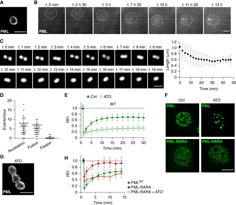
Figure 1. PML NBs display hallmarks of LLPS and switch to gel-like features upon ATO exposure. A, STED analysis uncovers the typical spherical core-shell PML NB. Scale bar, 1 μm. B, Time-lapse analysis of PML NB apparition upon GFP-PMLWT expression in PmlKO MEFs. The white circle indicates the nucleus. Representative data from three independent experiments. Scale bar, 5 μm. C, PML NBs undergo fusion with spherical relaxation, similar to liquid-like droplets. Representative time-lapse of PML NB fusion (left) and quantification of effective viscosity (right). Mean values ± SD of n = 21 fusion events in independent cells from three independent experiments. Scale bar, 1 μm. D, Quantification of PML NB nucleation, fusion, and fission events over 1 hour. Mean value ± SD of n = 37 cells from five independent experiments. E, FRAP analyses of GFP-PMLWT dynamics at NBs in control (Ctrl) or ATO-treated MEFs (1 μmol/L, 30 minutes). Mean value ± SD. Number of NBs assessed Ctrl (n = 36), ATO (n = 28), from at least five independent experiments. WT, wild-type. F, Confocal analysis of PML NBs in GFP-PMLWT or GFP-PML-RARA MEFs treated with 1 μmol/L of ATO for 1 hour. Scale bar, 5 μm. G, STED analysis demonstrating incomplete PML NB fusion upon ATO in PMLKO MEFs s. Scale bar, 1 μm. H, Comparison between GFP-PMLWT and GFP-PML-RARA dynamics at NBs by FRAP. ATO treatment (1 μmol/L, 30 minutes) induces a liquid- to gel-like transition of GFP-PML-RARA. Mean value ± SD. Number of dots assessed PML (n = 36), PML-RARA (n = 20), PML-RARA+ATO (n = 37), from at least five independent experiments. RFI, Relative fluorescence intensity.
Figure 1. PML NBs display hallmarks of LLPS and switch to gel-like features upon ATO exposure. A, STED analysis uncovers the typical spherical core-shell PML NB. Scale bar, 1 μm. B, Time-lapse analysis of PML NB apparition upon GFP-PMLWT expression in PmlKO MEFs. The white circle indicates the nucleus. Representative data from three independent experiments. Scale bar, 5 μm. C, PML NBs undergo fusion with spherical relaxation, similar to liquid-like droplets. Representative time-lapse of PML NB fusion (left) and quantification of effective viscosity (right). Mean values ± SD of n = 21 fusion events in independent cells from three independent experiments. Scale bar, 1 μm. D, Quantification of PML NB nucleation, fusion, and fission events over 1 hour. Mean value ± SD of n = 37 cells from five independent experiments. E, FRAP analyses of GFP-PMLWT dynamics at NBs in control (Ctrl) or ATO-treated MEFs (1 μmol/L, 30 minutes). Mean value ± SD. Number of NBs assessed Ctrl (n = 36), ATO (n = 28), from at least five independent experiments. WT, wild-type. F, Confocal analysis of PML NBs in GFP-PMLWT or GFP-PML-RARA MEFs treated with 1 μmol/L of ATO for 1 hour. Scale bar, 5 μm. G, STED analysis demonstrating incomplete PML NB fusion upon ATO in PMLKO MEFs s. Scale bar, 1 μm. H, Comparison between GFP-PMLWT and GFP-PML-RARA dynamics at NBs by FRAP. ATO treatment (1 μmol/L, 30 minutes) induces a liquid- to gel-like transition of GFP-PML-RARA. Mean value ± SD. Number of dots assessed PML (n = 36), PML-RARA (n = 20), PML-RARA+ATO (n = 37), from at least five independent experiments. RFI, Relative fluorescence intensity.CC BY-NC-ND · PubMed Central
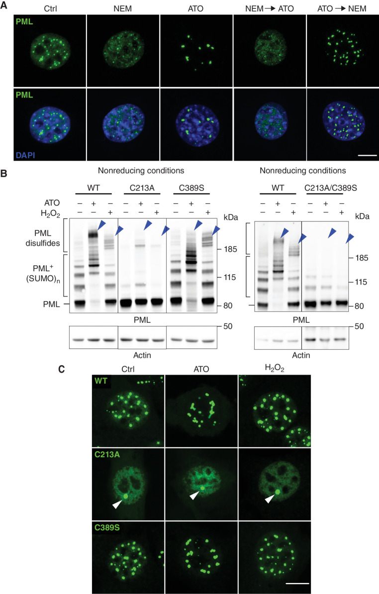
Figure 2. PML NB assembly depends on C213 in B2 domain, but not on C389-mediated intermolecular disulfide bonds. A, Confocal analysis of PML NB formation following cysteine alkylation with NEM (10 μmol/L, 1 hour) or ATO (1 μmol/L, 1 hour) used alone or sequentially in GFP-PMLWT MEFs. Scale bar, 5 μm. Ctrl, control. B, Western blot analysis (nonreducing conditions) of PML disulfide bond formation in MEFs expressing HA-PMLWT or PML cysteine mutants, treated or not with ATO (10 μmol/L, 1 hour) or H2O2 (500 μmol/L, 1 hour). Brackets: SUMO-conjugated or intermolecular disulfide-bound PML. Blue arrowheads, intermolecular disulfide-bound PML. Molecular weight (kDa). C, Confocal analysis of PML NBs upon ATO (1 μmol/L, 1 hour) or H2O2 (500 μmol/L, 1 hour) exposure in HA-PMLWT- or cysteine mutant–expressing MEFs. Arrowheads, single NB in HA-PMLC213A-expressing MEFs. Scale bar, 5 μm.
Figure 2. PML NB assembly depends on C213 in B2 domain, but not on C389-mediated intermolecular disulfide bonds. A, Confocal analysis of PML NB formation following cysteine alkylation with NEM (10 μmol/L, 1 hour) or ATO (1 μmol/L, 1 hour) used alone or sequentially in GFP-PMLWT MEFs. Scale bar, 5 μm. Ctrl, control. B, Western blot analysis (nonreducing conditions) of PML disulfide bond formation in MEFs expressing HA-PMLWT or PML cysteine mutants, treated or not with ATO (10 μmol/L, 1 hour) or H2O2 (500 μmol/L, 1 hour). Brackets: SUMO-conjugated or intermolecular disulfide-bound PML. Blue arrowheads, intermolecular disulfide-bound PML. Molecular weight (kDa). C, Confocal analysis of PML NBs upon ATO (1 μmol/L, 1 hour) or H2O2 (500 μmol/L, 1 hour) exposure in HA-PMLWT- or cysteine mutant–expressing MEFs. Arrowheads, single NB in HA-PMLC213A-expressing MEFs. Scale bar, 5 μm.CC BY-NC-ND · PubMed Central
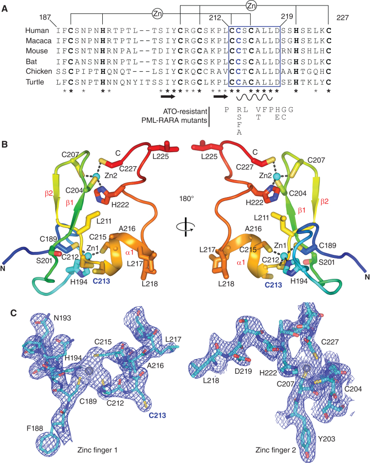
Figure 3. Uncovering PML B2 structure. A, Conservation of PML B2 box through evolution. Top, putative position of the two zinc fingers intertwined in a cross-brace. Bold, amino acids implicated in putative zinc coordination. The additional cysteine in position 213 is highlighted in blue. Predicted secondary structures of PML B-box-2 are depicted. Arrow, beta-sheet; loop, α1-helix; stars, evolutionary conserved amino acids (bold, identical; star custom, 1/6 variation only). The highly conserved α-helix is boxed in blue. Bottom, PML-RARA mutations from patients with therapy-resistant APL. B, Crystal structure of PML B2 monomer shown in a cartoon representation. PML B2 folds around two zinc fingers organized in a cross-brace. The C3H1 zinc coordination leaves the C213 free. Helix α1 (C213 to L218) exposes C213 toward the outside of the structure. Cyan sphere, zinc atoms; blue bold font, C213. C, Left, composite omit map contoured at 2 σ of a segment from PML B2 near zinc finger 1 at 2.1 Å. C213 appears as a free cysteine (blue bold font). Right, composite omit map contoured at 2 σ of a segment from PML B2 near zinc finger 2 at 2.1 Å. C227, not D219, coordinates Zn2+. Grey spheres, zinc atoms.
Figure 3. Uncovering PML B2 structure. A, Conservation of PML B2 box through evolution. Top, putative position of the two zinc fingers intertwined in a cross-brace. Bold, amino acids implicated in putative zinc coordination. The additional cysteine in position 213 is highlighted in blue. Predicted secondary structures of PML B-box-2 are depicted. Arrow, beta-sheet; loop, α1-helix; stars, evolutionary conserved amino acids (bold, identical; star custom, 1/6 variation only). The highly conserved α-helix is boxed in blue. Bottom, PML-RARA mutations from patients with therapy-resistant APL. B, Crystal structure of PML B2 monomer shown in a cartoon representation. PML B2 folds around two zinc fingers organized in a cross-brace. The C3H1 zinc coordination leaves the C213 free. Helix α1 (C213 to L218) exposes C213 toward the outside of the structure. Cyan sphere, zinc atoms; blue bold font, C213. C, Left, composite omit map contoured at 2 σ of a segment from PML B2 near zinc finger 1 at 2.1 Å. C213 appears as a free cysteine (blue bold font). Right, composite omit map contoured at 2 σ of a segment from PML B2 near zinc finger 2 at 2.1 Å. C227, not D219, coordinates Zn2+. Grey spheres, zinc atoms.CC BY-NC-ND · PubMed Central
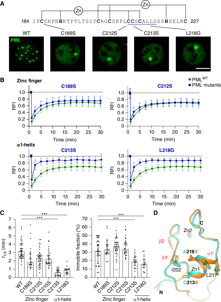
Figure 4. PML B2 α1-helix controls PML assembly and dynamics. A, Representative PML NB formation in MEFs expressing PML B2 mutants in Zn2+-coordinating cysteines or α1-helix (underlined in blue) residues. Scale bar, 5 μm. B, FRAP analyses of GFP-PML dynamics at NBs in WT- or mutant-expressing MEFs. Mean value ± SD. RFI, Relative fluorescence intensity. NBs assessed PMLWT (n = 36), PMLC189S (n = 25), PMLC212S (n = 42), PMLC213S (n = 24), PMLL218G (n = 20), from at least five independent experiments. C, Plot representation of B, with t1/2 (left) and immobile fraction (right) of PML mutants harboring mutations on the α1-helix or zinc finger. Each dot represents an individual NB. Median ± 95% confidence interval, statistical significance by comparison with GFP-PMLWT, Kruskal–Wallis test. ***, P ≤ 0.001 are displayed. P value for Zn-finger mutants, C189S = 0.0097, C212S = 0.0115, C213S = 0.028. D, Superposition of PML B2 crystal structure (cyan) with that of PMLC213A/A216V mutant (orange) demonstrates absence of misfolding.
Figure 4. PML B2 α1-helix controls PML assembly and dynamics. A, Representative PML NB formation in MEFs expressing PML B2 mutants in Zn2+-coordinating cysteines or α1-helix (underlined in blue) residues. Scale bar, 5 μm. B, FRAP analyses of GFP-PML dynamics at NBs in WT- or mutant-expressing MEFs. Mean value ± SD. RFI, Relative fluorescence intensity. NBs assessed PMLWT (n = 36), PMLC189S (n = 25), PMLC212S (n = 42), PMLC213S (n = 24), PMLL218G (n = 20), from at least five independent experiments. C, Plot representation of B, with t1/2 (left) and immobile fraction (right) of PML mutants harboring mutations on the α1-helix or zinc finger. Each dot represents an individual NB. Median ± 95% confidence interval, statistical significance by comparison with GFP-PMLWT, Kruskal–Wallis test. ***, P ≤ 0.001 are displayed. P value for Zn-finger mutants, C189S = 0.0097, C212S = 0.0115, C213S = 0.028. D, Superposition of PML B2 crystal structure (cyan) with that of PMLC213A/A216V mutant (orange) demonstrates absence of misfolding.CC BY-NC-ND · PubMed Central
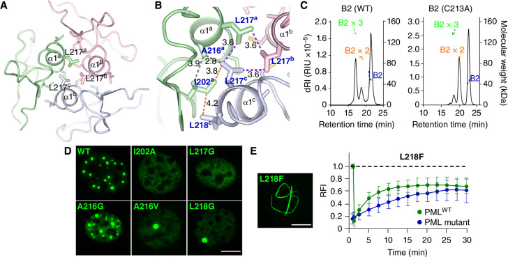
Figure 5. PML B2 mediates PML trimerization and is required for biological functions. A, Modeling of a PML B2 trimer. B2 monomers are colored in green, gray, and pink. Exponents refer to affiliation to a specific monomer. B, Close-up view of the PML B2 trimer model depicting hydrophobic interactions between the α1-helices. Residues involved in these hydrophobic interactions are shown in blue, linked by dashed lines. Distance between these key hydrophobic residues are in Å. C, SEC-MALs analysis showing a mix of monomeric, dimeric, and trimeric PML B2 (left) (dRI:differential refractive index, RIU:refractive index unit). The C213A mutant shifts toward monomeric and dimeric states (right). MBP-B2 molecular weight: 46.2 kDa. D, Representative PML NB formation in PML B2 mutants predicted to impair trimerization expressed in MEFs. Scale bar, 5 μm. E, GFP-PMLL218F yields filaments (left). Scale bar, 5 μm. FRAP analysis of the exchange rates of GFP-PMLL218F mutant compared with GFP-PMLWT (right). Mean value ± SD. NBs assessed PMLWT (n = 36) and PMLL218F (n = 24), from at least five independent experiments. RFI, Relative fluorescence intensity.
Figure 5. PML B2 mediates PML trimerization and is required for biological functions. A, Modeling of a PML B2 trimer. B2 monomers are colored in green, gray, and pink. Exponents refer to affiliation to a specific monomer. B, Close-up view of the PML B2 trimer model depicting hydrophobic interactions between the α1-helices. Residues involved in these hydrophobic interactions are shown in blue, linked by dashed lines. Distance between these key hydrophobic residues are in Å. C, SEC-MALs analysis showing a mix of monomeric, dimeric, and trimeric PML B2 (left) (dRI:differential refractive index, RIU:refractive index unit). The C213A mutant shifts toward monomeric and dimeric states (right). MBP-B2 molecular weight: 46.2 kDa. D, Representative PML NB formation in PML B2 mutants predicted to impair trimerization expressed in MEFs. Scale bar, 5 μm. E, GFP-PMLL218F yields filaments (left). Scale bar, 5 μm. FRAP analysis of the exchange rates of GFP-PMLL218F mutant compared with GFP-PMLWT (right). Mean value ± SD. NBs assessed PMLWT (n = 36) and PMLL218F (n = 24), from at least five independent experiments. RFI, Relative fluorescence intensity.CC BY-NC-ND · PubMed Central
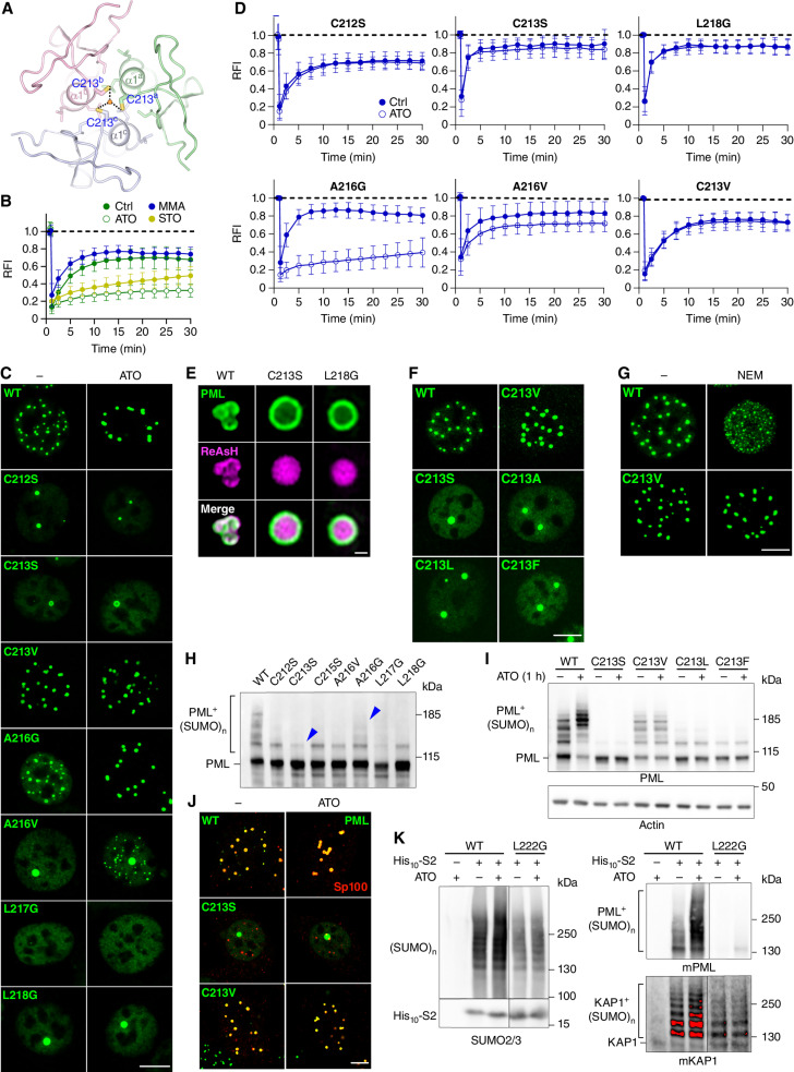
Figure 6. ATO binding to B2 trimer is responsible for ATO-induced phase transition. A, Modeling of PML B2 trimer around an arsenic atom. The key C213 residues are indicated in blue and the arsenic atom is shown as an orange sphere. The predicted distance between the arsenic atom and C213 represented by the dashed line is around 2.7 Å. B, FRAP analyses of GFP-PMLWT dynamics at NBs in untreated cells or cells treated with trivalent metalloid oxides: ATO (1 μmol/L, 30 minutes) or STO (1 μmol/L, 30 minutes) or divalent arsenic (MMA, 1 μmol/L, 30 minutes). Mean value ± SD, assessed NBs control (Ctrl; n = 36), MMA (n = 21), ATO (n = 28), STO (n = 33), from at least five independent experiments. RFI, relative fluorescence intensity. C, Representative confocal analyses of PML NB formation in PmlKO MEFs stably expressing GFP-PML B2 mutants, ± ATO (1 μmol/L, 1 hour). Scale bar, 5 μm. D, FRAP analyses of GFP-PML mutant–expressing MEFs treated or not with ATO (1 μmol/L, 30 minutes). Mean value ± SD. NBs assessed PMLC212S Ctrl (n = 42), PMLC212S ATO (n = 28), PMLC213S Ctrl (n = 24), PMLC213S ATO (n = 27), PMLL218G Ctrl (n = 20), PMLL218G ATO (n = 26), PMLA216G Ctrl (n = 40), PMLA216G ATO (n = 28), PMLA216V Ctrl (n = 43), PMLA216V ATO (n = 43), PMLC213V Ctrl (n = 32), PMLC213V ATO (n = 31), from at least five independent experiments. E, Representative images of red fluorescent arsenic (ReAsH) localization to NBs in GFP-PMLWT or mutant MEFs. Scale bar, 1 μm. F, Representative PML NB formation in PML C213 mutant–expressing MEFs. Scale bar, 5 μm. G, Confocal analysis of PML NB formation following cysteine alkylation with NEM (10 μmol/L, 1 hour) in GFP-PMLWT- or PMLC213V -expressing MEFs. Scale bar, 5 μm. H, Western blot analysis of basal PML sumoylation in GFP-PMLWT- or mutant-expressing MEFs (arrowheads highlight different sumoylation levels of PML mutants). I, Western blot analysis of PML sumoylation in GFP-PMLWT or mutant MEFs treated or not with ATO (1 μmol/L, 1 hour). J, Confocal analysis of PML and Sp100 localization in GFP-PMLWT- or C213 mutant–expressing MEFs treated or not with ATO (1 μmol/L, 1 hour). Scale bar, 5 μm. K, Pulldown of HIS10-SUMO2 conjugates from PmlWT or PmlL222G mESCs treated or not with ATO (1 μmol/L, 30 minutes). ATO-treated nontransduced PmlWT mESCs are shown as controls. Western blot with anti-SUMO2/3 (left), anti-mPML (right, top), or anti-KAP1 antibodies (right, bottom). Sumoylated species are indicated. Representative data of n = 3 independent experiments.
Figure 6. ATO binding to B2 trimer is responsible for ATO-induced phase transition. A, Modeling of PML B2 trimer around an arsenic atom. The key C213 residues are indicated in blue and the arsenic atom is shown as an orange sphere. The predicted distance between the arsenic atom and C213 represented by the dashed line is around 2.7 Å. B, FRAP analyses of GFP-PMLWT dynamics at NBs in untreated cells or cells treated with trivalent metalloid oxides: ATO (1 μmol/L, 30 minutes) or STO (1 μmol/L, 30 minutes) or divalent arsenic (MMA, 1 μmol/L, 30 minutes). Mean value ± SD, assessed NBs control (Ctrl; n = 36), MMA (n = 21), ATO (n = 28), STO (n = 33), from at least five independent experiments. RFI, relative fluorescence intensity. C, Representative confocal analyses of PML NB formation in PmlKO MEFs stably expressing GFP-PML B2 mutants, ± ATO (1 μmol/L, 1 hour). Scale bar, 5 μm. D, FRAP analyses of GFP-PML mutant–expressing MEFs treated or not with ATO (1 μmol/L, 30 minutes). Mean value ± SD. NBs assessed PMLC212S Ctrl (n = 42), PMLC212S ATO (n = 28), PMLC213S Ctrl (n = 24), PMLC213S ATO (n = 27), PMLL218G Ctrl (n = 20), PMLL218G ATO (n = 26), PMLA216G Ctrl (n = 40), PMLA216G ATO (n = 28), PMLA216V Ctrl (n = 43), PMLA216V ATO (n = 43), PMLC213V Ctrl (n = 32), PMLC213V ATO (n = 31), from at least five independent experiments. E, Representative images of red fluorescent arsenic (ReAsH) localization to NBs in GFP-PMLWT or mutant MEFs. Scale bar, 1 μm. F, Representative PML NB formation in PML C213 mutant–expressing MEFs. Scale bar, 5 μm. G, Confocal analysis of PML NB formation following cysteine alkylation with NEM (10 μmol/L, 1 hour) in GFP-PMLWT- or PMLC213V -expressing MEFs. Scale bar, 5 μm. H, Western blot analysis of basal PML sumoylation in GFP-PMLWT- or mutant-expressing MEFs (arrowheads highlight different sumoylation levels of PML mutants). I, Western blot analysis of PML sumoylation in GFP-PMLWT or mutant MEFs treated or not with ATO (1 μmol/L, 1 hour). J, Confocal analysis of PML and Sp100 localization in GFP-PMLWT- or C213 mutant–expressing MEFs treated or not with ATO (1 μmol/L, 1 hour). Scale bar, 5 μm. K, Pulldown of HIS10-SUMO2 conjugates from PmlWT or PmlL222G mESCs treated or not with ATO (1 μmol/L, 30 minutes). ATO-treated nontransduced PmlWT mESCs are shown as controls. Western blot with anti-SUMO2/3 (left), anti-mPML (right, top), or anti-KAP1 antibodies (right, bottom). Sumoylated species are indicated. Representative data of n = 3 independent experiments.CC BY-NC-ND · PubMed Central
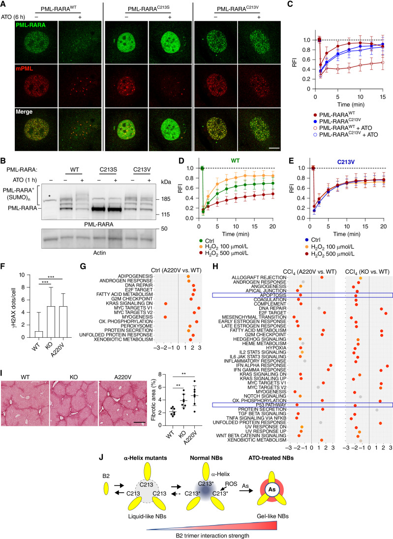
Figure 7. PML-RARA ATO sensitivity requires C213 and B2 controls in vivo responses to ROS. A, Confocal analysis of PmlWT MEFs stably expressing human GFP-PML-RARAWT or its B2 mutants treated or not with ATO (1 μmol/L, 6 hours). Scale bar, 5 μm. Different display settings for each genotype. B, Western blot analysis of PML-RARA sumoylation in Lin− hematopoietic progenitors expressing PML-RARA or the indicated mutants, treated or not with ATO (1 μmol/L, 1 hour). Representative data from three independent experiments. C, FRAP analysis of GFP-PML-RARA dynamics at NBs in GFP-PML-RARAWT- or GFP-PML-RARAC213V–expressing PmlKO MEFs treated or not with ATO (1 μmol/L, 30 minutes). Mean value ± SD. NBs assessed PML-RARAWT (n = 20), PML-RARAWT+ATO (n = 37), PML-RARAC213V (n = 42), PML-RARAC213V+ATO (n = 25), from at least five independent experiments. RFI, relative fluorescence intensity. D, FRAP analysis of GFP-PMLWT dynamics at NBs in MEFs treated or not with H2O2 (30 minutes). Mean value ± SD. NBs assessed PML (n = 35), H2O2 100 μmol/L (n = 27), H2O2 500 μmol/L (n = 32), from at least five independent experiments. Ctrl, control. E, Same as D with GFP-PMLC213V. Mean value ± SD. NBs assessed PMLC213V (n = 32), H2O2 100 μmol/L (n = 24), H2O2 500 μmol/L (n = 22), from at least five independent experiments. F, Box plot representing γH2AX dots counts assessed by immunofluorescence on liver tissues from three untreated mice. PmlWT (n = 633), PmlKO (n = 527), PmlA220V (n = 333). ***, P ≤ 0.001, unpaired t test. G, Gene-set enrichment analysis (GSEA) of differentially expressed genes from liver samples from untreated PmlWT or PmlA220V (equivalent of human PMLA216V) mice. Key pathways are boxed. Livers from n = 3 Pml or PmlA220V mice. H, GSEA of differentially expressed genes in liver samples from PmlKO or PmlA220V in comparison to PmlWT after an 18-hour CCl4 treatment. Key pathways are boxed. Livers from n = 3 mice of each genotype. I, Liver fibrosis following a 5-week CCl4 treatment detected by Picrosirius Red staining (left). Quantification of the fibrotic areas (right). Livers from n = 7 Pml, PmlKO, or PmlA220V mice. Scale bar, 400 μm. **, P ≤ 0.01, Mann–Whitney test. J, Model of PML NB liquid to gel-like transition controlled by the B2 α1 helix exposing C213 and hijacked by ATO. Hydrophobic-mediated trimerization of PML B2 (gray disk), regroups the three C213 in the center of the structure. The latter is impaired in α-helix mutants. In the physiologic states, this C213 triad behaves as a rheostat depending on their oxidation state (asterisks), fine-tuning the interactions within the trimer (light and dark gray disk). Arsenic binding crosslinks these cysteines, hijacking the ROS-rheostat to yield polymerization-induced gel-like transition.
Figure 7. PML-RARA ATO sensitivity requires C213 and B2 controls in vivo responses to ROS. A, Confocal analysis of PmlWT MEFs stably expressing human GFP-PML-RARAWT or its B2 mutants treated or not with ATO (1 μmol/L, 6 hours). Scale bar, 5 μm. Different display settings for each genotype. B, Western blot analysis of PML-RARA sumoylation in Lin− hematopoietic progenitors expressing PML-RARA or the indicated mutants, treated or not with ATO (1 μmol/L, 1 hour). Representative data from three independent experiments. C, FRAP analysis of GFP-PML-RARA dynamics at NBs in GFP-PML-RARAWT- or GFP-PML-RARAC213V–expressing PmlKO MEFs treated or not with ATO (1 μmol/L, 30 minutes). Mean value ± SD. NBs assessed PML-RARAWT (n = 20), PML-RARAWT+ATO (n = 37), PML-RARAC213V (n = 42), PML-RARAC213V+ATO (n = 25), from at least five independent experiments. RFI, relative fluorescence intensity. D, FRAP analysis of GFP-PMLWT dynamics at NBs in MEFs treated or not with H2O2 (30 minutes). Mean value ± SD. NBs assessed PML (n = 35), H2O2 100 μmol/L (n = 27), H2O2 500 μmol/L (n = 32), from at least five independent experiments. Ctrl, control. E, Same as D with GFP-PMLC213V. Mean value ± SD. NBs assessed PMLC213V (n = 32), H2O2 100 μmol/L (n = 24), H2O2 500 μmol/L (n = 22), from at least five independent experiments. F, Box plot representing γH2AX dots counts assessed by immunofluorescence on liver tissues from three untreated mice. PmlWT (n = 633), PmlKO (n = 527), PmlA220V (n = 333). ***, P ≤ 0.001, unpaired t test. G, Gene-set enrichment analysis (GSEA) of differentially expressed genes from liver samples from untreated PmlWT or PmlA220V (equivalent of human PMLA216V) mice. Key pathways are boxed. Livers from n = 3 Pml or PmlA220V mice. H, GSEA of differentially expressed genes in liver samples from PmlKO or PmlA220V in comparison to PmlWT after an 18-hour CCl4 treatment. Key pathways are boxed. Livers from n = 3 mice of each genotype. I, Liver fibrosis following a 5-week CCl4 treatment detected by Picrosirius Red staining (left). Quantification of the fibrotic areas (right). Livers from n = 7 Pml, PmlKO, or PmlA220V mice. Scale bar, 400 μm. **, P ≤ 0.01, Mann–Whitney test. J, Model of PML NB liquid to gel-like transition controlled by the B2 α1 helix exposing C213 and hijacked by ATO. Hydrophobic-mediated trimerization of PML B2 (gray disk), regroups the three C213 in the center of the structure. The latter is impaired in α-helix mutants. In the physiologic states, this C213 triad behaves as a rheostat depending on their oxidation state (asterisks), fine-tuning the interactions within the trimer (light and dark gray disk). Arsenic binding crosslinks these cysteines, hijacking the ROS-rheostat to yield polymerization-induced gel-like transition.CC BY-NC-ND · PubMed Central

Verification

Claims the paper makes

The claims and statistics of this paper haven’t been checked yet.

Run the check — free, no account needed. It reads the open-access text and checks the sources it cites.

A few free checks a day · papers already verified open instantly · results are public

Self-check

Percentages, sums and reported differences re-computed from the paper’s own numbers.

The self-check needs the paper’s full text.

Citations

Cites

Research works printed but not linked: 74

  1. Lallemand-Breitenbach, 2018, PML nuclear bodies: from architecture to function, Curr Opin Cell Biol, 52, 154, 10.1016/j.ceb.2018.03.011 · DOI
  2. Hsu, 2018, PML: regulation and multifaceted function beyond tumor suppression, Cell Biosci, 8, 5, 10.1186/s13578-018-0204-8 · DOI
  3. de The, 2017, Acute promyelocytic leukemia: a paradigm for oncoprotein-targeted cure, Cancer Cell, 32, 552, 10.1016/j.ccell.2017.10.002 · DOI
  4. Gurrieri, 2004, Mutations of the PML tumor suppressor gene in acute promyelocytic leukemia, Blood, 103, 2358, 10.1182/blood-2003-07-2200 · DOI
  5. Koken, 1995, The PML growth-suppressor has an altered expression in human oncogenesis, Oncogene, 10, 1315
  6. Mathews, 2010, Single-agent arsenic trioxide in the treatment of newly diagnosed acute promyelocytic leukemia: long-term follow-up data, J Clin Oncol, 28, 3866, 10.1200/JCO.2010.28.5031 · DOI
  7. Mathews, 2006, Single-agent arsenic trioxide in the treatment of newly diagnosed acute promyelocytic leukemia: durable remissions with minimal toxicity, Blood, 107, 2627, 10.1182/blood-2005-08-3532 · DOI
  8. Jeanne, 2010, PML/RARA oxidation and arsenic binding initiate the antileukemia response of As2O3, Cancer Cell, 18, 88, 10.1016/j.ccr.2010.06.003 · DOI
  9. Zhang, 2010, Arsenic trioxide controls the fate of the PML-RARalpha oncoprotein by directly binding PML, Science, 328, 240, 10.1126/science.1183424 · DOI
  10. Lallemand-Breitenbach, 2008, Arsenic degrades PML or PML-RARalpha through a SUMO-triggered RNF4/ubiquitin-mediated pathway, Nat Cell Biol, 10, 547, 10.1038/ncb1717 · DOI
  11. Banani, 2017, Biomolecular condensates: organizers of cellular biochemistry, Nat Rev Mol Cell Biol, 18, 285, 10.1038/nrm.2017.7 · DOI
  12. Shin, 2017, Spatiotemporal control of intracellular phase transitions using light-activated optoDroplets, Cell, 168, 159, 10.1016/j.cell.2016.11.054 · DOI
  13. Mehta, 2022, Liquid-liquid phase separation drives cellular function and dysfunction in cancer, Nat Rev Cancer, 22, 239, 10.1038/s41568-022-00444-7 · DOI
  14. Mann, 2021, RNA modulates physiological and neuropathological protein phase transitions, Neuron, 109, 2663, 10.1016/j.neuron.2021.06.023 · DOI
  15. Ahn, 2021, Phase separation drives aberrant chromatin looping and cancer development, Nature, 595, 591, 10.1038/s41586-021-03662-5 · DOI
  16. Chong, 2022, Tuning levels of low-complexity domain interactions to modulate endogenous oncogenic transcription, Mol Cell, 82, 2084, 10.1016/j.molcel.2022.04.007 · DOI
  17. Banani, 2016, Compositional control of phase-separated cellular bodies, Cell, 166, 651, 10.1016/j.cell.2016.06.010 · DOI
  18. Sahin, 2014, Oxidative stress-induced assembly of PML nuclear bodies controls sumoylation of partner proteins, J Cell Biol, 204, 931, 10.1083/jcb.201305148 · DOI
  19. Barroso-Gomila, 2021, Identification of proximal SUMO-dependent interactors using SUMO-ID, Nat Commun, 12, 6671, 10.1038/s41467-021-26807-6 · DOI
  20. Stadler, 1995, Transcriptional induction of the PML growth suppressor gene by interferons is mediated through an ISRE and a GAS element, Oncogene, 11, 2565
  21. Tessier, 2022, Exploration of nuclear body-enhanced sumoylation reveals that PML represses 2-cell features of embryonic stem cells, Nat Commun, 13, 5726, 10.1038/s41467-022-33147-6 · DOI
  22. Brangwynne, 2009, Germline P granules are liquid droplets that localize by controlled dissolution/condensation, Science, 324, 1729, 10.1126/science.1172046 · DOI
  23. Brangwynne, 2011, Active liquid-like behavior of nucleoli determines their size and shape in Xenopus laevis oocytes, Proc Natl Acad Sci U S A, 108, 4334, 10.1073/pnas.1017150108 · DOI
  24. Kim, 2019, Nuclear speckle fusion via long-range directional motion regulates speckle morphology after transcriptional inhibition, J Cell Sci, 132, jcs226563, 10.1242/jcs.226563 · DOI
  25. Weidtkamp-Peters, 2008, Dynamics of component exchange at PML nuclear bodies, J Cell Sci, 121, 2731, 10.1242/jcs.031922 · DOI
  26. Geoffroy, 2010, Arsenic-induced SUMO-dependent recruitment of RNF4 into PML nuclear bodies, Mol Biol Cell, 21, 4227, 10.1091/mbc.e10-05-0449 · DOI
  27. Daniel, 1993, PML protein expression in hematopoietic and acute promyelocytic leukemia cells, Blood, 82, 1858, 10.1182/blood.V82.6.1858.1858 · DOI
  28. Echaniz-Laguna, 2012, Mitochondrial myopathy caused by arsenic trioxide therapy, Blood, 119, 4272, 10.1182/blood-2011-10-385138 · DOI
  29. Flora, 1999, Arsenic-induced oxidative stress and its reversibility following combined administration of N-acetylcysteine and meso 2,3- dimercaptosuccinic acid in rats, Clin Exp Pharmacol Physiol, 26, 865, 10.1046/j.1440-1681.1999.03157.x · DOI
  30. Han, 2007, Direct evidence of arsenic(III)-carbonate complexes obtained using electrochemical scanning tunneling microscopy, Anal Chem, 79, 3615, 10.1021/ac062244t · DOI
  31. Reymond, 2001, The tripartite motif family identifies cell compartments, EMBO J, 20, 2140, 10.1093/emboj/20.9.2140 · DOI · Paper page
  32. Weerapana, 2010, Quantitative reactivity profiling predicts functional cysteines in proteomes, Nature, 468, 790, 10.1038/nature09472 · DOI · Paper page
  33. Wu, 2021, Actinomycin D targets NPM1c-primed mitochondria to restore PML-driven senescence in AML therapy, Cancer Discov, 11, 3198, 10.1158/2159-8290.CD-21-0177 · DOI
  34. Lallemand-Breitenbach, 2001, Promyelocytic leukemia, a unique model to design treatments targeting oncogenes, Médecine/Sciences, 17, 14, 10.4267/10608/1783 · DOI
  35. Goto, 2011, Missense mutations in PML-RARA are critical for the lack of responsiveness to arsenic trioxide treatment, Blood, 118, 1600, 10.1182/blood-2011-01-329433 · DOI
  36. Zhu, 2014, Resistance to arsenic therapy in acute promyelocytic leukemia, N Engl J Med, 370, 1864, 10.1056/NEJMc1316382 · DOI
  37. Lehmann-Che, 2014, Resistance to therapy in acute promyelocytic leukemia, N Engl J Med, 371, 1170, 10.1056/NEJMc1409040 · DOI
  38. Iaccarino, 2016, Mutations affecting both the rearranged and the unrearranged PML alleles in refractory acute promyelocytic leukaemia, Br J Haematol, 172, 909, 10.1111/bjh.13910 · DOI
  39. Wagner, 2016, Mechanism of B-box 2 domain-mediated higher-order assembly of the retroviral restriction factor TRIM5alpha, Elife, 5, e16309, 10.7554/eLife.16309 · DOI
  40. Stoll, 2019, Structure of KAP1 tripartite motif identifies molecular interfaces required for retroelement silencing, Proc Natl Acad Sci U S A, 116, 15042, 10.1073/pnas.1901318116 · DOI
  41. Wang, 2015, Methylated arsenic metabolites bind to PML protein but do not induce cellular differentiation and PML-RARalpha protein degradation, Oncotarget, 6, 25646, 10.18632/oncotarget.4662 · DOI
  42. Muller, 1998, Trivalent antimonials induce degradation of the PML-RAR oncoprotein and reorganization of the promyelocytic leukemia nuclear bodies in acute promyelocytic leukemia NB4 cells, Blood, 92, 4308, 10.1182/blood.V92.11.4308 · DOI
  43. Lallemand-Breitenbach, 2001, Role of promyelocytic leukemia (PML) sumolation in nuclear body formation, 11S proteasome recruitment, and As(2)O(3)-induced PML or PML/retinoic acid receptor alpha degradation, J Exp Med, 193, 1361, 10.1084/jem.193.12.1361 · DOI
  44. Koken, 1994, The t(15;17) translocation alters a nuclear body in a RA-reversible fashion, EMBO J, 13, 1073, 10.1002/j.1460-2075.1994.tb06356.x · DOI
  45. Niwa-Kawakita, 2017, PML is a ROS sensor activating p53 upon oxidative stress, J Exp Med, 214, 3197, 10.1084/jem.20160301 · DOI · Paper page
  46. Gentric, 2019, PML-regulated mitochondrial metabolism enhances chemosensitivity in human ovarian cancers, Cell Metab, 29, 156, 10.1016/j.cmet.2018.09.002 · DOI
  47. Alhazmi, 2020, The promyelocytic leukemia protein isoform PML1 is an oncoprotein and a direct target of the antioxidant sulforaphane (SFN), Biochim Biophys Acta Mol Cell Res, 1867, 118707, 10.1016/j.bbamcr.2020.118707 · DOI
  48. Guo, 2014, A cellular system that degrades misfolded proteins and protects against neurodegeneration, Mol Cell, 55, 15, 10.1016/j.molcel.2014.04.030 · DOI
  49. Rerolle, 2023, The PML hub: an emerging actor of leukemia therapies, J Exp Med, 220, e20221213, 10.1084/jem.20221213 · DOI
  50. Guo, 2014, Control of antioxidative response by the tumor suppressor protein PML through regulating Nrf2 activity, Mol Biol Cell, 25, 2485, 10.1091/mbc.e13-11-0692 · DOI
  51. Zhong, 1999, A role for PML and the nuclear body in genomic stability, Oncogene, 18, 7941, 10.1038/sj.onc.1203367 · DOI
  52. Shen, 2006, The mechanisms of PML-nuclear body formation, Mol Cell, 24, 331, 10.1016/j.molcel.2006.09.013 · DOI
  53. Shen, 2013, Arsenic binding to proteins, Chem Rev, 113, 7769, 10.1021/cr300015c · DOI
  54. Zhang, 2015, Systematic identification of arsenic-binding proteins reveals that hexokinase-2 is inhibited by arsenic, Proc Natl Acad Sci U S A, 112, 15084, 10.1073/pnas.1521316112 · DOI
  55. Ablain, 2014, Activation of a promyelocytic leukemia-tumor protein 53 axis underlies acute promyelocytic leukemia cure, Nat Med, 20, 167, 10.1038/nm.3441 · DOI
  56. Vitaliano-Prunier, 2014, Clearance of PML/RARA-bound promoters suffice to initiate APL differentiation, Blood, 124, 3772, 10.1182/blood-2014-03-561852 · DOI
  57. Dagher, 2021, JAK2V617F myeloproliferative neoplasm eradication by a novel interferon/arsenic therapy involves PML, J Exp Med, 218, e20201268, 10.1084/jem.20201268 · DOI
  58. Datta, 2020, Tumor suppressors having oncogenic functions: the double agents, Cells, 10, 46, 10.3390/cells10010046 · DOI
  59. Carracedo, 2012, A metabolic prosurvival role for PML in breast cancer, J Clin Invest, 122, 3088, 10.1172/JCI62129 · DOI
  60. Zhu, 2002, How acute promyelocytic leukemia revived arsenic, Nat Rev Cancer, 2, 705, 10.1038/nrc887 · DOI
  61. de The, 2010, Acute promyelocytic leukaemia: novel insights into the mechanisms of cure, Nat Rev Cancer, 10, 775, 10.1038/nrc2943 · DOI
  62. Madeira, 2022, Search and sequence analysis tools services from EMBL-EBI in 2022, Nucleic Acids Res, 50, W276, 10.1093/nar/gkac240 · DOI
  63. Zhu, 2005, A sumoyla­tion site in PML/RARA is essential for leukemic transformation, Cancer Cell, 7, 143, 10.1016/j.ccr.2005.01.005 · DOI
  64. Garcia-Jove Navarro, 2019, RNA is a critical element for the sizing and the composition of phase-separated RNA-protein condensates, Nat Commun, 10, 3230, 10.1038/s41467-019-11241-6 · DOI
  65. Sprague, 2004, Analysis of binding reactions by fluorescence recovery after photobleaching, Biophys J, 86, 3473, 10.1529/biophysj.103.026765 · DOI
  66. Evans, 2013, How good are my data and what is the resolution?, Acta Crystallogr D Biol Crystallogr, 69, 1204, 10.1107/S0907444913000061 · DOI
  67. Kabsch, 2010, Xds, Acta Crystallogr D Biol Crystallogr, 66, 125, 10.1107/S0907444909047337 · DOI · Paper page
  68. Winn, 2011, Overview of the CCP4 suite and current developments, Acta Crystallogr D Biol Crystallogr, 67, 235, 10.1107/S0907444910045749 · DOI · Paper page
  69. McCoy, 2007, Phaser crystallographic software, J Appl Crystallogr, 40, 658, 10.1107/S0021889807021206 · DOI · Paper page
  70. Emsley, 2004, Coot: model-building tools for molecular graphics, Acta Crystallogr D Biol Crystallogr, 60, 2126, 10.1107/S0907444904019158 · DOI · Paper page
  71. Liebschner, 2019, Macromolecular structure determination using X-rays, neutrons and electrons: recent developments in Phenix, Acta Crystallogr D Struct Biol, 75, 861, 10.1107/S2059798319011471 · DOI · Paper page
  72. Hashimoto, 2015, Electroporation enables the efficient mRNA delivery into the mouse zygotes and facilitates CRISPR/Cas9-based genome editing, Sci Rep, 5, 11315, 10.1038/srep11315 · DOI
  73. Carvalho, 2010, A framework for oligonucleotide microarray preprocessing, Bioinformatics, 26, 2363, 10.1093/bioinformatics/btq431 · DOI
  74. Ritchie, 2015, limma powers differential expression analyses for RNA-sequencing and microarray studies, Nucleic Acids Res, 43, e47, 10.1093/nar/gkv007 · DOI · Paper page

Metadata

Authors
Bercier, P., Wang, Q. Q., Zang, N., Zhang, J., Yang, C., Maimaitiyiming, Y., Abou-Ghali, M., Berthier, C., Wu, C., Niwa-Kawakita, M., Dirami, T., Geoffroy, M.-C., Ferhi, O., Quentin, S., Benhenda, S., Ogra, Y., Gueroui, Z., Zhou, C., Naranmandura, H., de Thé, H., & Lallemand-Breitenbach, V.
Affiliations
  • Centre National de la Recherche Scientifique (FR)
  • First Affiliated Hospital Zhejiang University (CN)
  • Sir Run Run Shaw Hospital (CN)
  • 1Center for Interdisciplinary Research in Biology (CIRB), Collège de France, CNRS, INSERM, Université PSL, Paris, France.
  • Chiba University (JP)
Corresponding author
  • Zhou, Chun
  • Naranmandura, Hua
  • de Thé, Hugues
  • Lallemand-Breitenbach, Valérie
Venue
Cancer Discovery
Published
2023-09-01
Volume
13
Issue
12
Pages
2548-2565
ISSN
  • 2159-8274 (ISSN-L)
  • 2159-8290
OpenAlex
W4386346642
Cited by
45
References
4
Citations by year
2026: 14 · 2025: 15 · 2024: 14 · 2023: 2
FWCI
4.00
Type
journal article
Language
en
Open access
Locations
  • Cancer Discovery (publishedVersion, cc-by-nc-nd)
  • PubMed Central (publishedVersion, cc-by-nc-nd)
  • PubMed (publishedVersion)
  • HAL (submittedVersion, public-domain)
Funders
  • Institut National de la Santé et de la Recherche Médicale
  • Institut des sciences biologiques
  • Collège de France
  • Alliance Nationale pour les Sciences de la Vie et de la Santé (PCSI C22028DS)
  • Fondation du Collège de France
  • European Research Council (785917&#x2013, PML-THERAPY)
  • Sjöbergstiftelsen (Awards 2018)
  • National Natural Science Foundation of China (82170143, 31971125)
  • Zhejiang University Student Science and Technology Innovation Activity Plan (2020R01006)
  • Fondation ARC pour la Recherche sur le Cancer
Topics
  • Life Sciences › Biochemistry, Genetics and Molecular Biology › Molecular Biology › Retinoids in leukemia and cellular processes
  • Health Sciences › Medicine › Oncology › Cancer-related Molecular Pathways
  • Life Sciences › Biochemistry, Genetics and Molecular Biology › Molecular Biology › Protein Structure and Dynamics
Keywords
PML nuclear bodies, PML-RARA fusion, acute promyelocytic leukemia, arsenic trioxide, liquid-liquid phase separation, arsenic resistance, oxidative stress response, protein structure
MeSH terms
Arsenic Trioxide, Promyelocytic Leukemia Nuclear Bodies, Arsenic, Arsenicals, Cysteine, Humans, Oxides, Leukemia, Promyelocytic, Acute, Oncogene Proteins, Oncogene Proteins, Fusion

About these checks

Registry, retraction, Cites and statistics results are computed, not judged, with one exception: a printed reference that no index lists is read once by a model before it counts as a citation error. Claims are extracted by a model and kept only when their quote appears verbatim in the text; each cited source is then checked, with a model’s help, for whether it says what the claim relies on. Those verdicts can be wrong — read the claim’s quote and the cited source before relying on one.

Statistics and arithmetic are recomputed, not judged.

Confidence bands are shown only for fields calibrated on our annotated test set. None is calibrated yet, so no band is shown.

What counts as a claim ›How we measure confidence ›How we check this ›

Are you an author, or do you see a problem with this page? Request a review