

The DOI matches its Crossref record; 52 of the 52 references it cites were found in a registry or an index.
Nonischemic Left Ventricular Scar as a Substrate of Life-Threatening Ventricular Arrhythmias and Sudden Cardiac Death in Competitive Athletes
Open accessAbstract
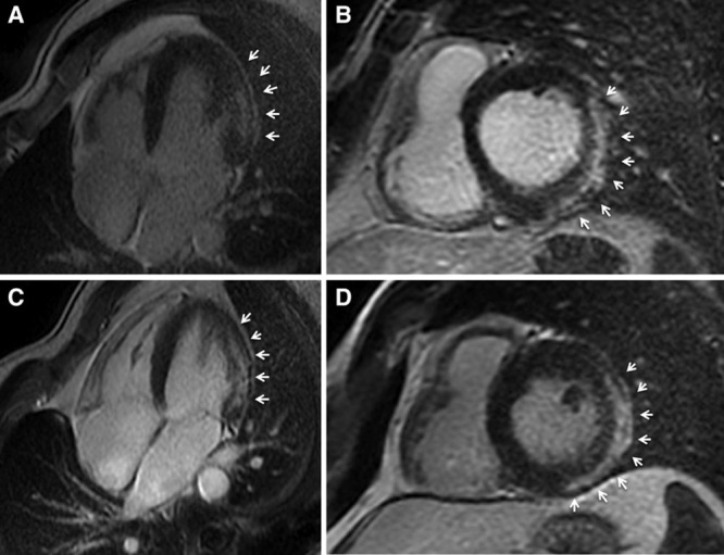
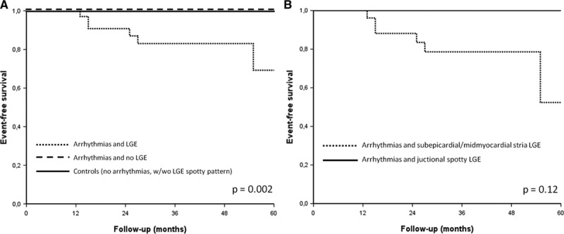
Background—: The clinical profile and arrhythmic outcome of competitive athletes with isolated nonischemic left ventricular (LV) scar as evidenced by contrast-enhanced cardiac magnetic resonance remain to be elucidated. Methods and Results—: We compared 35 athletes (80% men, age: 14–48 years) with ventricular arrhythmias and isolated LV subepicardial/midmyocardial late gadolinium enhancement (LGE) on contrast-enhanced cardiac magnetic resonance (group A) with 38 athletes with ventricular arrhythmias and no LGE (group B) and 40 healthy control athletes (group C). A stria LGE pattern with subepicardial/midmyocardial distribution, mostly involving the lateral LV wall, was found in 27 (77%) of group A versus 0 controls (group C; P <0.001), whereas a spotty pattern of LGE localized at the junction of the right ventricle to the septum was respectively observed in 11 (31%) versus 10 (25%; P =0.52). All athletes with stria pattern showed ventricular arrhythmias with a predominant right bundle branch block morphology, 13 of 27 (48%) showed ECG repolarization abnormalities, and 5 of 27 (19%) showed echocardiographic hypokinesis of the lateral LV wall. The majority of athletes with no or spotty LGE pattern had ventricular arrhythmias with a predominant left bundle branch block morphology and no ECG or echocardiographic abnormalities. During a follow-up of 38±25 months, 6 of 27 (22%) athletes with stria pattern experienced malignant arrhythmic events such as appropriate implantable cardiac defibrillator shock (n=4), sustained ventricular tachycardia (n=1), or sudden death (n=1), compared with none of athletes with no or LGE spotty pattern and controls. Conclusions—: Isolated nonischemic LV LGE with a stria pattern may be associated with life-threatening arrhythmias and sudden death in the athlete. Because of its subepicardial/midmyocardial location, LV scar is often not detected by echocardiography.
Overview
The claims and statistics of this paper haven’t been checked yet.
Run the check — free, no account needed. It reads the open-access text and checks the sources it cites.
Integrity signals
Registry
Cites
Statistics
Figures
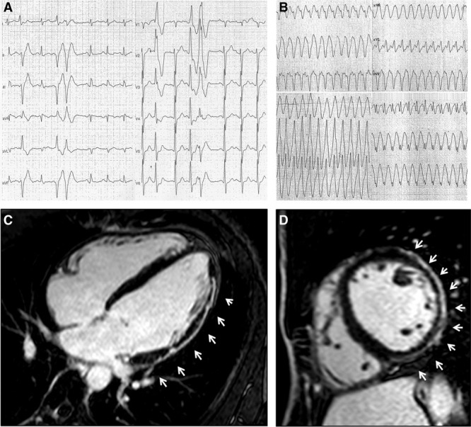
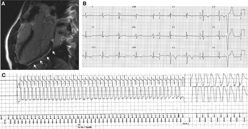
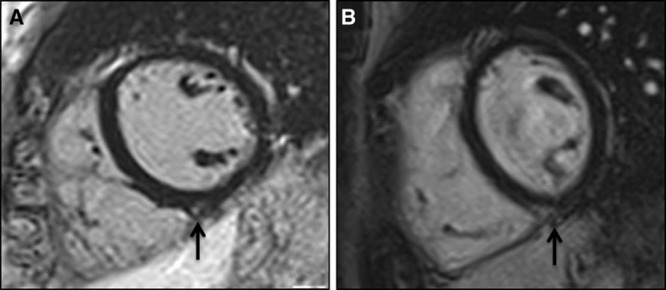
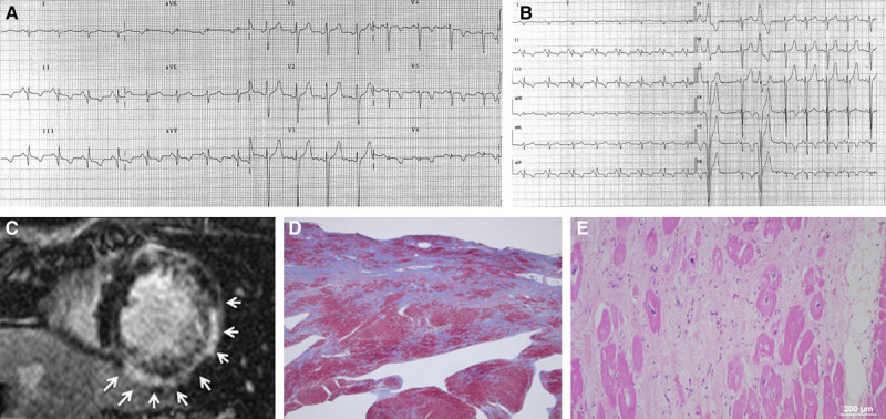
Verification
Claims the paper makes
The claims and statistics of this paper haven’t been checked yet.
Run the check — free, no account needed. It reads the open-access text and checks the sources it cites.
Claims other papers make about this one
Each is checked against this paper’s own text: does it say what the citing paper says it does?
The cited source says what the claim attributes to it.
In the citing paper
“Zorzi et al8 found athletes who presented with ventricular arrhythmia and were subsequently found to have nonischemic lateral LV wall fibrosis on CMR were more likely to experience malignant arrhythmic events than those without fibrosis.”
From Ventricular Arrhythmia and Cardiac Fibrosis in Endurance Experienced Athletes (VENTOUX) (2025) · cites this paper as [8]
Self-check
Percentages, sums and reported differences re-computed from the paper’s own numbers.
The self-check needs the paper’s full text.
Citations
Cites
Caforio, A. L. P., Pankuweit, S., Arbustini, E., Basso, C., Gimeno-Blanes, J., Felix, S. B., Fu, M., Heliö, T., Heymans, S., Jahns, R., et al. (2013) · European Heart Journal · doi:10.1093/eurheartj/eht210 · Cited by 3,479
Self-citationCardiovascular Magnetic Resonance in Myocarditis: A JACC White Paper
Friedrich, M. G., Sechtem, U. P., Schulz‐Menger, J., Holmvang, G., Alakija, P., Cooper, L. T., White, J. A., Abdel‐Aty, H., Gutberlet, M., Prasad, S. K., et al. (2009) · Journal of the American College of Cardiology · doi:10.1016/j.jacc.2009.02.007 · Cited by 2,523
Right Ventricular Cardiomyopathy and Sudden Death in Young People
Thiene, G., Nava, A. K., Corrado, D., Rossi, L., & PENNELLI, N. (1988) · New England Journal of Medicine · doi:10.1056/nejm198801213180301 · Cited by 1,614
Self-citationCorrado, D., Basso, C., Pavei, A., Michieli, P., Schiavon, M., & Thiene, G. (2006) · JAMA · doi:10.1001/jama.296.13.1593 · Cited by 1,559
Self-citationDoes sports activity enhance the risk of sudden death in adolescents and young adults?
Corrado, D., Basso, C., Rizzoli, G., Schiavon, M., & Thiene, G. (2003) · Journal of the American College of Cardiology · doi:10.1016/j.jacc.2003.03.002 · Cited by 1,311
Self-citationArrhythmogenic right ventricular cardiomyopathy
Basso, C., Corrado, D., Marcus, G. M., Nava, A. K., & Thiene, G. (2009) · The Lancet · doi:10.1016/s0140-6736(09)60256-7 · Cited by 938
Self-citationDelayed enhancement cardiovascular magnetic resonance assessment of non-ischaemic cardiomyopathies
Mahrholdt, H., Wagner, A., Judd, R. M., Sechtem, U. P., & Kim, R. J. (2005) · European Heart Journal · doi:10.1093/eurheartj/ehi258 · Cited by 914
Arrhythmogenic Right Ventricular Cardiomyopathy: Dysplasia, Dystrophy, or Myocarditis?
Basso, C., Thiene, G., Corrado, D., Angelini, A., Nava, A., & Valente, M. (1996) · Circulation · doi:10.1161/01.cir.94.5.983 · Cited by 890
Self-citationExercise-induced right ventricular dysfunction and structural remodelling in endurance athletes
La Gerche, A., Burns, A. T., Mooney, D. J., Inder, W. J., Taylor, A. J., Bogaert, J., MacIsaac, A. I., Heidbüchel, H., & Prior, D. L. (2012) · European Heart Journal · doi:10.1093/eurheartj/ehr397 · Cited by 776 · cited 2×
“T-wave inversion in V4-V6±1/aVL 7 (20) 0 .”
Left-Dominant Arrhythmogenic Cardiomyopathy
Sen‐Chowdhry, S., Syrris, P., Prasad, S., Hughes, S., Merrifield, R. D., Ward, D., Pennell, D. J., & McKenna, W. J. (2008) · Journal of the American College of Cardiology · doi:10.1016/j.jacc.2008.09.019 · Cited by 664
Cited by
Sorted by citations
2020 ESC guidelines on sports cardiology and exercise in patients with cardiovascular disease
Pelliccia, A., Sharma, S., Gati, S., Bäck, M., Börjesson, M., Caselli, S., Collet, J.-P., Corrado, D., Drezner, J. A., Halle, M., et al. (2021) · European Heart Journal · doi:10.1093/eurheartj/ehaa605 · Cited by 1,958
Self-citationMyocarditis and inflammatory cardiomyopathy: current evidence and future directions
Tschöpe, C., Ammirati, E., Bozkurt, B., Caforio, A. L. P., Cooper, L. T., Felix, S. B., Hare, J. M., Heidecker, B., Heymans, S., Hübner, N., et al. (2021) · Nature Reviews Cardiology · doi:10.1038/s41569-020-00435-x · Cited by 1,217
Arrhythmogenic Right Ventricular Cardiomyopathy
Corrado, D., Link, M. S., & Calkins, H. G. (2017) · New England Journal of Medicine · doi:10.1056/nejmra1509267 · Cited by 756
Self-citationDiagnosis of arrhythmogenic cardiomyopathy: The Padua criteria
Corrado, D., Marra, M. P., Zorzi, A., Beffagna, G., Cipriani, A., Lazzari, M. D., Migliore, F., Pilichou, K., Rampazzo, A., Rigato, I., et al. (2020) · International Journal of Cardiology · doi:10.1016/j.ijcard.2020.06.005 · Cited by 587
Self-citationCorrado, D., Basso, C., & Judge, D. P. (2017) · Circulation Research · doi:10.1161/circresaha.117.309345 · Cited by 474
Self-citation
Metadata
- Authors
- Zorzi, A., Perazzolo Marra, M., Rigato, I., De Lazzari, M., Susana, A., Niero, A., Pilichou, K., Migliore, F., Rizzo, S., Giorgi, B., De Conti, G., Sarto, P., Serratosa, L., Patrizi, G., De Maria, E., Pelliccia, A., Basso, C., Schiavon, M., Bauce, B., Iliceto, S., Thiene, G., & Corrado, D.
- Affiliations
- University of Padua (IT)
- From the Department of Cardiac, Thoracic, and Vascular Sciences (A.Z., M.P.M., I.R., M.D.L., A.S., A.N., K.P., F.M., S.R., C.B., B.B., S.I., G.T., D.C.) and Division of Radiology, Department of Medicine (B.G.), University of Padova, Padova, Italy; Department of Radiology, Azienda Ospedaliera di Padova, Padova, Italy (G.D.C.); Center for Sports Medicine, Treviso, Italy (P.S.); Unidad de Cardiologica del Deporte, Hospital Universitario Quiron, Madrid, Spain (L.S.); U.O. Cardiologia, Ospedale B...
- Corresponding author
- Zorzi, Alessandro
- ORCID
- Venue
- Circulation: Arrhythmia and Electrophysiology
- Published
- 2016-07
- Volume
- 9
- Issue
- 7
- ISSN
- 1941-3084 (ISSN-L)
- 1941-3149
- PMC
- PMC4956679
- OpenAlex
- W2464247829
- Cited by
- 326
- References
- 52
- Citations by year
- 2026: 21 · 2025: 28 · 2024: 44 · 2023: 47 · 2022: 32 · 2021: 44 · 2020: 50 · 2019: 27 · 2018: 20 · 2017: 12 · 2016: 1
- FWCI
- 16.47
- Type
- journal article
- Language
- en
- Open access
- hybrid
- doi.org
- Licence
- cc-by-nc-nd
- Locations
- Circulation Arrhythmia and Electrophysiology (publishedVersion, cc-by-nc-nd)
- Europe PMC (PubMed Central) (publishedVersion, cc-by-nc-nd)
- PubMed Central (publishedVersion, cc-by-nc-nd)
- PubMed (publishedVersion)
- Padua Research Archive (University of Padova) (submittedVersion, cc-by-nc-nd)
- Funders
- Università degli Studi di Padova (CPDA133979/13)
- Regione del Veneto
- Topics
- Health Sciences › Medicine › Cardiology and Cardiovascular Medicine › Cardiovascular Effects of Exercise
- Health Sciences › Medicine › Cardiology and Cardiovascular Medicine › Cardiomyopathy and Myosin Studies
- Health Sciences › Medicine › Cardiology and Cardiovascular Medicine › Cardiac electrophysiology and arrhythmias
- Keywords
- late gadolinium enhancement, sudden cardiac death, ventricular arrhythmias, competitive athletes, right bundle branch block, left bundle branch block, implantable cardioverter-defibrillator shocks, sustained ventricular tachycardia, repolarization abnormalities, cardiac magnetic resonance
- MeSH terms
- Adolescent, Adult, Cicatrix, Contrast Media, Female, Humans, Magnetic Resonance Imaging, Male, Meglumine, Middle Aged, Organometallic Compounds, Case-Control Studies, Death, Sudden, Cardiac, Tachycardia, Ventricular, Athletes
About these checks
Registry, retraction, Cites and statistics results are computed, not judged, with one exception: a printed reference that no index lists is read once by a model before it counts as a citation error. Claims are extracted by a model and kept only when their quote appears verbatim in the text; each cited source is then checked, with a model’s help, for whether it says what the claim relies on. Those verdicts can be wrong — read the claim’s quote and the cited source before relying on one.
Statistics and arithmetic are recomputed, not judged.
Confidence bands are shown only for fields calibrated on our annotated test set. None is calibrated yet, so no band is shown.
What counts as a claim ›How we measure confidence ›How we check this ›
Are you an author, or do you see a problem with this page? Request a review
