

The DOI matches its Crossref record; 3 of the 3 references it cites were found in a registry or an index.
Computed Tomography Imaging Measurements as a Surrogate for Clinical Outcomes After Surgical Management of Acute Type A Aortic Dissection: A Systematic Review
Open accessAbstract
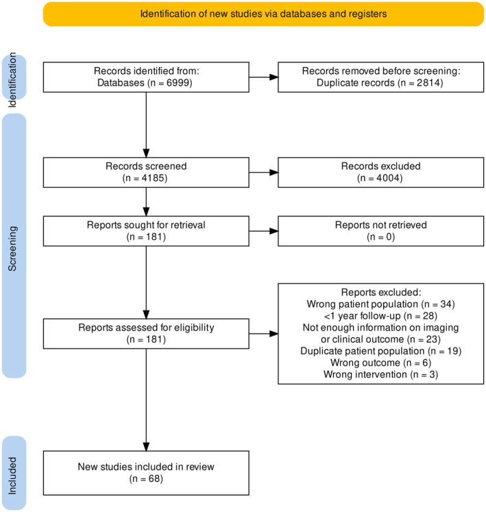
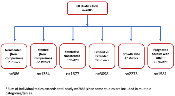
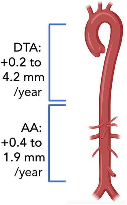
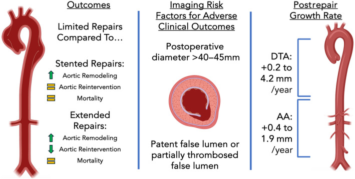
Background: Postoperative computed tomography imaging surveillance is an essential component of care after acute type A aortic dissection (ATAAD) repair. Prognostic imaging factors after ATAAD repair have not been systematically reviewed. Methods and Results: We performed a systematic review to summarize postoperative computed tomography measurements including aortic diameter, cross‐sectional area, volume, growth rate, and false lumen thrombosis in addition mid‐ to long‐term clinical outcomes after ATAAD repair. Searches were conducted in Medline, Embase, and CENTRAL in October 2022. Studies were included if they reported clinical outcomes such as mortality or aortic reintervention after 1 year and included aforementioned computed tomography findings. Studies of chronic aortic dissection and studies of exclusive patient populations such as those with connective tissue diseases were excluded. Risk of bias was assessed with the Newcastle‐Ottawa Scale. Searches retrieved 6999 articles. Sixty‐eight studies met inclusion criteria (7885 patients). Extended repairs were associated with improved false lumen thrombosis, decreased aortic growth rate, and decreased rates of reintervention but not improved survival. Growth rates of the aorta post‐ATAAD repair were highest in the descending thoracic aorta. The most frequent prognostic imaging factors reported were a patent/partially thrombosed false lumen and postoperative aortic diameter >40 to 45 mm. Conclusions: Established measurements of positive aortic remodeling, including complete false lumen thrombosis and stabilization of postoperative aortic diameter and growth are the most studied prognostic indicators for improved clinical outcomes after ATAAD repair. Growth rate of the aorta remains significant after ATAAD repair. Future studies should prospectively evaluate and compare prognostic factors for improved surveillance and management.
Overview
The claims and statistics of this paper haven’t been checked yet.
Run the check — free, no account needed. It reads the open-access text and checks the sources it cites.
Integrity signals
Registry
Cites
Statistics
Figures
Verification
Claims the paper makes
The claims and statistics of this paper haven’t been checked yet.
Run the check — free, no account needed. It reads the open-access text and checks the sources it cites.
Self-check
Percentages, sums and reported differences re-computed from the paper’s own numbers.
The self-check needs the paper’s full text.
Citations
Cites
The PRISMA 2020 statement: an updated guideline for reporting systematic reviews
Page, M. J., McKenzie, J. E., Bossuyt, P. M., Boutron, I., Hoffmann, T. C., Mulrow, C. D., Shamseer, L., Tetzlaff, J. M., Akl, E. A., Brennan, S. E., et al. (2021) · Systematic Reviews · doi:10.1186/s13643-021-01626-4 · Cited by 13,874
PRESS Peer Review of Electronic Search Strategies: 2015 Guideline Statement
McGowan, J., Sampson, M., Salzwedel, D. M., Cogo, E., Foerster, V., & Lefebvre, C. (2016) · Journal of Clinical Epidemiology · doi:10.1016/j.jclinepi.2016.01.021 · Cited by 5,700
Isselbacher, E. M., Preventza, O., Hamilton Black, J., Augoustides, J. G., Beck, A. W., Bolen, M. A., Braverman, A. C., Bray, B. E., Brown-Zimmerman, M. M., Chen, E. P., et al. (2022) · Circulation · doi:10.1161/cir.0000000000001106 · Cited by 2,082
Research works printed but not linked: 92
- 10.1093/ejcts/ezz071 · DOI
- 10.1016/j.jtcvs.2023.04.012 · DOI
- 10.1093/ejcts/ezv351 · DOI
- 10.1002/14651858.CD013149.pub2 · DOI
- 10.1111/jebm.12427 · DOI
- 10.1002/cl2.1230 · DOI · Paper page
- 10.5606/tgkdc.dergisi.2019.16879 · DOI
- 10.1016/j.jtcvs.2022.08.040 · DOI
- 10.1016/j.athoracsur.2015.07.060 · DOI
- 10.5090/kjtcs.2013.46.1.33 · DOI
- 10.5761/atcs.oa.15-00015 · DOI
- 10.1016/j.ejcts.2011.05.063 · DOI
- 10.1111/jocs.12608 · DOI
- 10.1093/ejcts/ezr311 · DOI
- 10.1016/j.athoracsur.2009.06.055 · DOI
- 10.1093/icvts/ivz154 · DOI
- 10.1532/hsf.1708 · DOI
- 10.21037/jtd.2018.06.148 · DOI
- 10.1016/j.athoracsur.2009.11.054 · DOI
- 10.1016/j.jtcvs.2006.07.043 · DOI
- 10.1016/j.jtcvs.2012.06.047 · DOI
- 10.1093/ejcts/ezx504 · DOI
- 10.1016/j.athoracsur.2018.05.067 · DOI
- 10.1093/ejcts/ezx131 · DOI
- 10.1093/ejcts/ezy381 · DOI
- 10.1093/ejcts/ezac443 · DOI
- 10.1093/icvts/ivy094 · DOI
- 10.1016/j.athoracsur.2010.06.087 · DOI
- 10.1016/j.athoracsur.2008.03.037 · DOI
- 10.1093/ejcts/ezw340 · DOI
- 10.1093/ejcts/ezu173 · DOI
- 10.1093/ejcts/ezs157 · DOI
- 10.1016/j.athoracsur.2022.03.054 · DOI
- 10.1016/j.jtcvs.2008.05.052 · DOI
- 10.1177/1708538115627728 · DOI
- 10.1007/s00268-020-05374-9 · DOI
- 10.1055/s-0036-1571813 · DOI
- 10.1016/j.ijcard.2018.01.010 · DOI
- 10.1016/j.ejvs.2018.05.023 · DOI
- 10.1093/icvts/ivw044 · DOI
- 10.1016/j.athoracsur.2010.07.077 · DOI
- 10.21037/jovs.2018.04.09 · DOI
- 10.1016/j.athoracsur.2005.04.004 · DOI
- 10.1093/ejcts/ezaa411 · DOI
- 10.1016/j.jtcvs.2015.03.068 · DOI
- 10.1016/j.athoracsur.2008.09.032 · DOI
- 10.1016/j.jtcvs.2018.11.145 · DOI
- 10.1016/j.athoracsur.2019.12.027 · DOI
- 10.1016/j.jcct.2016.10.005 · DOI
- 10.1016/j.athoracsur.2017.08.037 · DOI
- 10.1510/icvts.2006.132233 · DOI
- 10.1016/j.jtcvs.2014.02.077 · DOI
- 10.1016/j.jtcvs.2013.10.058 · DOI
- 10.1016/j.jacc.2007.03.064 · DOI
- 10.1016/j.jtcvs.2009.12.007 · DOI
- 10.1016/j.athoracsur.2022.05.070 · DOI
- 10.1161/CIRCULATIONAHA.110.015081 · DOI
- 10.1093/ejcts/ezx223 · DOI
- 10.1016/j.jtcvs.2014.02.003 · DOI
- 10.1016/j.athoracsur.2008.11.061 · DOI
- 10.1016/j.jtcvs.2020.02.087 · DOI
- 10.1016/j.jtcvs.2012.05.067 · DOI
- 10.21037/jtd-20-2356 · DOI
- 10.1093/ejcts/ezv374 · DOI
- 10.1016/j.jtcvs.2013.10.047 · DOI
- 10.1016/j.jtcvs.2020.10.136 · DOI
- 10.1016/j.xjtc.2022.05.013 · DOI
- 10.1093/ejcts/ezv044 · DOI
- 10.1093/icvts/ivaa095 · DOI
- 10.1016/j.jvs.2016.09.056 · DOI
- 10.1186/s13019-014-0140-6 · DOI
- 10.1093/icvts/ivy243 · DOI
- 10.1016/j.athoracsur.2007.03.084 · DOI
- 10.1093/ejcts/ezs121 · DOI
- 10.21037/acs.2016.05.06 · DOI
- 10.1093/eurheartj/ehad148 · DOI
- 10.1016/j.athoracsur.2023.03.037 · DOI
- 10.1161/CIRCULATIONAHA.107.702720 · DOI
- 10.1016/j.xjon.2022.02.025 · DOI
- 10.1016/j.athoracsur.2018.01.071 · DOI
- 10.1111/jocs.14446 · DOI
- 10.3390/jcdd9100349 · DOI
- 10.1001/jamanetworkopen.2018.1281 · DOI
- 10.1016/j.jtcvs.2019.10.125 · DOI
- 10.1016/j.jvs.2014.01.042 · DOI
- 10.1016/j.athoracsur.2020.04.064 · DOI
- 10.1016/j.jvs.2017.05.128 · DOI
- 10.1093/ejcts/ezab317 · DOI
- 10.1016/j.athoracsur.2023.11.014 · DOI
- 10.1016/j.jacc.2021.08.058 · DOI
- 10.1016/j.athoracsur.2021.04.010 · DOI
- 10.5195/jmla.2021.939 · DOI
Metadata
- Authors
- Vo, T. X., Rahmouni, K., Visintini, S., Guo, M. H., Ouzounian, M., Chu, M. W. A., Boodhwani, M., Appoo, J. J., Tucker, K., Al‐Atassi, T., & (CTAC), T. C. T. A. C.
- Affiliations
- Ottawa Heart Institute (CA)
- University of Toronto (CA)
- Western University (CA)
- University of Calgary (CA)
- University of Oxford (GB)
- Corresponding author
- Vo, Thin X.
- ORCID
- Venue
- Journal of the American Heart Association
- Published
- 2024-09-17
- Volume
- 13
- Issue
- 18
- Pages
- e034496-e034496
- ISSN
- 2047-9980 (ISSN-L)
- PMC
- PMC11935611
- OpenAlex
- W4402369427
- Cited by
- 2
- References
- 3
- Citations by year
- 2026: 2
- FWCI
- 0.15
- Type
- journal article
- Language
- en
- Open access
- gold
- dx.doi.org
- Licence
- cc-by-nc-nd
- Locations
- Journal of the American Heart Association (publishedVersion, cc-by-nc-nd)
- PubMed Central (publishedVersion, cc-by-nc-nd)
- PubMed (publishedVersion)
- DOAJ (DOAJ: Directory of Open Access Journals) (submittedVersion, cc-by-sa)
- Funders
- Heart and Stroke Foundation of Canada
- Edwards Lifesciences
- University of Ottawa
- Topics
- Health Sciences › Medicine › Cardiology and Cardiovascular Medicine › Aortic Disease and Treatment Approaches
- Health Sciences › Medicine › Surgery › Aortic aneurysm repair treatments
- Life Sciences › Biochemistry, Genetics and Molecular Biology › Genetics › Connective tissue disorders research
- Keywords
- acute type A aortic dissection, aortic dissection repair, postoperative computed tomography, aortic remodeling, false lumen thrombosis, aortic diameter, aortic growth rate, aortic cross-sectional area, aortic volume, aortic reintervention, mortality
- MeSH terms
- Computed Tomography Angiography, Acute Disease, Aortic Dissection, Aorta, Thoracic, Aortic Aneurysm, Humans, Prognosis, Tomography, X-Ray Computed, Treatment Outcome, Aortic Aneurysm, Thoracic, Blood Vessel Prosthesis Implantation
About these checks
Registry, retraction, Cites and statistics results are computed, not judged, with one exception: a printed reference that no index lists is read once by a model before it counts as a citation error. Claims are extracted by a model and kept only when their quote appears verbatim in the text; each cited source is then checked, with a model’s help, for whether it says what the claim relies on. Those verdicts can be wrong — read the claim’s quote and the cited source before relying on one.
Statistics and arithmetic are recomputed, not judged.
Confidence bands are shown only for fields calibrated on our annotated test set. None is calibrated yet, so no band is shown.
What counts as a claim ›How we measure confidence ›How we check this ›
Are you an author, or do you see a problem with this page? Request a review
How to cite
Cite CoChat for the checks on this page, and the paper for its own findings.
- This page
- CoChat. (2026). Citation check: Computed Tomography Imaging Measurements as a Surrogate for Clinical Outcomes After Surgical Management of Acute Type A Aortic Dissection: A Systematic Review. https://app.cochat.ai/papers/10.1161/jaha.124.034496
- The paper
- Vo, T. X., Rahmouni, K., Visintini, S., Guo, M. H., Ouzounian, M., Chu, M. W. A., Boodhwani, M., Appoo, J. J., Tucker, K., Al‐Atassi, T., & (CTAC), T. C. T. A. C. (2024). Computed Tomography Imaging Measurements as a Surrogate for Clinical Outcomes After Surgical Management of Acute Type A Aortic Dissection: A Systematic Review. Journal of the American Heart Association. https://doi.org/10.1161/jaha.124.034496
