

The DOI matches its Crossref record; 66 of the 66 references it cites were found in a registry or an index.
Vortioxetine disinhibits pyramidal cell function and enhances synaptic plasticity in the rat hippocampus
Open accessAbstract
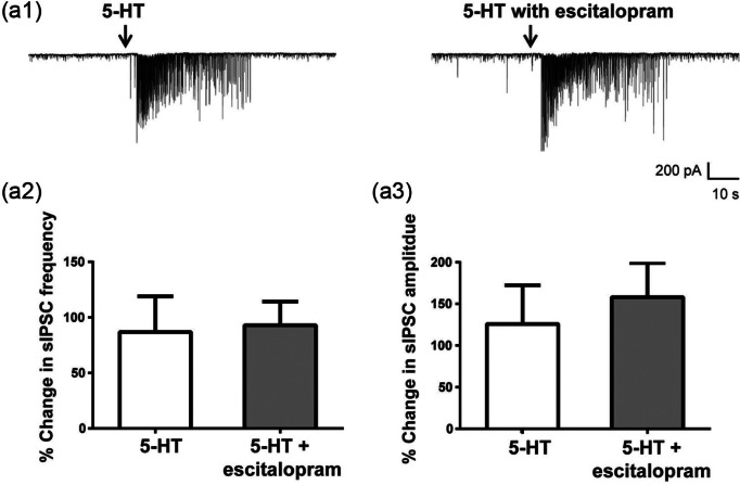
Vortioxetine, a novel antidepressant with multimodal action, is a serotonin (5-HT) 3 , 5-HT 7 and 5-HT 1D receptor antagonist, a 5-HT 1B receptor partial agonist, a 5-HT 1A receptor agonist and a 5-HT transporter (SERT) inhibitor. Vortioxetine has been shown to improve cognitive performance in several preclinical rat models and in patients with major depressive disorder. Here we investigated the mechanistic basis for these effects by studying the effect of vortioxetine on synaptic transmission, long-term potentiation (LTP), a cellular correlate of learning and memory, and theta oscillations in the rat hippocampus and frontal cortex. Vortioxetine was found to prevent the 5-HT-induced increase in inhibitory post-synaptic potentials recorded from CA1 pyramidal cells, most likely by 5-HT 3 receptor antagonism. Vortioxetine also enhanced LTP in the CA1 region of the hippocampus. Finally, vortioxetine increased fronto-cortical theta power during active wake in whole animal electroencephalographic recordings. In comparison, the selective SERT inhibitor escitalopram showed no effect on any of these measures. Taken together, our results indicate that vortioxetine can increase pyramidal cell output, which leads to enhanced synaptic plasticity in the hippocampus. Given the central role of the hippocampus in cognition, these findings may provide a cellular correlate to the observed preclinical and clinical cognition-enhancing effects of vortioxetine.
Overview
The claims and statistics of this paper haven’t been checked yet.
Free: it reads the open-access text and checks the first 5 claims that cite a source we can check. Create a free account to check every claim.
Integrity signals
Registry
Cites
Statistics
Figures




Verification
Claims the paper makes
The claims and statistics of this paper haven’t been checked yet.
Free: it reads the open-access text and checks the first 5 claims that cite a source we can check. Create a free account to check every claim.
Claims other papers make about this one
Each is checked against this paper’s own text: does it say what the citing paper says it does?
In the citing paper
“In rodents, vortioxetine increases hippocampal synaptic plasticity and dendritic spine maturation [101, 102].”
From Beyond the serotonin deficit hypothesis: Communicating a neuroplasticity framework of major depressive disorder (2024) · cites this paper as [102]
In the citing paper
“Preclinical studies have shown that vortioxetine may influence learning and memory processes by improving hippocampal synaptic plasticity and increasing the output of pyramidal cells [89].”
From Cognitive remission: a novel objective for the treatment of major depression? (2016) · cites this paper as [89]
Self-check
Percentages, sums and reported differences re-computed from the paper’s own numbers.
The self-check needs the paper’s full text.
Citations
Cites
EEG alpha and theta oscillations reflect cognitive and memory performance: a review and analysis
Klimesch, W. (1999) · Brain Research Reviews · doi:10.1016/s0165-0173(98)00056-3 · Cited by 6,889
Millan, M. J., Agid, Y., Brüne, M., Bullmore, E. T., Carter, C. S., Clayton, N. S., Connor, R., Davis, S., Deakin, B., DeRubeis, R. J., et al. (2012) · Nature Reviews Drug Discovery · doi:10.1038/nrd3628 · Cited by 1,292
Gating of human theta oscillations by a working memory task
Raghavachari, S., Kahana, M. J., Rizzuto, D. S., Caplan, J. B., Kirschen, M. P., Bourgeois, B., Madsen, J. R., & Lisman, J. E. (2001) · The Journal of Neuroscience · doi:10.1523/jneurosci.21-09-03175.2001 · Cited by 792 · cited 2×
“…oscillations are predominantly driven via hippocampal entorhinal-cortical projections (Young and McNaughton, 2009) and have been linked to cognitive functions in preclinical as well as clinical studies (Basar et al., 2000; Caplan et al., 2001; Klimesch, 1999; Raghavachari et al., 2001, 2006).”
Bang-Andersen, B., Ruhland, T., Jørgensen, M., Smith, G., Frederiksen, K., Jensen, K. G., Zhong, H., Nielsen, S. M., Hogg, S., Mørk, A., et al. (2011) · Journal of Medicinal Chemistry · doi:10.1021/jm101459g · Cited by 606
Andrade, R., & Nicoll, R. A. (1987) · The Journal of Physiology · doi:10.1113/jphysiol.1987.sp016862 · Cited by 552
Sanchez, C., Asin, K. E., & Artigas, F. (2015) · Pharmacology & Therapeutics · doi:10.1016/j.pharmthera.2014.07.001 · Cited by 547
Owens, M. J., Knight, D. L., & Nemeroff, C. B. (2001) · Biological Psychiatry · doi:10.1016/s0006-3223(01)01145-3 · Cited by 516
Conradi, H. J., Ormel, J., & de Jonge, P. (2011) · Psychological Medicine · doi:10.1017/s0033291710001911 · Cited by 515
Katona, C., Hansen, T., & Olsen, C. K. (2012) · International Clinical Psychopharmacology · doi:10.1097/yic.0b013e3283542457 · Cited by 445
Role of serotonin in memory impairment.
Buhot, M.-C., Martin, S., & Segu, L. (2000) · Annals of Medicine · doi:10.3109/07853890008998828 · Cited by 419
Research works printed but not linked: 4
- Adell A, 2010, IDrugs, 13, 900
- Maire M, 2013, J Cogn Behav Psychother, 13, 133
- Tokarski K, 2011, J Physiol Pharmacol, 62, 535
- Westrich L, 2012, Int J Psychiatry Clin Pract, 16, 47
Cited by
Sanchez, C., Asin, K. E., & Artigas, F. (2015) · Pharmacology & Therapeutics · doi:10.1016/j.pharmthera.2014.07.001 · Cited by 547
Cognitive remission: a novel objective for the treatment of major depression?
Bortolato, B., Miskowiak, K. W., Köhler, C. A., Maes, M., Fernandes, B. S., Berk, M., & Carvalho, A. F. (2016) · BMC Medicine · doi:10.1186/s12916-016-0560-3 · Cited by 252
Baune, B. T., Brignone, M., & Larsen, K. G. (2018) · International Journal of Neuropsychopharmacology · doi:10.1093/ijnp/pyx070 · Cited by 140
Vortioxetine: a novel antidepressant for the treatment of major depressive disorder
Gonda, X., Sharma, S. R., & Tarazi, F. I. (2019) · Expert Opinion on Drug Discovery · doi:10.1080/17460441.2019.1546691 · Cited by 123
Vortioxetine: A review of the pharmacology and clinical profile of the novel antidepressant
Sowa-Kućma, M., Pańczyszyn-Trzewik, P., Misztak, P., Jaeschke, R. R., Sendek, K., Styczeń, K., Datka, W., & Koperny, M. (2017) · Pharmacological Reports · doi:10.1016/j.pharep.2017.01.030 · Cited by 86
Metadata
- Authors
- Dale, E., Zhang, H., Leiser, S. C., Xiao, Y., Lu, D., Yang, C. R., Plath, N., & Sanchez, C.
- Affiliations
- Lundbeck Research USA, Paramus, NJ, USA
- Shanghai Chempartner Co. Ltd., Shanghai, China
- Lundbeck (Denmark) (DK)
- Corresponding author
- Dale, Elena
- Venue
- Journal of Psychopharmacology
- Publisher
- SAGE Publications
- Published
- 2014-08-13
- Volume
- 28
- Issue
- 10
- Pages
- 891-902
- ISSN
- 0269-8811 (ISSN-L)
- 1461-7285
- PMC
- PMC4230848
- OpenAlex
- W2135657370
- Cited by
- 93
- References
- 66
- Citations by year
- 2026: 3 · 2025: 5 · 2024: 6 · 2023: 8 · 2022: 4 · 2021: 7 · 2020: 6 · 2019: 5 · 2018: 8 · 2017: 14 · 2016: 11 · 2015: 14 · 2014: 2
- FWCI
- 9.94
- Type
- journal article
- Language
- en
- Update notices
- correction (2014-11-06)
- Open access
- hybrid
- doi.org
- Licence
- cc-by-nc
- journals.sagepub.com/page/policies/text-and-data-mining-license (tdm)
- Locations
- Journal of Psychopharmacology (publishedVersion, cc-by-nc)
- Europe PMC (PubMed Central) (publishedVersion, cc-by-nc)
- PubMed Central (publishedVersion, cc-by-nc)
- PubMed (publishedVersion)
- Topics
- Health Sciences › Medicine › Pharmacology (medical) › Treatment of Major Depression
- Life Sciences › Neuroscience › Cellular and Molecular Neuroscience › Neuroscience and Neuropharmacology Research
- Life Sciences › Neuroscience › Cellular and Molecular Neuroscience › Neurotransmitter Receptor Influence on Behavior
- Keywords
- vortioxetine, serotonin receptors, 5-HT3 receptor antagonism, synaptic plasticity, long-term potentiation, CA1 pyramidal neurons, inhibitory postsynaptic potentials, theta oscillations, serotonin transporter inhibition, escitalopram, cognitive enhancement, major depressive disorder, antidepressants
- MeSH terms
- Vortioxetine, Animals, Antidepressive Agents, Frontal Lobe, Synaptic Transmission, Neuronal Plasticity, Piperazines, Serotonin, Serotonin Antagonists, Sulfides, Theta Rhythm, Citalopram, Pyramidal Cells, Rats, Inhibitory Postsynaptic Potentials, CA1 Region, Hippocampal
About these checks
Registry, retraction, Cites and statistics results are computed, not judged, with one exception: a printed reference that no index lists is read once by a model before it counts as a citation error. Claims are extracted by a model and kept only when their quote appears verbatim in the text; each cited source is then checked, with a model’s help, for whether it says what the claim relies on. Those verdicts can be wrong — read the claim’s quote and the cited source before relying on one.
Statistics and arithmetic are recomputed, not judged.
Confidence bands are shown only for fields calibrated on our annotated test set. None is calibrated yet, so no band is shown.
What counts as a claim ›How we measure confidence ›How we check this ›
Are you an author, or do you see a problem with this page? Request a review
How to cite
Cite CoChat for the checks on this page, and the paper for its own findings.
- This page
- CoChat. (2026). Citation check: Vortioxetine disinhibits pyramidal cell function and enhances synaptic plasticity in the rat hippocampus. https://app.cochat.ai/papers/10.1177/0269881114543719
- The paper
- Dale, E., Zhang, H., Leiser, S. C., Xiao, Y., Lu, D., Yang, C. R., Plath, N., & Sanchez, C. (2014). Vortioxetine disinhibits pyramidal cell function and enhances synaptic plasticity in the rat hippocampus. Journal of Psychopharmacology. https://doi.org/10.1177/0269881114543719
Data sources
Bibliographic data: Semantic Scholar, OpenAlex, Crossref.
Contains information from the Semantic Scholar Academic Graph Datasets, made available under the ODC Attribution License (ODC-BY 1.0).
Semantic Scholar’s data is described in: Kinney, R., et al. (2023). The Semantic Scholar Open Data Platform. arXiv:2301.10140.
