

The DOI matches its Crossref record; 57 of the 57 references it cites were found in a registry or an index.
Predicting Language Function Post-Stroke: A Model-Based Structural Connectivity Approach
Open accessAbstract
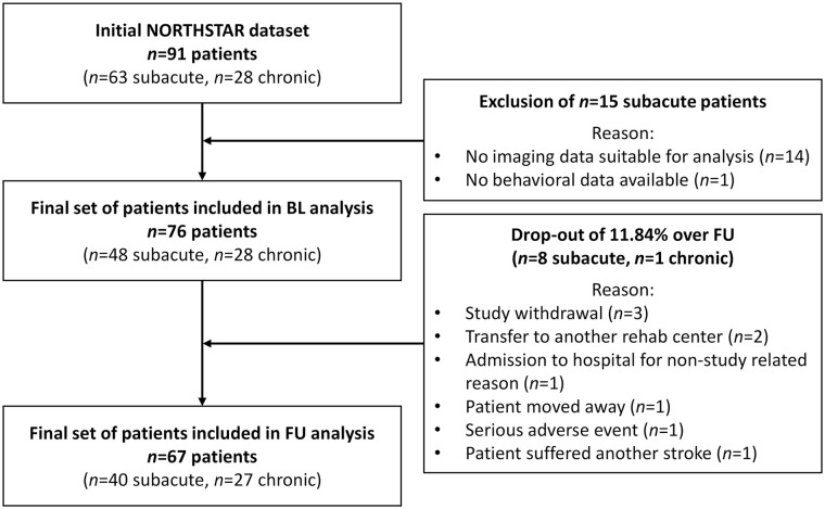
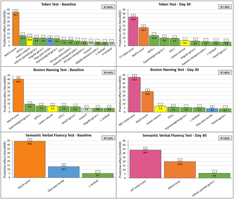
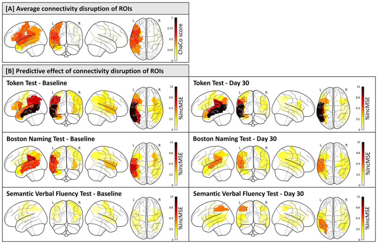
Background: The prediction of post-stroke language function is essential for the development of individualized treatment plans based on the personal recovery potential of aphasic stroke patients. Objective: To establish a framework for integrating information on connectivity disruption of the language network based on routinely collected clinical magnetic resonance (MR) images into Random Forest modeling to predict post-stroke language function. Methods: Language function was assessed in 76 stroke patients from the Non-Invasive Repeated Therapeutic Stimulation for Aphasia Recovery trial, using the Token Test (TT), Boston Naming Test (BNT), and Semantic Verbal Fluency (sVF) Test as primary outcome measures. Individual infarct masks were superimposed onto a diffusion tensor imaging tractogram reference set to calculate Change in Connectivity scores of language-relevant gray matter regions as estimates of structural connectivity disruption. Multivariable Random Forest models were derived to predict language function. Results: Random Forest models explained moderate to high amount of variance at baseline and follow-up for the TT (62.7% and 76.2%), BNT (47.0% and 84.3%), and sVF (52.2% and 61.1%). Initial language function and non-verbal cognitive ability were the most important variables to predict language function. Connectivity disruption explained additional variance, resulting in a prediction error increase of up to 12.8% with variable omission. Left middle temporal gyrus (12.8%) and supramarginal gyrus (9.8%) were identified as among the most important network nodes. Conclusion: Connectivity disruption of the language network adds predictive value beyond lesion volume, initial language function, and non-verbal cognitive ability. Obtaining information on connectivity disruption based on routine clinical MR images constitutes a significant advancement toward practical clinical application.
Overview
The claims and statistics of this paper haven’t been checked yet.
Free: it reads the open-access text and checks the first 5 claims that cite a source we can check. Create a free account to check every claim.
Integrity signals
Registry
Cites
Statistics
Figures
Verification
Claims the paper makes
The claims and statistics of this paper haven’t been checked yet.
Free: it reads the open-access text and checks the first 5 claims that cite a source we can check. Create a free account to check every claim.
Claims other papers make about this one
Each is checked against this paper’s own text: does it say what the citing paper says it does?
The cited source doesn’t support the claim as written.
In the citing paper
“In this study, 18 studies (19–29, 31–37) used supervised machine learning algorithms, and 1 study (30) used unsupervised machine learning algorithms”
From Machine learning models in post-stroke aphasia: a scoping review (2026) · cites this paper as [26]
Checked by a reasoning model — may be wrong.
Experimental — accuracy not yet measuredThe source backs part of the claim, not all of it — or backs it for a different population, condition or comparison.
In the citing paper
“The analysis results showed that the machine learning algorithms used in post-stroke aphasia models were mainly supervised methods, including random forests, neural networks, and support vector machines.”
From Machine learning models in post-stroke aphasia: a scoping review (2026) · cites this paper as [26]
Experimental — accuracy not yet measuredThe source backs part of the claim, not all of it — or backs it for a different population, condition or comparison.
In the citing paper
“Seven studies (21, 23, 26, 31, 32, 35, 36) primarily relied on error metrics, such as root mean square error and mean absolute error, combined with indicators, such as the Pearson correlation coefficient and coefficient of determination, to provide a more comprehensive assessment.”
From Machine learning models in post-stroke aphasia: a scoping review (2026) · cites this paper as [26]
Experimental — accuracy not yet measuredThe source backs part of the claim, not all of it — or backs it for a different population, condition or comparison.
In the citing paper
“three studies (26, 32, 34) predict the long-term language function outcome of patients”
From Machine learning models in post-stroke aphasia: a scoping review (2026) · cites this paper as [26]
Experimental — accuracy not yet measuredThe source backs part of the claim, not all of it — or backs it for a different population, condition or comparison.
In the citing paper
“In terms of language function and rehabilitation outcome prediction, most studies remain in the performance Validation stage and lack prospective clinical utility studies (20, 26, 29, 32, 34)”
From Machine learning models in post-stroke aphasia: a scoping review (2026) · cites this paper as [26]
Experimental — accuracy not yet measured
5 more claims about this paper
Create a free account to see every claim other papers make about this one.
Self-check
Percentages, sums and reported differences re-computed from the paper’s own numbers.
The self-check needs the paper’s full text.
Citations
Cites
Breiman, L. (2001) · Machine Learning · doi:10.1023/a:1010933404324 · Cited by 133,538 · cited 6×
“38 Variable importance is reported as the percentage increase of the MSE (%IncMSE), with a higher %IncMSE specifying a larger role of the predictor variable in explaining the model variance.”
BackgroundMethodInfluentialTzourio-Mazoyer, N., Landeau, B., Papathanassiou, D., Crivello, F., Etard, O., Delcroix, N., Mazoyer, B., & Joliot, M. (2002) · NeuroImage · doi:10.1006/nimg.2001.0978 · Cited by 16,319 · cited 1×
“34 To minimize the probability of type II statistical errors, we pre-selected 13 language-relevant GM regions of the left hemisphere (Table 1).”
MethodThe cortical organization of speech processing
Hickok, G., & Poeppel, D. (2007) · Nature Reviews Neuroscience · doi:10.1038/nrn2113 · Cited by 5,495
Price, C. J. (2012) · NeuroImage · doi:10.1016/j.neuroimage.2012.04.062 · Cited by 2,159 · cited 1×
“35-37 Respective right-hemispheric homologues of pre-defined ROIs were additionally included.”
BackgroundTombaugh, T. (1999) · Archives of Clinical Neuropsychology · doi:10.1016/s0887-6177(97)00095-4 · Cited by 1,439
Structure and Function of the Human Insula
Uddin, L. Q., Nomi, J. S., Hébert-Seropian, B., Ghaziri, J., & Boucher, O. (2017) · Journal of Clinical Neurophysiology · doi:10.1097/wnp.0000000000000377 · Cited by 1,262
Normative Data and Screening Power of a Shortened Version of the Token Test
De Renzi, E., & Faglioni, P. (1978) · Cortex · doi:10.1016/s0010-9452(78)80006-9 · Cited by 1,121 · cited 1×
“26 Receptive language function was assessed with a sentence comprehension test (shortened version of the Token Test, TT), 29 and language expression was measured with a picture naming test (Boston Naming Test, BNT), 30 and a Semantic Verbal Fluency (sVF) Test.”
MethodWhat do verbal fluency tasks measure? Predictors of verbal fluency performance in older adults
Shao, Z., Janse, E., Visser, K., & Meyer, A. S. (2014) · Frontiers in Psychology · doi:10.3389/fpsyg.2014.00772 · Cited by 905 · cited 1×
“48 Our sVF models did not demonstrate any major brain-structural predictors, which may be attributed to the hybrid test character and the recruitment of brain regions beyond our pre-selected language-specific ROIs.”
BackgroundSynthStrip: skull-stripping for any brain image
Hoopes, A., Mora, J. S., Dalca, A. V., Fischl, B., & Hoffmann, M. (2022) · NeuroImage · doi:10.1016/j.neuroimage.2022.119474 · Cited by 488 · cited 1×
“32 Brain-extracted scans were normalized to the MNI152 template, using a linear rigid body and affine normalization, followed by a non-linear transformation using the ANTs SyN (Advanced Normalization Tools, Symmetric Image Normalization) algorithm.”
MethodPoststroke Aphasia Frequency, Recovery, and Outcomes: A Systematic Review and Meta-Analysis
Flowers, H. L., Skoretz, S. A., Silver, F. L., Rochon, E., Fang, J., Flamand-Roze, C., & Martino, R. (2016) · Archives of Physical Medicine and Rehabilitation · doi:10.1016/j.apmr.2016.03.006 · Cited by 432
Research works printed but not linked: 4
- Hickok G, 2007, Nature, 8, 393
- Tombaugh TN., 1999, Arch Clin Neuropsychol, 14, 167
- Breiman L., Machine Learning, 2001, 5
- Liaw A, 2002, R News, 2, 18
Cited by
Navigating the Complexity of Bilingual Aphasia: Current Insights and Future Directions
Russell-Meill, M., Marte, M. J., Carpenter, E., & Kiran, S. (2025) · Brain Sciences · doi:10.3390/brainsci15090989 · Cited by 12
“…accuracy; lesion topology derived from structural MRI further improves performance [144,145]; network-level connectivity from diffusion MRI or tractography adds additional gains [146]; and, in some cases, functional measures from resting-state fMRI can outperform behavioral predictors [147].”
Desjardins, M.-È., Marcotte, K., Bedetti, C., Houzé, B., Brisebois, A., Descoteaux, M., Désautels, A., & Brambati, S. M. (2026) · Cortex · doi:10.1016/j.cortex.2026.01.004 · Cited by 4
Machine learning models in post-stroke aphasia: a scoping review
Li, X., Song, H., Guo, N., Kang, C., Gong, X., Ji, X., & Zheng, J. (2026) · Frontiers in Neurology · doi:10.3389/fneur.2026.1806856
“Seven studies (21, 23, 26, 31, 32, 35, 36) primarily relied on error metrics, such as root mean square error and mean absolute error, combined with indicators, such as the Pearson correlation coefficient and coefficient of determination, to provide a more comprehensive assessment.”
InfluentialHuang, Y., Chai, L., Ni, J., Xiong, X., Zuo, S., Lin, B., & Huang, J. (2026) · Aphasiology · doi:10.1080/02687038.2026.2649848
Tractography-Based Predictors of Poststroke Aphasia: A Systematic Review
Narimani, M., Kiani, I., Ghorani, H., Seraj, P., Cho, A., Tsang, D. A., Lalwani, K., Hillis, A. E., Lakhani, D. A., Dmytriw, A. A., et al. (2026) · American Journal of Neuroradiology · doi:10.3174/ajnr.a9333
Metadata
- Authors
- Hildesheim, F. E., Ophey, A., Zumbansen, A., Funck, T., Schuster, T., Jamison, K. W., Kuceyeski, A., & Thiel, A.
- Affiliations
- Jewish General Hospital (CA)
- University of Cologne (DE)
- University of Ottawa (CA)
- Forschungszentrum Jülich (DE)
- McGill University (CA)
- Weill Cornell Medicine (US)
- Corresponding author
- Thiel, Alexander
- ORCID
- Venue
- Neurorehabilitation and Neural Repair
- Publisher
- SAGE Publications
- Published
- 2024-04-11
- Volume
- 38
- Issue
- 6
- Pages
- 447-459
- ISSN
- 0888-4390 (ISSN-L)
- 1545-9683
- 1552-6844
- PMC
- PMC11097606
- OpenAlex
- W4394720352
- Cited by
- 8
- References
- 56
- Citations by year
- 2026: 7 · 2025: 1
- FWCI
- 1.94
- Type
- journal article
- Language
- en
- Open access
- hybrid
- doi.org
- Licence
- Locations
- Neurorehabilitation and neural repair (publishedVersion, cc-by-nc)
- PubMed Central (publishedVersion, cc-by-nc)
- PubMed (publishedVersion)
- JuSER (Forschungszentrum Jülich) (submittedVersion, other-oa)
- JuSER Publikationsportal
- Funders
- Lady Davis Institute for Medical Research (CLIPP#2014)
- Canadian Institutes of Health Research (CIHR MOP#125954)
- Brain Canada Foundation & Canadian Partnership for Stroke Recovery (Platform Support Grant (CanStim))
- Topics
- Life Sciences › Neuroscience › Cognitive Neuroscience › Neurobiology of Language and Bilingualism
- Health Sciences › Medicine › Radiology, Nuclear Medicine and Imaging › Advanced Neuroimaging Techniques and Applications
- Health Sciences › Medicine › Rehabilitation › Stroke Rehabilitation and Recovery
- Keywords
- aphasia, language network, diffusion tensor tractography, random forest prediction, Boston Naming Test, Token Test, semantic fluency, middle temporal gyrus, supramarginal gyrus, lesion volume, personalized treatment planning
- MeSH terms
- Adult, Aged, Aphasia, Female, Humans, Language, Magnetic Resonance Imaging, Male, Middle Aged, Stroke, Diffusion Tensor Imaging
About these checks
Registry, retraction, Cites and statistics results are computed, not judged, with one exception: a printed reference that no index lists is read once by a model before it counts as a citation error. Claims are extracted by a model and kept only when their quote appears verbatim in the text; each cited source is then checked, with a model’s help, for whether it says what the claim relies on. Those verdicts can be wrong — read the claim’s quote and the cited source before relying on one.
Statistics and arithmetic are recomputed, not judged.
Confidence bands are shown only for fields calibrated on our annotated test set. None is calibrated yet, so no band is shown.
What counts as a claim ›How we measure confidence ›How we check this ›
Are you an author, or do you see a problem with this page? Request a review
How to cite
Cite CoChat for the checks on this page, and the paper for its own findings.
- This page
- CoChat. (2026). Citation check: Predicting Language Function Post-Stroke: A Model-Based Structural Connectivity Approach. https://app.cochat.ai/papers/10.1177/15459683241245410
- The paper
- Hildesheim, F. E., Ophey, A., Zumbansen, A., Funck, T., Schuster, T., Jamison, K. W., Kuceyeski, A., & Thiel, A. (2024). Predicting Language Function Post-Stroke: A Model-Based Structural Connectivity Approach. Neurorehabilitation and Neural Repair. https://doi.org/10.1177/15459683241245410
Data sources
Bibliographic data: Semantic Scholar, OpenAlex, Crossref.
Contains information from the Semantic Scholar Academic Graph Datasets, made available under the ODC Attribution License (ODC-BY 1.0).
Semantic Scholar’s data is described in: Kinney, R., et al. (2023). The Semantic Scholar Open Data Platform. arXiv:2301.10140.
