

The DOI matches its Crossref record; 1 of the 1 references it cites were found in a registry or an index.
The Cliff Sign: A New Radiographic Sign of Hip Instability
Open accessAbstract
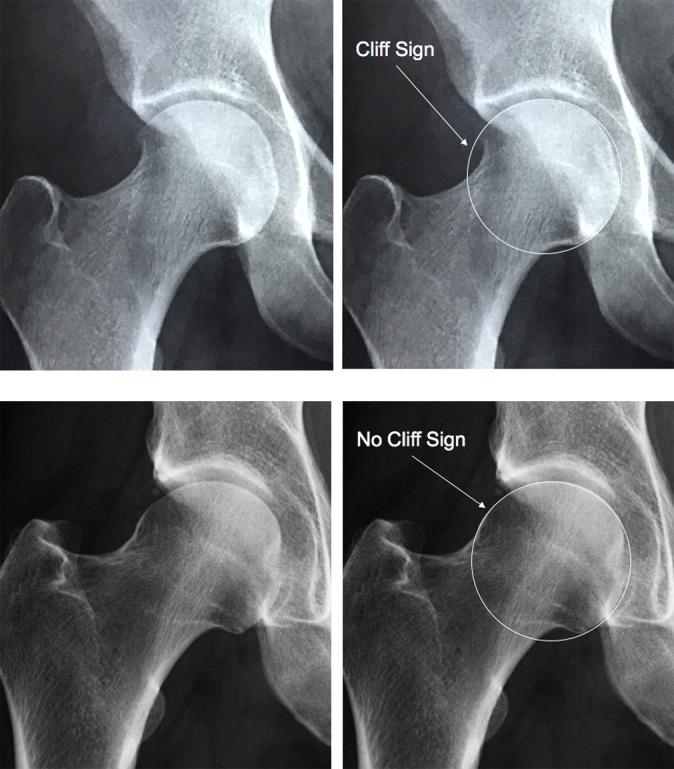
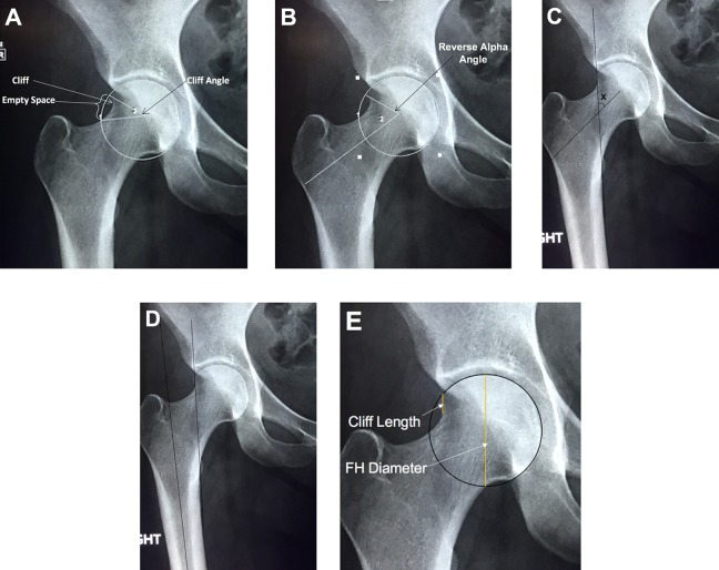
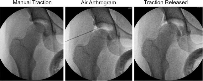
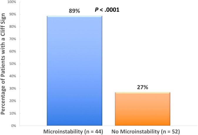
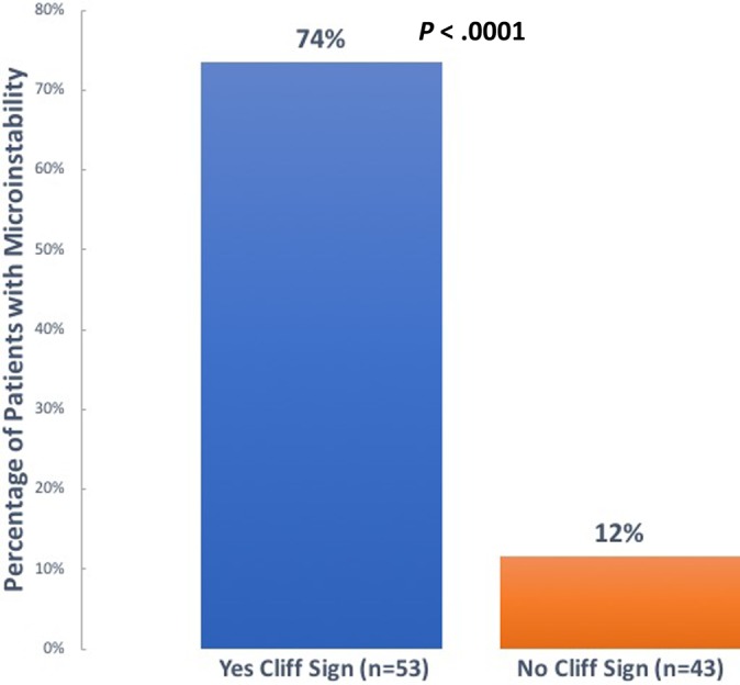
Background: The preoperative diagnosis of hip microinstability is challenging. Although physical examination maneuvers and magnetic resonance imaging findings associated with microinstability have been described, there are limited reports of radiographic features. In patients with microinstability, we observed a high incidence of a steep drop-off on the lateral edge of the femoral head, which we have named the “cliff sign.” Purpose: (1) To determine the relationship of the cliff sign and associated measurements with intraoperative microinstability and (2) to determine the interobserver reliability of these measurements. Study Design: Cohort study (diagnosis); Level of evidence, 2. Methods: A total of 115 consecutive patients who underwent hip arthroscopy were identified. Patients with prior hip surgery, Legg-Calve-Perthes disease, fractures, pigmented villonodular synovitis, or synovial chondromatosis were excluded, resulting in the inclusion of 96 patients in the study. A perfect circle around the femoral head was created on anteroposterior pelvis radiographs. If the lateral femoral head did not completely fill the perfect circle, it was considered a positive cliff sign. Five additional measurements relating to the cliff sign were calculated. The diagnosis of microinstability was made intraoperatively by the (1) amount of traction required to distract the hip, (2) lack of hip reduction after initial traction release following joint venting, or (3) intraoperative findings consistent with hip microinstability. Continuous variables were analyzed through use of unpaired t tests and discrete variables with Fisher exact tests. Interobserver reliability (n = 3) was determined for each measurement. Results: Overall, 89% (39/44) of patients with microinstability had a cliff sign, compared with 27% of patients (14/52) without instability ( P < .0001). Conversely, 74% of patients with a cliff sign had microinstability, while only 12% of patients without a cliff sign had instability ( P < .0001). In women younger than 32 years with a cliff sign, 100% (20/20) were diagnosed with instability. No differences were found in any of the 5 additional measurements. Excellent interobserver reliability was found for the presence of a cliff sign and the cliff angle measurement. Conclusion: We have identified a radiographic finding, the cliff sign, that is associated with the intraoperative diagnosis of hip microinstability and has excellent interobserver reliability. Results showed that 100% of young women with a cliff sign had intraoperative microinstability. The cliff sign may be useful in the preoperative diagnosis of hip microinstability.
Overview
The claims and statistics of this paper haven’t been checked yet.
Run the check — free, no account needed. It reads the open-access text and checks the sources it cites.
Integrity signals
Registry
Cites
Statistics
Figures
Verification
Claims the paper makes
The claims and statistics of this paper haven’t been checked yet.
Run the check — free, no account needed. It reads the open-access text and checks the sources it cites.
Claims other papers make about this one
Each is checked against this paper’s own text: does it say what the citing paper says it does?
The cited source says what the claim attributes to it.
In the citing paper
“Much previous research has debated the morphological characteristics to diagnose hip instability; however, the optimal evaluation method of dynamic hip instability remains unclear [2, 4, 17, 20, 22, 23].”
From Femoral head translation in borderline and definite dysplastic hips during weight‐bearing: 2D/3D image registration analysis (2023) · cites this paper as [17]
Self-check
Percentages, sums and reported differences re-computed from the paper’s own numbers.
The self-check needs the paper’s full text.
Citations
Cites
Intraclass correlations: Uses in assessing rater reliability.
Shrout, P. E., & Fleiss, J. L. (1979) · Psychological Bulletin · doi:10.1037/0033-2909.86.2.420 · Cited by 24,532
Research works printed but not linked: 30
- 10.1016/j.arthro.2011.08.288 · DOI
- 10.1177/0363546510386002 · DOI
- 10.1016/j.arthro.2012.07.009 · DOI
- 10.1016/j.arthro.2012.04.057 · DOI
- 10.1177/0363546513499154 · DOI · Paper page
- 10.1007/s00167-016-4411-8 · DOI
- Fleiss J, 1986, The Design and Analysis of Clinical Experiments
- 10.1054/arth.2002.27674 · DOI
- 10.1177/2325967117740121 · DOI
- 10.1177/0363546515620391 · DOI
- 10.1007/s00167-016-4377-6 · DOI
- 10.1093/jhps/hnv017 · DOI · Paper page
- 10.1016/j.arthro.2015.06.005 · DOI
- 10.1016/j.arthro.2013.07.261 · DOI
- 10.1016/j.arthro.2007.08.024 · DOI
- 10.1007/s00167-012-2262-5 · DOI
- 10.1007/s00167-013-2591-z · DOI
- 10.1016/j.arthro.2015.12.049 · DOI
- 10.1177/0363546511412161 · DOI
- 10.1177/2325967114561962 · DOI
- 10.1007/s00167-012-2091-6 · DOI
- 10.1177/0363546517697296 · DOI
- 10.1007/s00167-016-4342-4 · DOI
- 10.1016/j.csm.2010.12.008 · DOI
- 10.1016/j.arthro.2011.01.007 · DOI
- 10.1302/0301-620X.97B4.34638 · DOI
- 10.1016/j.arth.2017.02.067 · DOI
- 10.1016/j.arthro.2016.01.049 · DOI
- 10.1007/s11999-016-5137-0 · DOI
- 10.1177/0363546515608162 · DOI
Cited by
Sorted by citations
Sakai, S., Kutsuna, T., Kono, K., Kinoshita, T., Mashima, N., & Takao, M. (2023) · Journal of Experimental Orthopaedics · doi:10.1186/s40634-023-00707-8 · Cited by 16
“In addition, we evaluated the breakdown in Shenton’s line, Cliff sign and femoro-epiphyseal acetabular roof (FEAR) index [2, 17, 20, 22, 23].”
BackgroundMethod
Metadata
- Authors
- Packer, J. D., Cowan, J. B., Rebolledo, B. J., Shibata, K. R., Riley, G. M., Finlay, A. K., & Safran, M. R.
- Affiliations
- University of Maryland, Baltimore (US)
- Stanford Medicine (US)
- Scripps Clinic (US)
- Corresponding author
- Safran, Marc R.
- Venue
- Orthopaedic Journal of Sports Medicine
- Publisher
- SAGE Publications
- Published
- 2018-11-01
- Volume
- 6
- Issue
- 11
- Pages
- 2325967118807176-2325967118807176
- ISSN
- 2325-9671 (ISSN-L)
- PMC
- PMC6240982
- OpenAlex
- W2757738087
- Cited by
- 56
- References
- 1
- Citations by year
- 2026: 8 · 2025: 10 · 2024: 11 · 2023: 3 · 2022: 12 · 2021: 3 · 2020: 7 · 2019: 2
- FWCI
- 3.12
- Type
- journal article
- Language
- en
- Open access
- gold
- doi.org
- Licence
- Locations
- Orthopaedic Journal of Sports Medicine (publishedVersion, cc-by-nc-nd)
- Europe PMC (PubMed Central) (publishedVersion, cc-by-nc-nd)
- PubMed Central (publishedVersion, cc-by-nc-nd)
- PubMed (publishedVersion)
- DOAJ (DOAJ: Directory of Open Access Journals) (submittedVersion, cc-by-sa)
- Topics
- Health Sciences › Medicine › Surgery › Hip disorders and treatments
- Physical Sciences › Engineering › Biomedical Engineering › Lower Extremity Biomechanics and Pathologies
- Health Sciences › Medicine › Surgery › Shoulder Injury and Treatment
- Keywords
- hip microinstability, femoral head, radiographic findings, hip arthroscopy, intraoperative diagnosis, inter-rater reliability, hip reduction, young women, preoperative diagnosis, femoral neck stress fracture, femoral head resorption
About these checks
Registry, retraction, Cites and statistics results are computed, not judged, with one exception: a printed reference that no index lists is read once by a model before it counts as a citation error. Claims are extracted by a model and kept only when their quote appears verbatim in the text; each cited source is then checked, with a model’s help, for whether it says what the claim relies on. Those verdicts can be wrong — read the claim’s quote and the cited source before relying on one.
Statistics and arithmetic are recomputed, not judged.
Confidence bands are shown only for fields calibrated on our annotated test set. None is calibrated yet, so no band is shown.
What counts as a claim ›How we measure confidence ›How we check this ›
Are you an author, or do you see a problem with this page? Request a review
