

The DOI matches its Crossref record; 61 of the 61 references it cites were found in a registry or an index.
Raman imaging at biological interfaces: applications in breast cancer diagnosis
Open accessAbstract
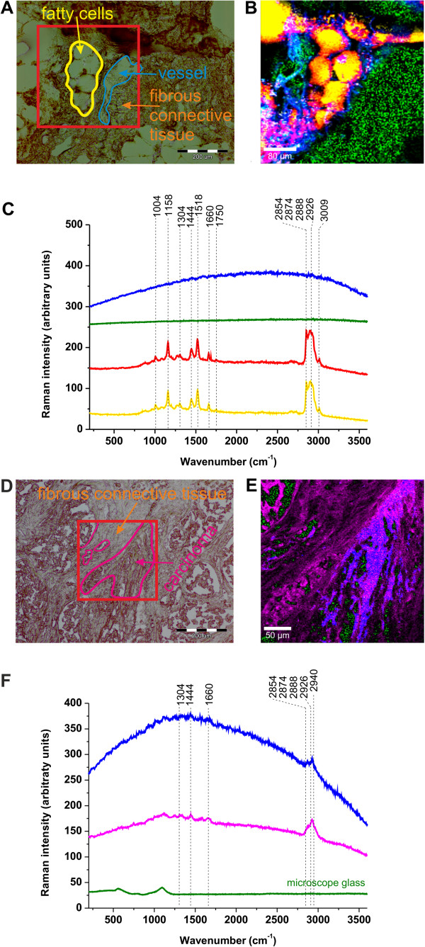
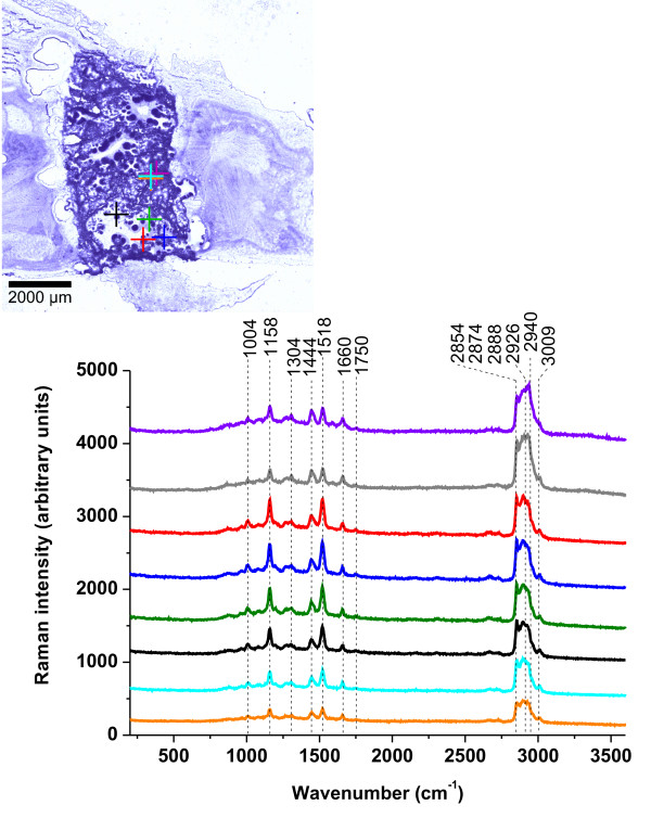
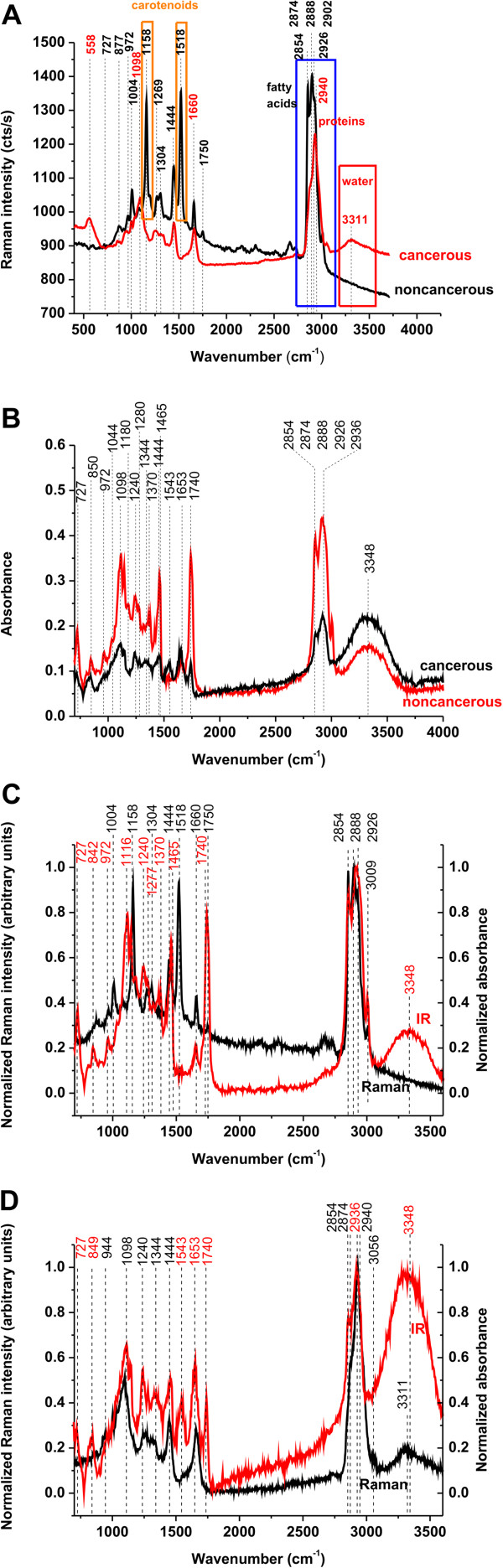
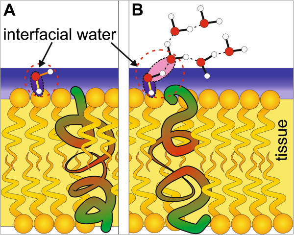
Background: One of the most important areas of Raman medical diagnostics is identification and characterization of cancerous and noncancerous tissues. The methods based on Raman scattering has shown significant potential for probing human breast tissue to provide valuable information for early diagnosis of breast cancer. A vibrational fingerprint from the biological tissue provides information which can be used to identify, characterize and discriminate structures in breast tissue, both in the normal and cancerous environment. Results: The paper reviews recent progress in understanding structure and interactions at biological interfaces of the human tissue by using confocal Raman imaging and IR spectroscopy. The important differences between the noncancerous and cancerous human breast tissues were found in regions characteristic for vibrations of carotenoids, fatty acids, proteins, and interfacial water. Particular attention was paid to the role played by unsaturated fatty acids and their derivatives as well as carotenoids and interfacial water. Conclusions: We demonstrate that Raman imaging has reached a clinically relevant level in regard to breast cancer diagnosis applications. The results presented in the paper may have serious implications on understanding mechanisms of interactions in living cells under realistically crowded conditions of biological tissue.
Overview
The claims and statistics of this paper haven’t been checked yet.
Run the check — free, no account needed. It reads the open-access text and checks the sources it cites.
Integrity signals
Registry
Cites
Statistics
Figures
Verification
Claims the paper makes
The claims and statistics of this paper haven’t been checked yet.
Run the check — free, no account needed. It reads the open-access text and checks the sources it cites.
Claims other papers make about this one
Each is checked against this paper’s own text: does it say what the citing paper says it does?
The source backs part of the claim, not all of it — or backs it for a different population, condition or comparison.
In the citing paper
“a spectral window where Raman bands from biological molecules and tissues are minimized 13, 14”
From Design and evaluation of Raman reporters for the Raman-silent region (2022) · cites this paper as [14]
Experimental — accuracy not yet measuredThe check wasn’t confident either way, so it gives no verdict — never disputed.
In the citing paper
“this imaging technique can suffer from poor signal-to-noise ratio, as the complexity of biological tissues can lead to overlapping of Raman bands from tissues and the Raman reporter molecule utilized.”
From Design and evaluation of Raman reporters for the Raman-silent region (2022) · cites this paper as [14]
Experimental — accuracy not yet measured
Self-check
Percentages, sums and reported differences re-computed from the paper’s own numbers.
The self-check needs the paper’s full text.
Citations
Cites
Two-photon laser scanning fluorescence microscopy
Denk, W., Strickler, J. H., & Webb, W. W. (1990) · Science · doi:10.1126/science.2321027 · Cited by 9,488
Single Molecule Detection Using Surface-Enhanced Raman Scattering (SERS)
Kneipp, K. D., Wang, Y., Kneipp, H., Perelman, L. T., Itzkan, I., Dasari, R. R., & Feld, M. S. (1997) · Physical Review Letters · doi:10.1103/physrevlett.78.1667 · Cited by 6,824
Nanoparticles with Raman Spectroscopic Fingerprints for DNA and RNA Detection
Cao, Y. C., Jin, R. M., & Mirkin, C. A. (2002) · Science · doi:10.1126/science.297.5586.1536 · Cited by 2,977
The Gap Junction Communication Channel
Kumar, N. M., & Gilula, N. B. (1996) · Cell · doi:10.1016/s0092-8674(00)81282-9 · Cited by 1,929
Ten-Year Risk of False Positive Screening Mammograms and Clinical Breast Examinations
Elmore, J. G., Barton, M. B., Moceri, V. M., Polk, S., Arena, P. J., & Fletcher, S. W. (1998) · New England Journal of Medicine · doi:10.1056/nejm199804163381601 · Cited by 1,051
Wavelength-Scanned Surface-Enhanced Raman Excitation Spectroscopy
McFarland, A. D., Young, M. A., Dieringer, J. A., & Duyne, R. P. V. (2005) · The Journal of Physical Chemistry B · doi:10.1021/jp050508u · Cited by 958
Analysis of cancers missed at screening mammography.
Bird, R. E., Wallace, T., & Yankaskas, B. C. (1992) · Radiology · doi:10.1148/radiology.184.3.1509041 · Cited by 821
Plasmon-Sampled Surface-Enhanced Raman Excitation Spectroscopy
Haynes, C. L., & Duyne, R. P. V. (2003) · The Journal of Physical Chemistry B · doi:10.1021/jp027749b · Cited by 692
Characterisation and identification of bacteria using SERS
Jarvis, R. M., & Goodacre, R. (2008) · Chemical Society Reviews · doi:10.1039/b705973f · Cited by 372
Tip‐Enhanced Raman Spectroscopy of Single RNA Strands: Towards a Novel Direct‐Sequencing Method
Bailo, E., & Deckert, V. (2008) · Angewandte Chemie International Edition · doi:10.1002/anie.200704054 · Cited by 333
Cited by
Sorted by citations
Applications of Raman spectroscopy in cancer diagnosis
Auner, G. W., Koya, S. K., Huang, C., Broadbent, B., Trexler, M., Auner, Z., Elias, A., Mehne, K. C., & Brusatori, M. (2018) · Cancer and Metastasis Reviews · doi:10.1007/s10555-018-9770-9 · Cited by 389
Vibrational Spectroscopy Fingerprinting in Medicine: from Molecular to Clinical Practice
Bălan, V., Mihai, C. T., Cojocaru, F.-D., Urîtu, C. M., Dodi, G., Botezat, D., & Gardikiotis, I. (2019) · Materials · doi:10.3390/ma12182884 · Cited by 370
Abramczyk, H., Surmacki, J. M., Kopeć, M., Olejnik, A. K., Lubecka, K., & Fabianowska‐Majewska, K. (2015) · The Analyst · doi:10.1039/c4an01875c · Cited by 219
Self-citationTuteja, S. K., Chen, R., Kukkar, M., Song, C. K., Mutreja, R., Singh, S. B., Paul, A. K., Lee, H., Kim, K., Deep, A., et al. (2016) · Biosensors and Bioelectronics · doi:10.1016/j.bios.2016.07.052 · Cited by 177
Raman spectroscopy and machine learning for the classification of breast cancers
Zhang, L., Li, C., Peng, D., Yi, X., He, S., Liu, F., Zheng, X., Huang, W. E., Zhao, L., & Huang, X. (2021) · Spectrochimica Acta Part A Molecular and Biomolecular Spectroscopy · doi:10.1016/j.saa.2021.120300 · Cited by 166
Metadata
- Authors
- Surmacki, J., Musial, J., Kordek, R., & Abramczyk, H.
- Affiliations
- Lodz University of Technology (PL)
- Corresponding author
- Abramczyk, Halina
- Venue
- Molecular Cancer
- Published
- 2013-05-24
- Volume
- 12
- Issue
- 1
- Pages
- 48-48
- ISSN
- 1476-4598 (ISSN-L)
- PMC
- PMC3681552
- OpenAlex
- W2132590101
- Cited by
- 143
- References
- 61
- Citations by year
- 2026: 5 · 2025: 8 · 2024: 8 · 2023: 9 · 2022: 6 · 2021: 15 · 2020: 7 · 2019: 19 · 2018: 11 · 2017: 12 · 2016: 17 · 2015: 13 · 2014: 12 · 2013: 1
- FWCI
- 8.69
- Type
- journal article
- Language
- en
- Open access
- gold
- doi.org
- Licence
- cc-by
- Locations
- Molecular Cancer (publishedVersion, cc-by)
- Europe PMC (PubMed Central) (publishedVersion, cc-by)
- PubMed Central (publishedVersion, cc-by)
- PubMed (publishedVersion)
- Funders
- Uniwersytet Łódzki
- Topics
- Life Sciences › Biochemistry, Genetics and Molecular Biology › Biophysics › Spectroscopy Techniques in Biomedical and Chemical Research
- Health Sciences › Medicine › Radiology, Nuclear Medicine and Imaging › Optical Imaging and Spectroscopy Techniques
- Physical Sciences › Physics and Astronomy › Atomic and Molecular Physics, and Optics › Spectroscopy and Quantum Chemical Studies
- Keywords
- Raman imaging, breast cancer diagnosis, biointerfaces, confocal Raman microscopy, infrared spectroscopy, breast tissue, carotenoids, unsaturated fatty acids, interfacial water, protein vibrations, early cancer detection
- MeSH terms
- Breast, Breast Neoplasms, Diagnostic Imaging, Female, Humans, Spectrum Analysis, Raman
About these checks
Registry, retraction, Cites and statistics results are computed, not judged, with one exception: a printed reference that no index lists is read once by a model before it counts as a citation error. Claims are extracted by a model and kept only when their quote appears verbatim in the text; each cited source is then checked, with a model’s help, for whether it says what the claim relies on. Those verdicts can be wrong — read the claim’s quote and the cited source before relying on one.
Statistics and arithmetic are recomputed, not judged.
Confidence bands are shown only for fields calibrated on our annotated test set. None is calibrated yet, so no band is shown.
What counts as a claim ›How we measure confidence ›How we check this ›
Are you an author, or do you see a problem with this page? Request a review
