

The DOI matches its Crossref record; 43 of the 43 references it cites were found in a registry or an index.
Toll-like receptor 4 signaling promotes epithelial-mesenchymal transition in human hepatocellular carcinoma induced by lipopolysaccharide
Open accessAbstract
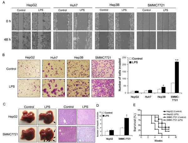
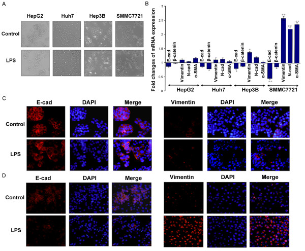
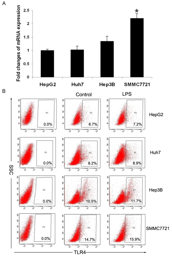
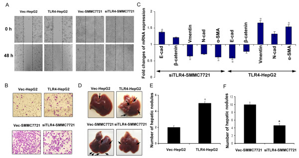
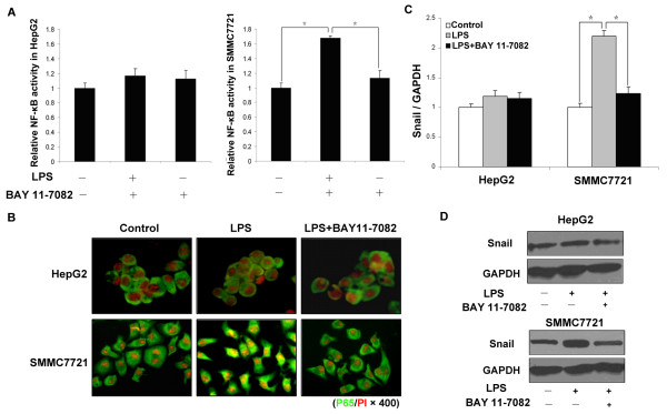
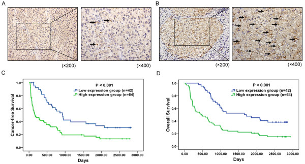
Background: The endotoxin level in the portal and peripheral veins of hepatocellular carcinoma (HCC) patients is higher and lipopolysaccharide (LPS), a cell wall constituent of gram-negative bacteria, has been reported to inhibit tumor growth. However, in this study, we found that LPS-induced toll-like receptor 4 (TLR4) signaling was involved in tumor invasion and survival, and the molecular mechanism was investigated, Methods: Four HCC cell lines and a splenic vein metastasis of the nude mouse model were used to study the invasion ability of LPS-induced HCC cells and the epithelia-mesenchymal transition (EMT) in vitro and in vivo. A total of 106 clinical samples from HCC patients were used to evaluate TLR4 expression and analyze its association with clinicopathological characteristics Results: The in vitro and in vivo experiments demonstrated that LPS could significantly enhance the invasive potential and induce EMT in HCC cells with TLR4 dependent. Further studies showed that LPS could directly activate nuclear factor kappa B (NF-κB) signaling through TLR4 in HCC cells. Interestingly, blocking NF-κB signaling significantly inhibited transcription factor Snail expression and thereby inhibited EMT occurrence. High expression of TLR4 in HCC tissues was strongly associated with both poor cancer-free survival and overall survival in patients. Conclusions: Our results indicate that TLR4 signaling is required for LPS-induced EMT, tumor cell invasion and metastasis, which provide molecular insights for LPS-related pathogenesis and a basis for developing new strategies against metastasis in HCC.
Overview
The claims and statistics of this paper haven’t been checked yet.
Run the check — free, no account needed. It reads the open-access text and checks the sources it cites.
Integrity signals
Registry
Cites
Statistics
Figures
Verification
Claims the paper makes
The claims and statistics of this paper haven’t been checked yet.
Run the check — free, no account needed. It reads the open-access text and checks the sources it cites.
Claims other papers make about this one
Each is checked against this paper’s own text: does it say what the citing paper says it does?
In the citing paper
“In addition, TLR4 activation in HCC cell lines by LPS enhances their invasive potential and induces the epithelial–mesenchymal transition83.”
From The gut microbiome and liver cancer: mechanisms and clinical translation (2017) · cites this paper as [83]
Self-check
Percentages, sums and reported differences re-computed from the paper’s own numbers.
The self-check needs the paper’s full text.
Citations
Cites
Akira, S., & Takeda, K. (2004) · Nature reviews. Immunology · doi:10.1038/nri1391 · Cited by 8,312
Toll-like receptor control of the adaptive immune responses
Iwasaki, A., & Medzhitov, R. M. (2004) · Nature Immunology · doi:10.1038/ni1112 · Cited by 4,166
Cano, A., Pérez‐Moreno, M., Rodrigo, I., Locascio, A., Blanco, M. J., Barrio, M. G. D., Portillo, F., & Nieto, M. A. (2000) · Nature Cell Biology · doi:10.1038/35000025 · Cited by 3,672
Batlle, E., Sancho, E., Francí, C., Dominguez-Sola, D., Monfar, M., Baulida, J., & de Herreros, A. G. (2000) · Nature Cell Biology · doi:10.1038/35000034 · Cited by 2,730
Larue, L., & Bellacosa, A. (2005) · Oncogene · doi:10.1038/sj.onc.1209091 · Cited by 1,247
Huber, M. A., Azoitei, N., Baumann, B., Grünert, S., Sommer, A., Pehamberger, H., Kraut, N., Beug, H., & Wirth, T. (2004) · Journal of Clinical Investigation · doi:10.1172/jci21358 · Cited by 1,049
Intravital imaging of cell movement in tumours
Condeelis, J. S., & Segall, J. E. (2003) · Nature reviews. Cancer · doi:10.1038/nrc1231 · Cited by 930
Stabilization of Snail by NF-κB Is Required for Inflammation-Induced Cell Migration and Invasion
Wu, Y., Deng, J., Rychahou, P. G., Qiu, S., Evers, B. M., & Zhou, B. P. (2009) · Cancer Cell · doi:10.1016/j.ccr.2009.03.016 · Cited by 814
Prospective analysis of risk factors for hepatocellular carcinoma in patients with liver cirrhosis
Velázquez, R. F., Rodríguez, M., Navascués, C., Linares, A., Pérez, R., Sotorríos, N. G., Martínez, I., & Rodrigo, L. (2003) · Hepatology · doi:10.1053/jhep.2003.50093 · Cited by 457
Toll-Like Receptor Signaling in the Liver
Schwabe, R. F., Seki, E., & Brenner, D. A. (2006) · Gastroenterology · doi:10.1053/j.gastro.2006.01.038 · Cited by 411
Cited by
Sorted by citations
Items 101–163 of 163
Banday, M. M., Kumar, A., Vestal, G. D., Sethi, J. S., Patel, K., O’Neill, E. B., Finan, J. E., Cheng, F., Lin, M., Davis, N. M., et al. (2021) · The Journal of Heart and Lung Transplantation · doi:10.1016/j.healun.2021.02.014 · Cited by 12
Yang, X. F., Zong, C., Feng, C., Zhang, C., Smirnov, A., Sun, G., Shao, C., Zhang, L., Hou, X., Liu, W., et al. (2023) · Advanced Science · doi:10.1002/advs.202300424 · Cited by 12
Self-citationNonalcoholic Fatty Liver Disease and the Intestinal Microbiome: An Inseparable Link
Effenberger, M., Grander, C., Grabherr, F., & Tilg, H. (2023) · Journal of Clinical and Translational Hepatology · doi:10.14218/jcth.2023.00069 · Cited by 14
Goyal, H., Fatima, K., & Kaur, J. (2025) · Cancer Cell International · doi:10.1186/s12935-025-03840-9 · Cited by 11
Impact of variability in cell cycle periodicity on cell population dynamics
Nowak, C. M., Quarton, T., & Bleris, L. G. (2023) · PLoS Computational Biology · doi:10.1371/journal.pcbi.1011080 · Cited by 11
Prognostic role of TLR4 and TLR2 in hepatocellular carcinoma
Kairaluoma, V., Kemi, N., Huhta, H. I., Pohjanen, V., & Helminen, O. (2021) · Acta Oncologica · doi:10.1080/0284186x.2021.1877346 · Cited by 11
Li, N., Zhu, Q., Yang, C., Li, F., Zhou, Z.-H., Lv, Y., Sang, J., Han, Q., & Liu, Z. (2015) · Tumor Biology · doi:10.1007/s13277-015-4423-x · Cited by 11
Lü, Y., Feng, X., Wang, Z., Zou, M., Xu, Z., Liu, Q.-J., Chen, W., Ding, J., & Liu, H. (2025) · Precision Clinical Medicine · doi:10.1093/pcmedi/pbaf020 · Cited by 12
Molecular mechanisms of melatonin in the reversal of LPS-induced EMT in peritoneal mesothelial cells
Shi, S., Zhang, Y., Wen, W., Zhao, Y., & Sun, L. (2016) · Molecular Medicine Reports · doi:10.3892/mmr.2016.5744 · Cited by 10
Othaim, A. A., Al-Hawary, S. I. S., Alsaab, H. O., Almalki, S. G., Najm, M. A. A., Hjazi, A. M., Alsalamy, A., Almulla, A. F., & Alizadeh, H. (2023) · Frontiers in Genetics · doi:10.3389/fgene.2023.1280051 · Cited by 10
Zhang, J., Xu, M., Gao, H. J., Guo, J., Guo, Y., Zou, M., & Wu, X. (2018) · Oncology Letters · doi:10.3892/ol.2018.7778 · Cited by 10
Shen, X. T., Zhao, D., Wang, F., Li, X., Sun, K., Du, H., Song, S., Nie, L., & Wang, C. (2026) · BMC Cancer · doi:10.1186/s12885-025-15325-z · Cited by 9
Self-citationCommon Denominator of MASLD and Some Non-Communicable Diseases
Ferenc, K., Jarmakiewicz-Czaja, S., Sokal-Dembowska, A., Stasik, K., & Filip, R. (2024) · Current Issues in Molecular Biology · doi:10.3390/cimb46070399 · Cited by 9
Gowing, S. D., Cool-Lartigue, J., Spicer, J. D., SEELY, A. J. E., & Ferri, L. E. (2020) · Clinical & Experimental Metastasis · doi:10.1007/s10585-020-10018-2 · Cited by 9
Lu, W., Aihaiti, A., Abudukeranmu, P., Liu, Y., & Gao, H. (2024) · Journal of Translational Medicine · doi:10.1186/s12967-024-05320-6 · Cited by 8
Self-citationCitral-Enriched Fraction of Lemon Essential Oil Mitigates LPS-Induced Hepatocyte Injuries
Gasparro, R., Pucci, M., Costanzo, E., Urzì, O., Tinnirello, V., Moschetti, M., Conigliaro, A., Raimondo, S., Corleone, V., Fontana, S., et al. (2023) · Biology · doi:10.3390/biology12121535 · Cited by 8
The stemness of hepatocytes is maintained by high levels of lipopolysaccharide via YAP1 activation
Shao, C., Yang, X. F., Jing, Y., Hou, X., Huang, Y., Zong, C., Gao, L., Liu, W., Jiang, J., Ye, F., et al. (2021) · Stem Cell Research & Therapy · doi:10.1186/s13287-021-02421-7 · Cited by 8
Self-citationEpigenetic Regulation of Toll-Like Receptor Signaling: Implications for Cancer Development
Boi, S. K., & Elsawa, S. F. (2013) · Medical Epigenetics · doi:10.1159/000353684 · Cited by 8
Lollo, V. D., Canciello, A., Peserico, A., Orsini, M., Russo, V., Cerveró-Varona, A., Dufrusine, B., Khatib, M. E., Curini, V., Mauro, A., et al. (2023) · iScience · doi:10.1016/j.isci.2023.107582 · Cited by 7
Could intratumoural microbiota be key to unlocking treatment responses in hepatocellular carcinoma?
Yu, K. L., & Shen, S. (2024) · European Journal of Cancer · doi:10.1016/j.ejca.2024.115195 · Cited by 8
Fu, K., Shao, L., Mei, L., Li, H., Feng, Y., Tian, W., Huan, Y., & Cao, R. (2021) · Theriogenology · doi:10.1016/j.theriogenology.2021.10.001 · Cited by 7
Microbiota-derived metabolites in tumorigenesis: mechanistic insights and therapeutic implications
Yang, S.-F., Chen, X., & Pan, Y. (2025) · Frontiers in Pharmacology · doi:10.3389/fphar.2025.1598009 · Cited by 6
Epigenetic Changes of the Immune System with Role in Tumor Development
Florea, I. D., & Karaoulani, C. (2018) · Methods in molecular biology · doi:10.1007/978-1-4939-8751-1_11 · Cited by 6
A novel TLR4 accessory molecule drives hepatic oncogenesis through tumor-associated macrophages
Kim, D., Allen, C., Chung, D.-J., Meng, L., Zhang, X., Zhang, W., Ouyang, Y.-Q., Li, Z., & Hong, F. (2025) · Cancer Letters · doi:10.1016/j.canlet.2025.217543 · Cited by 6
Chirumbolo, S., & Bjørklund, G. (2016) · International Journal of Clinical Oncology · doi:10.1007/s10147-016-1072-z · Cited by 5
Strauss, P., Marti, H., Beisland, C., Scherer, A., Lysne, V., Leh, S. M., Flatberg, A., Koch, E., Beisvåg, V., Landolt, L. Z., et al. (2018) · International Journal of Molecular Sciences · doi:10.3390/ijms19030803 · Cited by 5
TLR4-Induced Inflammation Is a Key Promoter of Tumor Growth, Vascularization, and Metastasis
Ran, S., Bhattarai, N., Patel, R., & Volk-Draper, L. D. (2019) · IntechOpen eBooks · doi:10.5772/intechopen.85195 · Cited by 6
Lu, J., Lin, L., Sun, Y., Liu, D., & Gong, Z. (2022) · Biomedicines · doi:10.3390/biomedicines10010095 · Cited by 5
Unveiling hidden players: the role of intratumoral microbiota in gastrointestinal cancer dynamics
Tang, W., Li, F., Zheng, H., Zhou, S., Li, C., Xu, X., & Fu, J. (2025) · Journal of Cancer Research and Clinical Oncology · doi:10.1007/s00432-025-06401-9 · Cited by 5
Interactions between human microbiome, liver diseases, and immunosuppression after liver transplant
Peruhova, M., Peshevska-Sekulovska, M., & Velikova, T. V. (2021) · World Journal of Immunology · doi:10.5411/wji.v11.i2.11 · Cited by 4
TLR AND CANCER: THE ENIGMATIC CROSS TALK
Fehri, E., Ennaifer, E., Ardhaoui, M., Tounsi, H., Rhouma, R. B. H., & Boubaker, S. (2024) · IntechOpen eBooks · doi:10.5772/intechopen.1004101 · Cited by 3
Mahmud, S., Hamza, A., Lee, Y.-B., Min, J., Islam, R., Dogsom, O., & Park, J. (2023) · Biomolecules · doi:10.3390/biom14010006 · Cited by 2
The effect of detoxified Shigella sonnei lipopolysaccharide on the metastasis of B16 melanoma cells
Novikova, E. M., Chukhina, E. S., Razvalyayeva, N. A., Козырева, О. В., & Stepanenko, R. N. (2025) · Immunologiya · doi:10.33029/1816-2134-2025-46-1-38-50 · Cited by 2
Microbiota, Inflammation, and Gut Barrier Dysfunction in HCC
Ram, A. K., Wright, G. A. K., & Vairappan, B. (2019) · IntechOpen eBooks · doi:10.5772/intechopen.89578 · Cited by 2
El-Sayed, E., & Hamdy, N. M. (2023) · Egyptian Journal of Food Science · doi:10.21608/ejfs.2023.213441.1166 · Cited by 2
Soft cell-derived hybrid microparticles with platelet decoys for enhanced cancer chemotherapy
Bie, N., Li, S., Sun, K., Li, J., Li, X., Zhang, X., Tian, M., Xie, Z., Xiao, Y., Zhang, Y., et al. (2026) · Acta Pharmaceutica Sinica B · doi:10.1016/j.apsb.2026.03.028 · Cited by 1
Self-citationThe Role of Epithelial–Mesenchymal Transition in Oral Inflammatory Diseases
Wang, Y., Yang, Y., Xu, C., Shi, P., Wang, J., Ding, Y., & Xie, X. (2026) · Journal of Cellular Physiology · doi:10.1002/jcp.70194 · Cited by 1
Czekay, R., & Higgins, P. J. (2025) · Springer eBooks · doi:10.1007/978-3-031-55254-0_7-1 · Cited by 1
Toll-like receptor 4 inhibition sensitizes non-small cell lung cancer to radiotherapy
Haroun, R., Rutihinda, C., Diallo, A. H., Ordoñez, J. P., Nassri, S., Shams, A., Pacheco, M. F. M., Saidi, N. E., Bouchard, L., Turgeon, G., et al. (2025) · Cancer Biology & Therapy · doi:10.1080/15384047.2025.2590881 · Cited by 1
Song, S., Wang, L., Li, L., Bai, S.-H., Zhu, X., Zhang, X.-L., Deng, C.-H., & Han, Z. (2026) · Cancer Letters · doi:10.1016/j.canlet.2026.218567 · Cited by 1
Self-citationGut metabolites: key factors in the cross-talk between the gut microbiota and tumor immunotherapy
Zeng, L., Ren, Y., Huang, H., Wang, Q., Li, H., Song, J., He, F., & Li, J. (2026) · Frontiers in Immunology · doi:10.3389/fimmu.2026.1882542 · Cited by 1
Hepatitis B Virus, Hepatitis C Virus, the Microbiome, and Hepatocellular Carcinoma
Chen, F., Freyer, C., Gong, L., & Zekry, A. (2019) · Elsevier eBooks · doi:10.1016/b978-0-323-54843-4.00004-0 · Cited by 1
Potential involvement of epithelial-mesenchymal transition in the pathogenesis of periodontitis
Abdulkareem, A. A. (2017) · University of Birmingham Institutional Research Archive (University of Birmingham) · Cited by 1
Novel insights into the role of bacterial gut microbiota in hepatocellular carcinoma
Chang, R., Zhang, L., Qiu, G., Xu, X., & Zhou, Y. (2018) · Cancer translational medicine · doi:10.4103/ctm.ctm_8_18
Kaur, K. K., Allahbadia, G. N., & Singh, M. (2023) · Liver Research - Open Journal · doi:10.17140/lroj-4-115
Role of microbiome and microbial extracellular vesicles in tumor microenvironment
Kim, Y. (2024) · Elsevier eBooks · doi:10.1016/b978-0-12-824040-3.00002-9
Therapeutic potential of kakkatin derivatives against hepatocellular carcinoma
Chahal, S., & Patial, V. (2025) · World Journal of Clinical Oncology · doi:10.5306/wjco.v16.i3.101686
Elmoneim, H. M. A. (2025) · Saudi Toxicology Journal · doi:10.70957/uqu.edu.sa/s.toxicology.s/stj.2025.2.3
Hsu, C., Jasim, S. A., Rasool, K. H., Malathi, H., Kaur, J., Jabir, M. S., Alhajlah, S., Kumar, A., Jawad, S. F., & Husseen, B. (2025) · Seminars in Oncology · doi:10.1016/j.seminoncol.2025.152344
Xu, W., Zhang, J., Zhang, X., Wang, X., Xie, Y., Min, S., Wang, X., & Lian, C. (2025) · Frontiers in Genetics · doi:10.3389/fgene.2025.1556366
Zhu, J., Yang, B., Zhai, X., Ni, F., Tao, D., & Tan, C. (2025) · BMC Women s Health · doi:10.1186/s12905-025-04001-w
Shao, C., Yang, X., Jing, Y., Hou, X., Huang, Y., Zong, C., Gao, L., Liu, W., Jiang, J., Ye, F., et al. (2021) · Figshare · doi:10.6084/m9.figshare.14767018
Self-citationShao, C., Yang, X., Jing, Y., Hou, X., Huang, Y., Zong, C., Gao, L., Liu, W., Jiang, J., Ye, F., et al. (2021) · Figshare · doi:10.6084/m9.figshare.14767021
Self-citationShao, C., Yang, X., Jing, Y., Hou, X., Huang, Y., Zong, C., Gao, L., Liu, W., Jiang, J., Ye, F., et al. (2021) · Figshare · doi:10.6084/m9.figshare.14767009
Self-citationShao, C., Yang, X., Jing, Y., Hou, X., Huang, Y., Zong, C., Gao, L., Liu, W., Jiang, J., Ye, F., et al. (2021) · Figshare · doi:10.6084/m9.figshare.14767027
Self-citationShao, C., Yang, X., Jing, Y., Hou, X., Huang, Y., Zong, C., Gao, L., Liu, W., Jiang, J., Ye, F., et al. (2021) · Figshare · doi:10.6084/m9.figshare.14767012
Self-citationShao, C., Yang, X., Jing, Y., Hou, X., Huang, Y., Zong, C., Gao, L., Liu, W., Jiang, J., Ye, F., et al. (2021) · Figshare · doi:10.6084/m9.figshare.14767024
Self-citationShao, C., Yang, X., Jing, Y., Hou, X., Huang, Y., Zong, C., Gao, L., Liu, W., Jiang, J., Ye, F., et al. (2021) · Figshare · doi:10.6084/m9.figshare.14767015
Self-citationThe role of gut microbiota and its metabolites in preventing oncogenesis
Li, Z., Yan, J., Zeng, Z., & Zhao, L. (2026) · Frontiers in Cell and Developmental Biology · doi:10.3389/fcell.2026.1790063
Deciphering the role of glycolipids as emerging therapeutic targets in EMT-driven cancer progression
Mukherjee, A., Paul, S., Datta, C., & Bhattacharjee, A. (2026) · Discover Oncology · doi:10.1007/s12672-026-04819-2
Impact of respiratory viral infections on tumor progression in Non-Small-Cell Lung Carcinoma (NSCLC)
Angrini, M. (2021) · theses.fr (ABES) · doi:10.70675/9c027d88z3675z44adza870z1802353e674a
Gut microbiota as predictors of immunotherapy response in hepatocellular carcinoma
Xia, S., Yang, Q., Liu, Y., Yan, A., Li, S., Xu, L., Guan, H., Xie, Y., & Hu, S. (2026) · Hepatoma Research · doi:10.20517/2394-5079.2026.10
Self-citation
Metadata
- Authors
- Jing, Y.-Y., Han, Z.-P., Sun, K., Zhang, S.-S., Hou, J., Liu, Y., Li, R., Gao, L., Zhao, X., Zhao, Q.-D., Wu, M.-C., & Wei, L.-X.
- Affiliations
- Second Military Medical University (CN)
- Venue
- BMC Medicine
- Published
- 2012-08-31
- Volume
- 10
- Issue
- 1
- Pages
- 98-98
- ISSN
- 1741-7015 (ISSN-L)
- PMC
- PMC3482562
- OpenAlex
- W1970916626
- Cited by
- 162
- References
- 43
- Citations by year
- 2026: 8 · 2025: 15 · 2024: 11 · 2023: 13 · 2022: 14 · 2021: 18 · 2020: 9 · 2019: 11 · 2018: 11 · 2017: 13 · 2016: 10 · 2015: 11 · 2014: 11 · 2013: 7
- FWCI
- 3.99
- Type
- journal article
- Language
- en
- Open access
- gold
- doi.org
- Licence
- Locations
- BMC Medicine (publishedVersion, cc-by)
- Europe PMC (PubMed Central) (publishedVersion, cc-by)
- PubMed Central (publishedVersion, cc-by)
- PubMed (publishedVersion)
- DOAJ (DOAJ: Directory of Open Access Journals) (submittedVersion)
- Funders
- National Natural Science Foundation of China (30921006, 81030041, 2008ZX10002-019, 81000970, SCMRC1102, 2011CB966200, 81101622, 31171321, 30901722)
- Science and Technology Commission of Shanghai Municipality (10411963100, 10ZR1439900, 11ZR1449500, 10ZR1439600)
- Science Fund for Creative Research Groups (30921006)
- Topics
- Life Sciences › Immunology and Microbiology › Immunology › Immune Response and Inflammation
- Life Sciences › Biochemistry, Genetics and Molecular Biology › Molecular Biology › TGF-β signaling in diseases
- Health Sciences › Medicine › Hepatology › Hepatocellular Carcinoma Treatment and Prognosis
- Keywords
- TLR4, hepatocellular carcinoma, epithelial-mesenchymal transition, lipopolysaccharide, NF-κB signaling, Snail transcription factor, tumor invasion, metastasis, endotoxin, tumor-free survival, overall survival
- MeSH terms
- Adolescent, Adult, Aged, Animals, Child, Endotoxins, Female, Carcinoma, Hepatocellular, Humans, Lipopolysaccharides, Male, Mice, Nude, Middle Aged, Signal Transduction, Gene Expression Profiling, Toll-Like Receptor 4, Mice, Young Adult, Epithelial-Mesenchymal Transition
About these checks
Registry, retraction, Cites and statistics results are computed, not judged, with one exception: a printed reference that no index lists is read once by a model before it counts as a citation error. Claims are extracted by a model and kept only when their quote appears verbatim in the text; each cited source is then checked, with a model’s help, for whether it says what the claim relies on. Those verdicts can be wrong — read the claim’s quote and the cited source before relying on one.
Statistics and arithmetic are recomputed, not judged.
Confidence bands are shown only for fields calibrated on our annotated test set. None is calibrated yet, so no band is shown.
What counts as a claim ›How we measure confidence ›How we check this ›
Are you an author, or do you see a problem with this page? Request a review
