

The DOI matches its Crossref record; 85 of the 86 references it cites were found in a registry or an index.
Neuroinflammation mediated by IL-1β increases susceptibility of dopamine neurons to degeneration in an animal model of Parkinson's disease
Open accessAbstract
Background: The etiology of Parkinson's disease (PD) remains elusive despite identification of several genetic mutations. It is more likely that multiple factors converge to give rise to PD than any single cause. Here we report that inflammation can trigger degeneration of dopamine (DA) neurons in an animal model of Parkinson's disease. Methods: We examined the effects of inflammation on the progressive 6-OHDA rat model of Parkinson's disease using immunohistochemistry, multiplex ELISA, and cell counting stereology. Results: We show that a non-toxic dose of lipopolysaccharide (LPS) induced secretion of cytokines and predisposed DA neurons to be more vulnerable to a subsequent low dose of 6-hydroxydopamine. Alterations in cytokines, prominently an increase in interleukin-1beta (IL-1β), were identified as being potential mediators of this effect that was associated with activation of microglia. Administration of an interleukin-1 receptor antagonist resulted in significant reductions in tumor necrosis factor-α and interferon-γ and attenuated the augmented loss of DA neurons caused by the LPS-induced sensitization to dopaminergic degeneration. Conclusion: These data provide insight into the etiology of PD and support a role for inflammation as a risk factor for the development of neurodegenerative disease.
Overview
The claims and statistics of this paper haven’t been checked yet.
Run the check — free, no account needed. It reads the open-access text and checks the sources it cites.
Integrity signals
Registry
Cites
Statistics
Figures
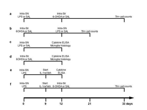
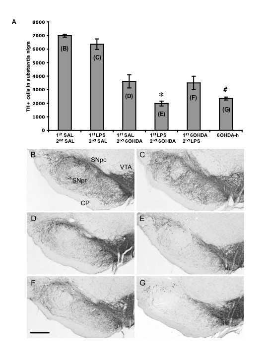
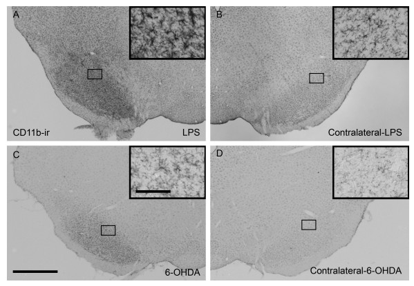
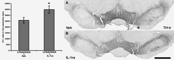
Verification
Claims the paper makes
The claims and statistics of this paper haven’t been checked yet.
Run the check — free, no account needed. It reads the open-access text and checks the sources it cites.
Claims other papers make about this one
Each is checked against this paper’s own text: does it say what the citing paper says it does?
The source backs part of the claim, not all of it — or backs it for a different population, condition or comparison.
In the citing paper
“In a different PD mouse model, blocking IL-1β signaling using an IL-1 receptor antagonist also significantly attenuated neurodegeneration.362,363”
From Role of neuroinflammation in neurodegeneration development (2023) · cites this paper as [362]
Experimental — accuracy not yet measured
Self-check
Percentages, sums and reported differences re-computed from the paper’s own numbers.
The self-check needs the paper’s full text.
Citations
Cites
The rat brain in stereotaxic coordinates
Palkovits, M. (1983) · Neuropeptides · doi:10.1016/0143-4179(83)90049-5 · Cited by 28,959
The Rat Brain in Stereotaxic Coordinates
Paxinos, G., & Watson, C. (1983) · Elsevier eBooks · doi:10.1016/c2009-0-63235-9 · Cited by 28,585
Mutation in the α-Synuclein Gene Identified in Families with Parkinson's Disease
Polymeropoulos, M. H., Lavedan, C., Leroy, E., Ide, S. E., Dehejia, A., Dutra, A., Pike, B., Root, H., Rubenstein, J., Boyer, R., et al. (1997) · Science · doi:10.1126/science.276.5321.2045 · Cited by 8,240
Mutations in the parkin gene cause autosomal recessive juvenile parkinsonism
Kitada, T., Asakawa, S., Hattori, N., Matsumine, H., Yamamura, Y., Minoshima, S., Yokochi, M., Mizuno, Y., & Shimizu, N. (1998) · Nature · doi:10.1038/33416 · Cited by 5,227
Microglia: a sensor for pathological events in the CNS
Kreutzberg, G. W. (1996) · Trends in Neurosciences · doi:10.1016/0166-2236(96)10049-7 · Cited by 4,396
Hereditary Early-Onset Parkinson's Disease Caused by Mutations in PINK1
Valente, E. M., Abou-Sleiman, P. M., Caputo, V., Muqit, M. M. K., Harvey, K., Gispert, S., Ali, Z., Del Turco, D., Bentivoglio, A. R., Healy, D. G., et al. (2004) · Science · doi:10.1126/science.1096284 · Cited by 3,481
Mutations in LRRK2 Cause Autosomal-Dominant Parkinsonism with Pleomorphic Pathology
Zimprich, A. A., Biskup, S., Leitner, P., Lichtner, P., Farrer, M. J., Lincoln, S., Kachergus, J. M., Hulihan, M., Uitti, R. J., Calne, D. B., et al. (2004) · Neuron · doi:10.1016/j.neuron.2004.11.005 · Cited by 3,088
West, M. J., Slomianka, L., & Gundersen, H. J. G. (1991) · The Anatomical Record · doi:10.1002/ar.1092310411 · Cited by 2,994
McGeer, P. L., Itagaki, S., Boyes, B. E., & McGeer, E. G. (1988) · Neurology · doi:10.1212/wnl.38.8.1285 · Cited by 2,990
Mutations in the DJ-1 Gene Associated with Autosomal Recessive Early-Onset Parkinsonism
Bonifati, V., Rizzu, P., van Baren, M. J., Schaap, O., Breedveld, G. J., Krieger, E., Dekker, M. C. J., Squitieri, F., Ibáñez, P., Joosse, M., et al. (2003) · Science · doi:10.1126/science.1077209 · Cited by 2,854
Cited by
Sorted by citations
Items 301–355 of 355
Is Chronic Systemic Inflammation a Determinant Factor in Developing Parkinson’s Disease?
Ugalde‐Muñiz, P., Pérez‐Hernández, J., & Chavarría, A. (2016) · InTech eBooks · doi:10.5772/62955 · Cited by 1
Growth factors and their receptors
Lazaldin, M. A. M., Lambuk, L., Ahmad, S., & Mohamud, R. (2023) · Elsevier eBooks · doi:10.1016/b978-0-443-18677-6.00005-1 · Cited by 1
Sakiyama, H., Baba, K., Kimura, Y., Ogawa, K., Nishiike, U., Hayakawa, H., Yoshida, M., Aguirre, C., Ikenaka, K., Nagano, S., et al. (2024) · Neurochemistry International · doi:10.1016/j.neuint.2024.105906 · Cited by 1
Sarbazi-Golezari, A., & Haghdoost-Yazdi, H. (2021) · Metabolic Brain Disease · doi:10.1007/s11011-021-00868-4 · Cited by 1
RetractedVitamin D and Parkinson's disease
Gonçalves, V., Scorza, F. A., de Almeida, A.-C. G., & Scorza, C. A. (2023) · Elsevier eBooks · doi:10.1016/b978-0-323-89835-5.00031-4 · Cited by 1
SIGNALING PATHWAYS INVOLVED IN IL-1BETA- INDUCED REGULATION OF MOR EXPRESSION IN HUMAN NEURONS By
Mohan, S., Hertfordshire, H., & Uk, E. (2008) · SHAREOK (University of Oklahoma; Oklahoma State University; Central Oklahoma University) · Cited by 1
Shafi, V. (2024) · Research Square · doi:10.21203/rs.3.rs-4232044/v1 · Cited by 1
Transcriptional pathobiology and multi-omics predictors for Parkinson’s diseasePreprint
Hu, R., Wang, R., Yuan, J., Lin, Z., Hutchins, E. D., Landin, B., Liao, Z., Liu, G., Scherzer, C. R., & Dong, X. (2024) · bioRxiv (Cold Spring Harbor Laboratory) · doi:10.1101/2024.06.18.599639 · Cited by 1
Effects of Alpha-Synuclein on Cellular Homeostasis
Vekrellis, K., Minakaki, G., & Emmanouilidou, E. (2011) · InTech eBooks · doi:10.5772/17637
Inflammation in Parkinson’s Disease: Causes and Consequences
Louise, M., Toulouse, A., & Yvonne, M. (2012) · InTech eBooks · doi:10.5772/18184
A Biomaterials Approach for an Ex-Vivo Multiple Sclerosis Model of Inflammatory Demyelination
Mathew, A. (2013) · ARAN (University of Galway Research Repository) (Ollscoil na Gaillimhe – University of Galway) · doi:10.13025/17941
Compensatory Mechanisms and T Cell Migration In Mouse Models of Dopaminergic Loss
Anderson, K. M. (2015) · DigitalCommons@UNMC
LRRK-2 as a Key Molecule Bridging Inflammation to Parkinson's Disease
Roca, V., & Puntel, M. (2013) · Advances in Neuroimmune Biology · doi:10.3233/nib-130067
Gomes, C. A., George, J., Chen, J., & Cunha, R. A. (2015) · Current topics in neurotoxicity · doi:10.1007/978-3-319-20273-0_5
Review Article Brain angiotensin and dopaminergic degeneration: relevance to Parkinson's disease
Labandeira-García, J. L., Rodríguez‐Pallares, J., Rodriguez-Perez, A. I., Garrido‐Gil, P., Valenzuela, R., & Guerra, M. J. (2012)
Effects of Anti-viral Acute Phase Response on Hippocampal Glutamate Regulation
Hunsberger, H. C. (2016) · The Research Repository @ WVU (West Virginia University) · doi:10.33915/etd.5839
Hyaluronan and CD44 control of cell fate
Woods, E. L. (2016) · ORCA Online Research @Cardiff (Cardiff University)
Functional facets of dopaminergic system and cancer. Part 1
Bocharov, E. V., Кучеряну, В. Г., & Bocharova, O. A. (2017) · Zhurnal «Patologicheskaia fiziologiia i eksperimental`naia terapiia» · doi:10.25557/0031-2991.2017.03.116-126
Hanamsagar, R., Cardona, S. M., Kielian, T., & Cardona, A. E. (2014) · Springer eBooks · doi:10.1007/978-1-4939-1429-6_5
Blood Biomarker and Physical Activity Correlations in Persons with Parkinson’s Disease
Morales, J. (2024) · The Boller Review · doi:10.18776/tcu/br/7/170
Astrocytes in multiple sclerosis
Piester, G., & Quintana, F. J. (2024) · Elsevier eBooks · doi:10.1016/b978-0-12-823848-6.00010-5
Animal Models for the Study of Neurological Diseases and Their Link to Sleep
Rubio, C., González-Sánchez, E., Lee, Á., Ponce-Juárez, A., Serrano‐García, N., & Rubio‐Osornio, M. (2025) · Biomedicines · doi:10.3390/biomedicines13082005
Choi, S.-Y., Nam, J.-H., Song, M.-S., Lee, J., Noh, K.-E., Oh, J.-S., Jung, N.-C., Song, J. Y., & Lim, D. (2025) · Frontiers in Bioscience-Landmark · doi:10.31083/fbl39570
Bowles, K. R., Pugh, D. A., Liu, Y., Patel, T., Renton, A. E., Bandrés‐Ciga, S., Gan‐Or, Z., Heutink, P., Siitonen, A., Bertelsen, S. E., et al. (2022) · Figshare · doi:10.6084/m9.figshare.20329492
Bowles, K. R., Pugh, D. A., Liu, Y., Patel, T., Renton, A. E., Bandrés‐Ciga, S., Gan‐Or, Z., Heutink, P., Siitonen, A., Bertelsen, S. E., et al. (2022) · Figshare · doi:10.6084/m9.figshare.20329513
Bowles, K. R., Pugh, D. A., Liu, Y., Patel, T., Renton, A. E., Bandrés‐Ciga, S., Gan‐Or, Z., Heutink, P., Siitonen, A., Bertelsen, S. E., et al. (2022) · Figshare · doi:10.6084/m9.figshare.20329501
Bowles, K. R., Pugh, D. A., Liu, Y., Patel, T., Renton, A. E., Bandrés‐Ciga, S., Gan‐Or, Z., Heutink, P., Siitonen, A., Bertelsen, S. E., et al. (2022) · Figshare · doi:10.6084/m9.figshare.20329528
Bowles, K. R., Pugh, D. A., Liu, Y., Patel, T., Renton, A. E., Bandrés‐Ciga, S., Gan‐Or, Z., Heutink, P., Siitonen, A., Bertelsen, S. E., et al. (2022) · Figshare · doi:10.6084/m9.figshare.20329489
Bowles, K. R., Pugh, D. A., Liu, Y., Patel, T., Renton, A. E., Bandrés‐Ciga, S., Gan‐Or, Z., Heutink, P., Siitonen, A., Bertelsen, S. E., et al. (2022) · Figshare · doi:10.6084/m9.figshare.20329525
Bowles, K. R., Pugh, D. A., Liu, Y., Patel, T., Renton, A. E., Bandrés‐Ciga, S., Gan‐Or, Z., Heutink, P., Siitonen, A., Bertelsen, S. E., et al. (2022) · Figshare · doi:10.6084/m9.figshare.20329483
Zhu, C., Bilousova, T. V., Focht, S., Jun, M., Elias, C. J., Melnik, M., Chandra, S., Campagna, J. J., Cohn, W., Hatami, A., et al. (2021) · Figshare · doi:10.6084/m9.figshare.14450653
Bowles, K. R., Pugh, D. A., Liu, Y., Patel, T., Renton, A. E., Bandrés‐Ciga, S., Gan‐Or, Z., Heutink, P., Siitonen, A., Bertelsen, S. E., et al. (2022) · Figshare · doi:10.6084/m9.figshare.20329531
Bowles, K. R., Pugh, D. A., Liu, Y., Patel, T., Renton, A. E., Bandrés‐Ciga, S., Gan‐Or, Z., Heutink, P., Siitonen, A., Bertelsen, S. E., et al. (2022) · Figshare · doi:10.6084/m9.figshare.20329516
Bowles, K. R., Pugh, D. A., Liu, Y., Patel, T., Renton, A. E., Bandrés‐Ciga, S., Gan‐Or, Z., Heutink, P., Siitonen, A., Bertelsen, S. E., et al. (2022) · Figshare · doi:10.6084/m9.figshare.20329498
Bowles, K. R., Pugh, D. A., Liu, Y., Patel, T., Renton, A. E., Bandrés‐Ciga, S., Gan‐Or, Z., Heutink, P., Siitonen, A., Bertelsen, S. E., et al. (2022) · Figshare · doi:10.6084/m9.figshare.20329504
Bowles, K. R., Pugh, D. A., Liu, Y., Patel, T., Renton, A. E., Bandrés‐Ciga, S., Gan‐Or, Z., Heutink, P., Siitonen, A., Bertelsen, S. E., et al. (2022) · Figshare · doi:10.6084/m9.figshare.20329522
Bowles, K. R., Pugh, D. A., Liu, Y., Patel, T., Renton, A. E., Bandrés‐Ciga, S., Gan‐Or, Z., Heutink, P., Siitonen, A., Bertelsen, S. E., et al. (2022) · Figshare · doi:10.6084/m9.figshare.20329519
Dabouz, R., Cheng, C. W. H., Abram, P., Omri, S., Cagnone, G., Sawmy, K. V., Joyal, J., Desjarlais, M., Olson, D. M., Weil, A. G., et al. (2020) · Figshare · doi:10.6084/m9.figshare.13297102
Bowles, K. R., Pugh, D. A., Liu, Y., Patel, T., Renton, A. E., Bandrés‐Ciga, S., Gan‐Or, Z., Heutink, P., Siitonen, A., Bertelsen, S. E., et al. (2022) · Figshare · doi:10.6084/m9.figshare.20329510
Bowles, K. R., Pugh, D. A., Liu, Y., Patel, T., Renton, A. E., Bandrés‐Ciga, S., Gan‐Or, Z., Heutink, P., Siitonen, A., Bertelsen, S. E., et al. (2022) · Figshare · doi:10.6084/m9.figshare.20329495
Extracellular ATP in neurodegenerative disorders
Boué‐Grabot, É., & Carracedo, S. (2026) · Elsevier eBooks · doi:10.1016/b978-0-443-30250-3.00021-7
Yadav, A., Vacchi, E., Pinton, S., Lazzarini, E., Pecoraro, M., Raimondi, A., Barile, L., Kaelin‐Lang, A., & Melli, G. (2026) · bioRxiv (Cold Spring Harbor Laboratory) · doi:10.64898/2026.03.10.709299 · Cited by 1
Charles‐Messance, H. (2018) · theses.fr (ABES) · doi:10.70675/674377b7z4bf8z451cz9328zbe6648512747
Characterising the role of alpha synuclein in Ferroptosis, in the context of Parkinson's Disease
Mahoney‐Sanchez, L. (2022) · theses.fr (ABES) · doi:10.70675/a7f080d0z9ffaz4e1ez9550z0e29c4958e66
Micro- and nanoplastics as environmental modifiers of neuroimmune dysfunction in Parkinson’s disease
Qian, S., Gao, F., & Wang, P. (2026) · Frontiers in Neuroscience · doi:10.3389/fnins.2026.1886999
Wang, M., Huang, H., Lan, X., Yin, A., Deng, Q., Zhong, Y., & Wang, M. (2026) · Neurological Sciences · doi:10.1007/s10072-026-09199-7
Fanarioti, Ε., Tsarouchi, M., Mountaki, C., Karathanos, V. Τ., Chroni, A., & Dermon, C. R. (2026) · Antioxidants · doi:10.3390/antiox15070906
Luque-Bolivar, A., Ruiz-Araujo, K., Aristizábal-Pachón, A. F., & González, J. (2026) · Frontiers in Cellular Neuroscience · doi:10.3389/fncel.2026.1870142
Heemann, F. (2026) · Psychoactives · doi:10.3390/psychoactives5040029
Lee, J. H., Jeong, J. H., Mun, B.-R., Jang, J. K., Choi, Y. M., Baek, J.-H., & Choi, W.-S. (2026) · Brain Research · doi:10.1016/j.brainres.2026.150555
Shehu, K., Onimisi, O. B., Bunza, H. A., Umar, S. M., Oyem, J. C., & Adeyeye, T. A. (2026) · Experimental Neurology · doi:10.1016/j.expneurol.2026.116029
Metadata
- Authors
- Koprich, J. B., Reske-Nielsen, C., Mithal, P., & Isacson, O.
- Affiliations
- Harvard University (US)
- ORCID
- Koprich, James B
- Venue
- Journal of Neuroinflammation
- Published
- 2008-02-27
- Volume
- 5
- Issue
- 1
- Pages
- 8-8
- ISSN
- 1742-2094 (ISSN-L)
- PMC
- PMC2292163
- OpenAlex
- W1990872391
- Cited by
- 355
- References
- 86
- Citations by year
- 2026: 14 · 2025: 15 · 2024: 27 · 2023: 18 · 2022: 30 · 2021: 24 · 2020: 22 · 2019: 15 · 2018: 21 · 2017: 15 · 2016: 21 · 2015: 22 · 2014: 16 · 2013: 19 · 2012: 14
- FWCI
- 13.05
- Type
- journal article
- Language
- en
- Open access
- gold
- doi.org
- Licence
- cc-by
- Locations
- Journal of Neuroinflammation (publishedVersion, cc-by)
- PubMed Central (publishedVersion, cc-by)
- PubMed (publishedVersion)
- Digital Access to Scholarship at Harvard (DASH) (Harvard University) (submittedVersion, cc-by)
- DOAJ (DOAJ: Directory of Open Access Journals) (submittedVersion)
- Funders
- National Institute of Neurological Disorders and Stroke (P50NS039793, NS-39793)
- Topics
- Health Sciences › Medicine › Clinical Neurology › Parkinson's Disease Mechanisms and Treatments
- Life Sciences › Neuroscience › Neurology › Neuroinflammation and Neurodegeneration Mechanisms
- Life Sciences › Biochemistry, Genetics and Molecular Biology › Molecular Biology › Nuclear Receptors and Signaling
- Keywords
- Parkinson's disease, neuroinflammation, IL-1β, dopaminergic neurons, dopaminergic neuron loss, lipopolysaccharide, 6-hydroxydopamine, microglial activation, interleukin-1 receptor antagonist, TNF-α, interferon-gamma, neurodegeneration, Parkinson's disease model
- MeSH terms
- Animals, Disease Models, Animal, Disease Susceptibility, Dopamine, Female, Lipopolysaccharides, Nerve Degeneration, Neuritis, Risk Factors, Substantia Nigra, Sympatholytics, Neuroimmunomodulation, Oxidopamine, Rats, Sprague-Dawley, Microglia, Antirheumatic Agents, Parkinsonian Disorders, Rats, Interleukin-1beta, Interleukin 1 Receptor Antagonist Protein
About these checks
Registry, retraction, Cites and statistics results are computed, not judged, with one exception: a printed reference that no index lists is read once by a model before it counts as a citation error. Claims are extracted by a model and kept only when their quote appears verbatim in the text; each cited source is then checked, with a model’s help, for whether it says what the claim relies on. Those verdicts can be wrong — read the claim’s quote and the cited source before relying on one.
Statistics and arithmetic are recomputed, not judged.
Confidence bands are shown only for fields calibrated on our annotated test set. None is calibrated yet, so no band is shown.
What counts as a claim ›How we measure confidence ›How we check this ›
Are you an author, or do you see a problem with this page? Request a review
