

The DOI matches its Crossref record; 70 of the 72 references it cites were found in a registry or an index.
JNK signaling is the shared pathway linking neuroinflammation, blood-brain barrier disruption and oligodendroglial apoptosis in the white matter injury of the immature brain
Open accessAbstract
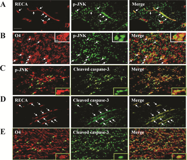
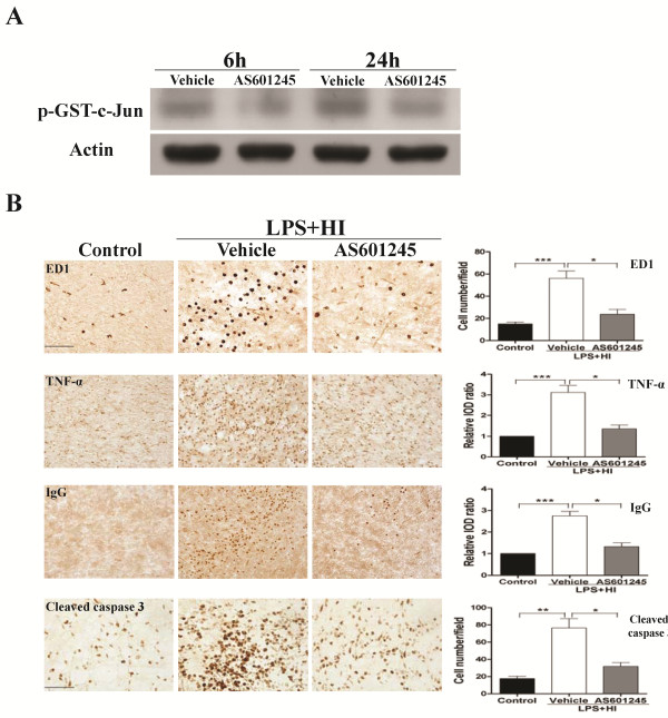
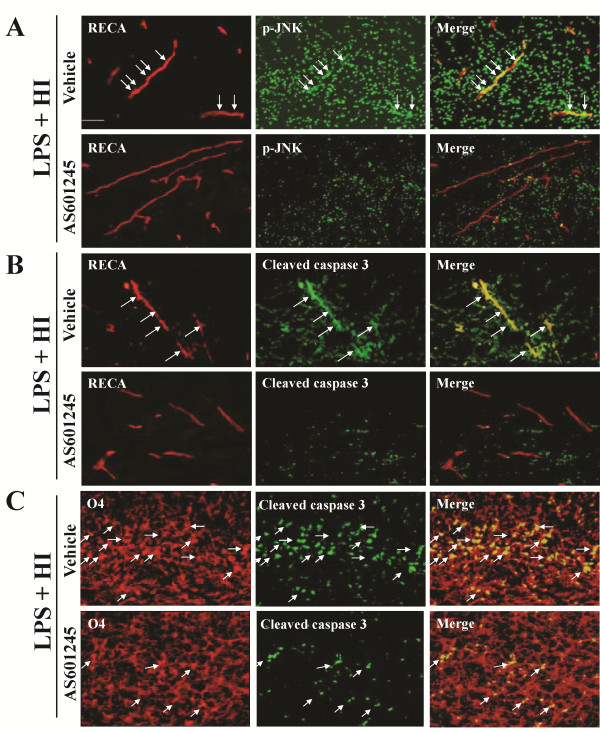
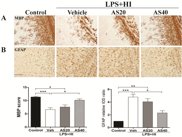
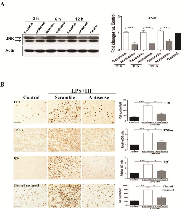
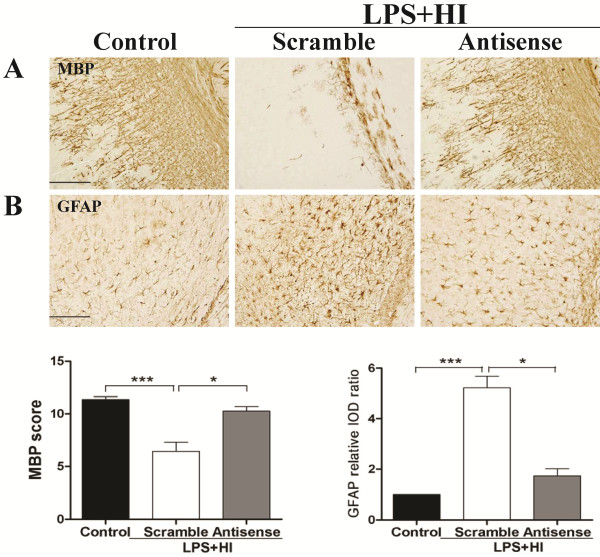
BACKGROUND: White matter injury is the major form of brain damage in very preterm infants. Selective white matter injury in the immature brain can be induced by lipopolysaccharide (LPS)-sensitized hypoxic-ischemia (HI) in the postpartum (P) day 2 rat pups whose brain maturation status is equivalent to that in preterm infants less than 30 weeks of gestation. Neuroinflammation, blood-brain barrier (BBB) damage and oligodendrocyte progenitor apoptosis may affect the susceptibility of LPS-sensitized HI in white matter injury. c-Jun N-terminal kinases (JNK) are important stress-responsive kinases in various forms of insults. We hypothesized that LPS-sensitized HI causes white matter injury through JNK activation-mediated neuroinflammation, BBB leakage and oligodendroglial apoptosis in the white matter of P2 rat pups. METHODS: P2 pups received LPS (0.05 mg/kg) or normal saline injection followed by 90-min HI. Immunohistochemistry and immunoblotting were used to determine microglia activation, TNF-α, BBB damage, cleaved caspase-3, JNK and phospho-JNK (p-JNK), myelin basic protein (MBP), and glial fibrillary acidic protein (GFAP) expression. Immunofluorescence was performed to determine the cellular distribution of p-JNK. Pharmacological and genetic approaches were used to inhibit JNK activity. RESULTS: P2 pups had selective white matter injury associated with upregulation of activated microglia, TNF-α, IgG extravasation and oligodendroglial progenitor apoptosis after LPS-sensitized HI. Immunohistochemical analyses showed early and sustained JNK activation in the white matter at 6 and 24 h post-insult. Immunofluorescence demonstrated upregulation of p-JNK in activated microglia, vascular endothelial cells and oligodendrocyte progenitors, and also showed perivascular aggregation of p-JNK-positive cells around the vessels 24 h post-insult. JNK inhibition by AS601245 or by antisense oligodeoxynucleotides (ODN) significantly reduced microglial activation, TNF-α immunoreactivity, IgG extravasation, and cleaved caspase-3 in the endothelial cells and oligodendrocyte progenitors, and also attenuated perivascular aggregation of p-JNK-positive cells 24 h post-insult. The AS601245 or JNK antisense ODN group had significantly increased MBP and decreased GFAP expression in the white matter on P11 than the vehicle or scrambled ODN group. CONCLUSIONS: LPS-sensitized HI causes white matter injury through JNK activation-mediated upregulation of neuroinflammation, BBB leakage and oligodendrocyte progenitor apoptosis in the immature brain.
Overview
The claims and statistics of this paper haven’t been checked yet.
Run the check — free, no account needed. It reads the open-access text and checks the sources it cites.
Integrity signals
Registry
Cites
Statistics
Figures
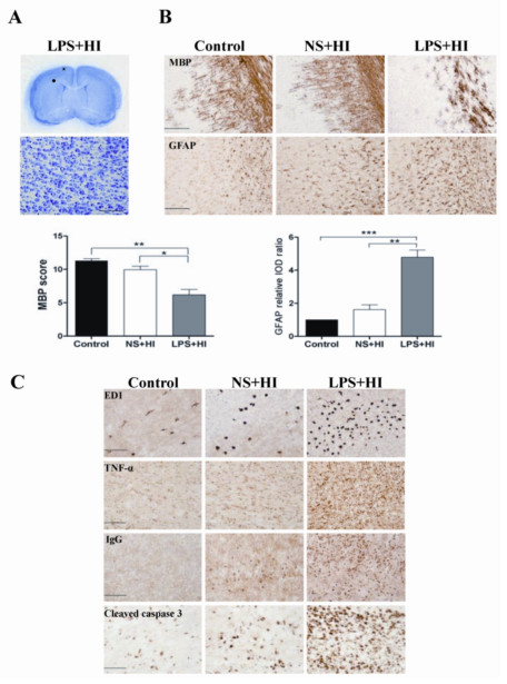
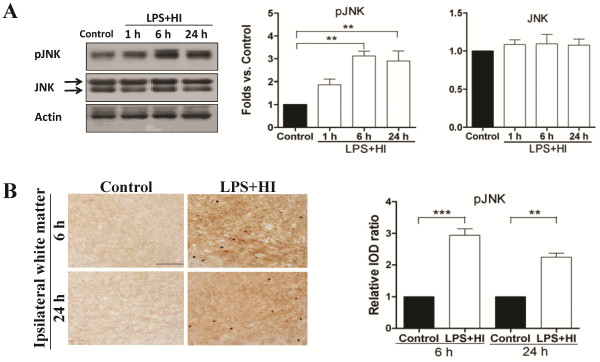
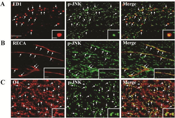
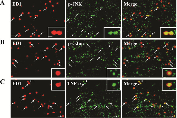
Verification
Claims the paper makes
The claims and statistics of this paper haven’t been checked yet.
Run the check — free, no account needed. It reads the open-access text and checks the sources it cites.
Claims other papers make about this one
Each is checked against this paper’s own text: does it say what the citing paper says it does?
In the citing paper
“Recently, the neuroinflammatory state was shown to activate the c-Jun N-terminal kinase (JNK) pathway not only in microglia but also in ECs, leading to microglial and endothelial activation with direct and microglial-induced BBB disruption, further cytokine production and perpetuation of the neuroinflammatory process (Tu et al., 2011; Wang et al., 2012).”
From The impact of microglial activation on blood-brain barrier in brain diseases (2014) · cites this paper as Wang et al., 2012
Self-check
Percentages, sums and reported differences re-computed from the paper’s own numbers.
The self-check needs the paper’s full text.
Citations
Cites
The rat brain in stereotaxic coordinates
Palkovits, M. (1983) · Neuropeptides · doi:10.1016/0143-4179(83)90049-5 · Cited by 28,959
The Rat Brain in Stereotaxic Coordinates
Paxinos, G., & Watson, C. (1983) · Elsevier eBooks · doi:10.1016/c2009-0-63235-9 · Cited by 28,585
AChE-stained horizontal sections of the rat brain in stereotaxic coordinates
Paxinos, G., WATSON, C. R. R., & Emson, P. C. (1980) · Journal of Neuroscience Methods · doi:10.1016/0165-0270(80)90021-7 · Cited by 4,514
Volpe, J. J. (1981) · PubMed · Cited by 2,859
Brain injury in premature infants: a complex amalgam of destructive and developmental disturbances
Volpe, J. J. (2008) · The Lancet Neurology · doi:10.1016/s1474-4422(08)70294-1 · Cited by 2,501
Kamata, H., Honda, S.-I., Maeda, S., Chang, L., Hirata, H., & Karin, M. (2005) · Cell · doi:10.1016/j.cell.2004.12.041 · Cited by 1,792
Signal transduction by tumor necrosis factor and its relatives.
Baud, V., & Karin, M. (2001) · Trends in Cell Biology · doi:10.1016/s0962-8924(01)02064-5 · Cited by 1,741
Stoll, B. J. (2004) · JAMA · doi:10.1001/jama.292.19.2357 · Cited by 1,535
Lehnardt, S., Massillon, L., Follett, P., Jensen, F. E., Ratan, R., Rosenberg, P. A., Volpe, J. J., & Vartanian, T. (2003) · Proceedings of the National Academy of Sciences · doi:10.1073/pnas.1432609100 · Cited by 1,052
Back, S. A., Luo, N. L., Borenstein, N. S., Levine, J. M., Volpe, J. J., & Kinney, H. C. (2001) · Journal of Neuroscience · doi:10.1523/jneurosci.21-04-01302.2001 · Cited by 936
Cited by
Sorted by citations
The role of inflammation in perinatal brain injury
Hagberg, H., Mallard, C. E., Ferriero, D. M., Vannucci, S. J., Levison, S. W., Vexler, Z. S., & Gressèns, P. (2015) · Nature Reviews Neurology · doi:10.1038/nrneurol.2015.13 · Cited by 852
Zhou, D., Huang, C., Lin, Z., Zhan, S., Kong, L., Fang, C., & Li, J. (2014) · Cellular Signalling · doi:10.1016/j.cellsig.2013.11.004 · Cited by 778
Self-citationThe impact of microglial activation on blood-brain barrier in brain diseases
da Fonseca, A. C. C., Matias, D., Garcia, C., Amaral, R., Geraldo, L. H., Freitas, C., & Lima, F. R. S. (2014) · Frontiers in Cellular Neuroscience · doi:10.3389/fncel.2014.00362 · Cited by 572
Role of Inflammation and Its Mediators in Acute Ischemic Stroke
Jin, R., Liu, L., Zhang, S., Nanda, A., & Li, G. (2013) · Journal of Cardiovascular Translational Research · doi:10.1007/s12265-013-9508-6 · Cited by 422
van Tilborg, E., de Theije, C. G. M., van Hal, M., Wagenaar, N., de Vries, L. S., Benders, M. J. N. L., Rowitch, D. H., & Nijboer, C. H. A. (2017) · Glia · doi:10.1002/glia.23256 · Cited by 321
Amin, F. U., Shah, S. A., Badshah, H., Khan, M., & Kim, M. O. (2017) · Journal of Nanobiotechnology · doi:10.1186/s12951-016-0227-4 · Cited by 161
Role of Oxidative Stress in Blood–Brain Barrier Disruption and Neurodegenerative Diseases
Kim, S., Jung, U. J., & Kim, S. R. (2024) · Antioxidants · doi:10.3390/antiox13121462 · Cited by 189
Impaired oligodendrocyte maturation in preterm infants: Potential therapeutic targets
van Tilborg, E., Heijnen, C. J., Benders, M. J. N. L., van Bel, F., Fleiss, B., Gressèns, P., & Nijboer, C. H. A. (2015) · Progress in Neurobiology · doi:10.1016/j.pneurobio.2015.11.002 · Cited by 159
Liu, X., Sui, B., & Sun, J. (2017) · Biomaterials · doi:10.1016/j.biomaterials.2017.01.006 · Cited by 140
Badshah, H., Ali, T., Rehman, S. U., Amin, F.-U., Ullah, F., Kim, T. H., & Kim, M. O. (2015) · Journal of Neuroimmune Pharmacology · doi:10.1007/s11481-015-9623-z · Cited by 125
Kao, T.-K., Chang, C.-Y., Ou, Y., Chen, W., Kuan, Y., Pan, H.-C., Liao, S., Guo-zhang, L., & Chen, C. (2013) · Experimental Neurology · doi:10.1016/j.expneurol.2013.04.010 · Cited by 116
Çelik, H., Küçükler, S., Çomaklı, S., Özdemir, S., Çağlayan, C., Yardım, A., & Kandemır, F. M. (2019) · NeuroToxicology · doi:10.1016/j.neuro.2019.11.004 · Cited by 112
Subedi, L., Venkatesan, R., & Kim, S. Y. (2017) · International Journal of Molecular Sciences · doi:10.3390/ijms18071423 · Cited by 109
Liao, B., Geng, L., Zhang, F., Shu, L., Wei, L., Yeung, P. K. K., Lam, K. S. L., Chung, S. K., Chang, J., Vanhoutte, P. M., et al. (2020) · European Heart Journal · doi:10.1093/eurheartj/ehaa207 · Cited by 107
Li, D., Liu, N., Zhao, H., Zhang, X., Kawano, H., Liu, L., Zhao, L., & Li, H.-P. (2017) · Journal of Neuroinflammation · doi:10.1186/s12974-017-0841-6 · Cited by 102
Wang, L.-Y., Tu, Y., Lin, Y., & Huang, C. (2016) · Journal of Neuroinflammation · doi:10.1186/s12974-015-0474-6 · Cited by 85
Self-citationKim, H. J., & Chuang, D. (2014) · Europe PMC (PubMed Central) · Cited by 79
Shah, F. A., Kury, L. A., Li, T., Zeb, A., Koh, P., Liu, F., Zhou, Q., Hussain, I., Khan, A., Jiang, Y., et al. (2019) · Frontiers in Pharmacology · doi:10.3389/fphar.2019.00663 · Cited by 76
Liao, Z., Cao, D., Han, X., Liu, C., Peng, J., Zuo, Z., Wang, F., & Li, Y. (2014) · Brain Research Bulletin · doi:10.1016/j.brainresbull.2014.07.001 · Cited by 74
Apolipoprotein A ‐ I : Insights from redox proteomics for its role in neurodegeneration
Keeney, J. T. R., Swomley, A. M., Förster, S., Harris, J. L., Rukhsana Sultana, A., & Butterfield, D. A. (2012) · PROTEOMICS - CLINICAL APPLICATIONS · doi:10.1002/prca.201200087 · Cited by 74
Polymorphonuclear Neutrophil in Brain Parenchyma After Experimental Intracerebral Hemorrhage
Zhao, X., Sun, G., Zhang, H., Ting, S., Shen, S., Gonzales, N. R., & Aronowski, J. A. (2014) · Translational Stroke Research · doi:10.1007/s12975-014-0341-2 · Cited by 70
Hu, Y., Wang, Z., Pan, S., Zhang, H., Fang, M., Jiang, H., Zhang, H., Gao, Z., Xu, K., Li, Z., et al. (2017) · Oncotarget · doi:10.18632/oncotarget.15780 · Cited by 69
Hase, Y., Craggs, L., Hase, M., Stevenson, W., Slade, J. Y., Lopez, D., Mehta, R., Chen, A., Liang, D., Oakley, A. E., et al. (2017) · Journal of Neuroinflammation · doi:10.1186/s12974-017-0850-5 · Cited by 68
Athanasoulis, C. A., Moncure, A. C., Greenfield, A. J., Ryan, J. A., & Dodson, T. F. (1980) · PubMed · Cited by 66
White Matter Injury After Intracerebral Hemorrhage
Fu, X., Zhou, G., Zhuang, J., Xu, C., Zhou, H., Peng, Y., Cao, Y., Zeng, H., Li, J., Yan, F., et al. (2021) · Frontiers in Neurology · doi:10.3389/fneur.2021.562090 · Cited by 65
Self-citationWang, L., Chang, Y., Chen, S., Tseng, C.-H., Tu, Y., Liao, N., Huang, C., & Ho, C.-J. (2014) · Journal of Neuroinflammation · doi:10.1186/s12974-014-0215-2 · Cited by 62
Self-citationCerebral Ischemic Preconditioning: the Road So Far…
Thushara Vijayakumar, N., Sangwan, A., Sharma, B., Majid, A., & Rajanikant, G. K. (2015) · Molecular Neurobiology · doi:10.1007/s12035-015-9278-z · Cited by 60
Zhang, X., Chen, X., Lin, J., Xiong, Y., Liao, W., & Wan, Q. (2016) · Brain Research · doi:10.1016/j.brainres.2016.11.001 · Cited by 59
Cao, Y., Ni, C., Li, Z., Li, L., Liu, Y., Wang, C., Zhong, Y., Cui, D., & Guo, X. (2014) · Neuroscience Letters · doi:10.1016/j.neulet.2014.12.018 · Cited by 58
Amin, F. U., Shah, S. A., & Kim, M. O. (2016) · Neurochemistry International · doi:10.1016/j.neuint.2016.04.001 · Cited by 57
Singh, A., Upadhayay, S., & Mehan, S. (2021) · Neurotoxicity Research · doi:10.1007/s12640-021-00401-6 · Cited by 53
Hao, Y., Xin, M., Feng, L., Wang, X., Wang, X., Ma, D., & Feng, J. (2020) · Frontiers in Neurology · doi:10.3389/fneur.2020.00812 · Cited by 56
Li, D., Li, X., Wu, J., Li, J., Zhang, L., Xiong, T., Tang, J., Qu, Y., & Mu, D. (2015) · PLoS ONE · doi:10.1371/journal.pone.0132998 · Cited by 53
RetractedHuang, F., Liu, X., Xu, S., Hu, S., Wang, S., Shi, D., Wang, K., Wang, Z., Lin, Q., Li, S., et al. (2022) · Frontiers in Nutrition · doi:10.3389/fnut.2021.805465 · Cited by 56
Deferoxamine reduces intracerebral hemorrhage-induced white matter damage in aged rats
Ni, W., Okauchi, M., Hatakeyama, T., Gu, Y., Keep, R. F., Xi, G., & Hua, Y. (2015) · Experimental Neurology · doi:10.1016/j.expneurol.2015.02.035 · Cited by 51
JNK Inhibitors As Anti-Inflammatory and Neuroprotective Agents
Graczyk, P. P. (2013) · Future Medicinal Chemistry · doi:10.4155/fmc.13.34 · Cited by 50
HUCBC s Increase Angiopoietin 1 and Induce Neurorestorative Effects after Stroke in T1 DM Rats
Yan, T., Venkat, P., Ye, X., Chopp, M., Zacharek, A., Ning, R., Cui, Y., Roberts, C. J., Kuzmin‐Nichols, N., Sanberg, C. D., et al. (2014) · CNS Neuroscience & Therapeutics · doi:10.1111/cns.12307 · Cited by 50
Roles of Macrophages and Their Interactions with Schwann Cells After Peripheral Nerve Injury
Wu, G., Wen, X., Kuang, R., Lui, K. H. W., He, B., Li, G., & Zhu, Z. (2023) · Cellular and Molecular Neurobiology · doi:10.1007/s10571-023-01442-5 · Cited by 49
Macrophage Polarization in IL-10 Treatment of Particle-Induced Inflammation and Osteolysis
Jiang, J., Jia, T., Gong, W., Ning, B., Wooley, P. H., & Yang, S. (2015) · American Journal Of Pathology · doi:10.1016/j.ajpath.2015.09.006 · Cited by 48
Adebiyi, O. E., Olopade, J. O., & Olayemi, F. O. (2018) · Brain and Behavior · doi:10.1002/brb3.1014 · Cited by 48
White Matter Injury in Early Brain Injury after Subarachnoid Hemorrhage
Pang, J., Peng, J., Yang, P., Li, K., Chen, L., Zhang, J. H., & Jiang, Y. Q. (2018) · Cell Transplantation · doi:10.1177/0963689718812054 · Cited by 44
Wei, L., Chen, Y., Yu, Q., Wang, Y., & Liu, C. (2017) · Brain Research · doi:10.1016/j.brainres.2017.12.039 · Cited by 43
Huang, F., Chen, Z., Chen, H., Lu, W., Xie, S., Meng, Q. H., Wu, Y., & Xia, D. (2018) · Toxicological Sciences · doi:10.1093/toxsci/kfy039 · Cited by 43
Zhang, J., Lin, W., Tang, M., Zhao, Y., Zhang, K., Wang, X., & Li, Y. (2019) · Psychoneuroendocrinology · doi:10.1016/j.psyneuen.2019.104580 · Cited by 44
Khan, M. S., Khan, A., Ahmad, S., Ahmad, R., Rehman, I. U. R., Ikram, M. A., & Kim, M. O. (2020) · Oxidative Medicine and Cellular Longevity · doi:10.1155/2020/5291852 · Cited by 43
RetractedLi, L., Alattar, A., Tan, Z., Shah, F. A., Ali, T., Alshaman, R., Koh, P., & Li, S. (2020) · Frontiers in Pharmacology · doi:10.3389/fphar.2020.01220 · Cited by 42
Glass, T. J. A., Chau, V., GRUNAU, R. E., Synnes, A. R., Guo, T., Duerden, E. G., Foong, J., Poskitt, K. J., & Miller, S. P. (2018) · The Journal of Pediatrics · doi:10.1016/j.jpeds.2017.12.041 · Cited by 40
Sesamin suppresses LPS-induced microglial activation via regulation of TLR4 expression
Udomruk, S., Kaewmool, C., Pothacharoen, P., Phitak, T., & Kongtawelert, P. (2018) · Journal of Functional Foods · doi:10.1016/j.jff.2018.08.020 · Cited by 39
Current Evidence on Cell Death in Preterm Brain Injury in Human and Preclinical Models
Truttmann, A. C., Ginet, V., & Puyal, J. (2020) · Frontiers in Cell and Developmental Biology · doi:10.3389/fcell.2020.00027 · Cited by 38
Li, C., Shi, L., Wang, Y., Peng, C., Wu, L., Zhang, Y., & Du, Z. (2021) · Environmental Toxicology · doi:10.1002/tox.23137 · Cited by 43
Chierto, E., Simon, A., Castoldi, F., Meffre, D., Cristinziano, G., Sapone, F., Carreté, A., Borderie, D., Étienne, F., Rannou, F., et al. (2018) · Molecular Neurobiology · doi:10.1007/s12035-018-1372-6 · Cited by 33
Chu, J. M. T., Lee, K. M., Wong, D. P. K., Wong, G. T. C., & Yue, K. K. M. (2016) · Metabolic Brain Disease · doi:10.1007/s11011-016-9849-3 · Cited by 34
Alam, M. S., Rallabandi, V. P. S., & Roy, P. K. (2016) · Frontiers in Neurology · doi:10.3389/fneur.2016.00094 · Cited by 33
Yu, H., Li, W., Cao, X., Wang, X., Zhao, Y., Song, L., Chen, J., wang, S., Chen, B., & Xu, Y. (2019) · European Journal of Pharmacology · doi:10.1016/j.ejphar.2019.172610 · Cited by 31
Silwedel, C., Haarmann, A., Fehrholz, M., Claus, H., Speer, C. P., & Glaser, K. (2019) · Journal of Neuroinflammation · doi:10.1186/s12974-019-1413-8 · Cited by 31
Zhang, M., Gong, J.-X., Wang, J.-L., Jiang, M., Li, L., Hu, Y., Qi, J., Zhang, L., Zhao, H., Cui, X., et al. (2016) · Molecular Neurobiology · doi:10.1007/s12035-015-9652-x · Cited by 27
Wong, S. Y., Tan, M. G. K., Banks, W. A., Wong, W. S. F., Wong, P. T. ‐., & Lai, M. K. P. (2016) · Journal of Neuroinflammation · doi:10.1186/s12974-016-0498-6 · Cited by 27
Yu, H., Zou, Z., Zhang, X., Peng, W., Chen, C., Ye, Y., Xu, J., & Wang, H. (2018) · International Journal of Molecular Sciences · doi:10.3390/ijms19020513 · Cited by 26
Choi, H., Lee, E., Han, M., An, S., & Park, J. (2019) · Frontiers in Neuroscience · doi:10.3389/fnins.2019.01350 · Cited by 24
The role of the endoplasmic reticulum stress in depression
Kowalczyk, M., Majsterek, I., Gałecki, P., & Talarowska, M. E. (2020) · Psychiatria Polska · doi:10.12740/pp/109130 · Cited by 23
Zhao, L., Guo, Y., Ji, X., & Zhang, M. (2014) · BMC Neuroscience · doi:10.1186/1471-2202-15-25 · Cited by 23
Cao, M., Tan, X., Jin, W., Zheng, H., Xu, W., Rui, Y., Li, L., Cao, J., Wu, X., Cui, G., et al. (2013) · Neurochemistry International · doi:10.1016/j.neuint.2013.01.025 · Cited by 23
Mi, X., Cao, Y., Li, Y., Li, Y., Hong, J., He, J., Liang, Y., Yang, N., Liu, T., Han, D., et al. (2021) · Frontiers in Aging Neuroscience · doi:10.3389/fnagi.2021.624387 · Cited by 22
Rosso, P., Nicolò, S. D., Carito, V., Fiore, M., Iannitelli, A., Moreno, S., & Tirassa, P. (2017) · CNS Neuroscience & Therapeutics · doi:10.1111/cns.12661 · Cited by 21
Huang, C.-T., & Tsai, Y.-J. (2016) · Journal of Nutritional Biochemistry · doi:10.1016/j.jnutbio.2015.11.009 · Cited by 22
Self-citationNanou, A., Bourbouli, M., Vetrano, S., Schaeper, U., Ley, S. C., & Kollias, G. A. (2019) · Cell Reports · doi:10.1016/j.celrep.2021.109168 · Cited by 19
Chu, J. M. T., Abulimiti, A., Wong, B. S., Zhao, G. D., Xiong, S. H., Zhao, M., Wang, Y., Chen, Y., Wang, J., Zhang, Y., et al. (2022) · Frontiers in Pharmacology · doi:10.3389/fphar.2022.846631 · Cited by 19
Yin, X., Zhao, J., Jiang, H., Li, L., Jiang, J., Xi, H., Peng, X., Yin, X., Shi, X., & Zhang, L. (2018) · Neonatology · doi:10.1159/000487220 · Cited by 17
Bhujbal, S. P., & Hah, J. (2025) · Archives of Pharmacal Research · doi:10.1007/s12272-025-01566-w · Cited by 17
Ball, B. K., Kuhn, M. K., Fleeman, R. M., Proctor, E. A., & Brubaker, D. K. (2024) · Scientific Reports · doi:10.1038/s41598-024-62155-3 · Cited by 16
JNK inhibitors: is there a future?
Cicenas, J. (2015) · MAP Kinase · doi:10.4081/mk.2015.5700 · Cited by 15
Canedo-Antelo, M., Serrano‐Regal, M. P., Manterola, A., Ruiz, A., Llavero, F., Mato, S., Zugaza, J. L., Pérez‐Cerdá, F., Matute, C., & Sánchez‐Gómez, M. V. (2018) · Frontiers in Molecular Neuroscience · doi:10.3389/fnmol.2018.00333 · Cited by 15
Favrais, G., Bokobza, C., Saliba, É., Chalon, S., & Gressèns, P. (2022) · Frontiers in Physiology · doi:10.3389/fphys.2022.881674 · Cited by 15
Yao, F., Zhang, M., & Chen, L. (2017) · Journal of Diabetes Research · doi:10.1155/2017/4504612 · Cited by 15
Плотников, М. Б., Анищенко, А. М., Khlebnikov, А. I., & Schepetkin, I. A. (2025) · Molecules · doi:10.3390/molecules30112353 · Cited by 13
Subedi, L., & Yumnam, S. (2021) · International Journal of Molecular Sciences · doi:10.3390/ijms22020965 · Cited by 13
Lorenzati, M., Boda, E., Parolisi, R., Bonato, M., Borsello, T., Herdegen, T., Buffo, A., & Vercelli, A. (2021) · Scientific Reports · doi:10.1038/s41598-021-86673-6 · Cited by 14
Gao, H., Findeis, E. L., Culmone, L., Powell, B., Landschoot‐Ward, J., Zacharek, A., Wu, T., Lü, M., Chopp, M., & Venkat, P. (2023) · Frontiers in Aging Neuroscience · doi:10.3389/fnagi.2023.1180913 · Cited by 13
Sarode, L. P., Ghatage, T., Mardhekar, V. Y., VERMA, B. L., Prakash, A., & Ugale, R. R. (2023) · Metabolic Brain Disease · doi:10.1007/s11011-023-01240-4 · Cited by 13
Huang, J. B., Yang, J., Zou, X., Zuo, S., Wang, J., Cheng, J., Zhu, H., Li, W., Shi, M., Zhao, G., et al. (2021) · Experimental Biology and Medicine · doi:10.1177/1535370221989955 · Cited by 14
Mao, F., Li, W.-J., Chen, H., Qian, L.-H., & Buzby, J. S. (2013) · Brain Research · doi:10.1016/j.brainres.2013.08.006 · Cited by 13
Erythropoietin reduces white matter damage in two-day-old rats exposed to hypoxic/ischemia injury
Ren, Q., Zhang, X., & Yang, J. (2016) · Neurological Research · doi:10.1080/01616412.2016.1242451 · Cited by 13
Shi, J., Liu, X., Xu, C., Ge, J., Ren, J., Wang, J., Song, X., Dai, S., Tao, W., & Lu, H. (2015) · Journal of Molecular Histology · doi:10.1007/s10735-015-9637-0 · Cited by 12
Zhang, Y., Liu, X., Xue, H., Liu, X., Dai, A., Song, Y., Ke, K., & Cao, M. (2016) · Journal of Molecular Neuroscience · doi:10.1007/s12031-016-0744-5 · Cited by 12
Martini, A. C., Forner, S., Koepp, J., & Rae, G. A. (2016) · Neuroscience Letters · doi:10.1016/j.neulet.2016.04.017 · Cited by 11
Udomruk, S., Wudtiwai, B., Shwe, T. H., Phitak, T., Pothacharoen, P., Phimphilai, M., & Kongtawelert, P. (2021) · Brain Research Bulletin · doi:10.1016/j.brainresbull.2021.04.012 · Cited by 10
Choi, J. Y., Yun, J., Hwang, C. J., Lee, H. P., Kim, H. D., Chun, H., Park, P., Choi, D. Y., Han, S., & Hong, J. T. (2019) · International Journal of Molecular Sciences · doi:10.3390/ijms20112632 · Cited by 8
JNK signaling provides a novel therapeutic target for Rett syndrome
Musi, C. A., Castaldo, A., Valsecchi, A. E., Cimini, S., Morello, N., Pizzo, R., Renieri, A., Meloni, I., Bonati, M. R., Giustetto, M., et al. (2021) · BMC Biology · doi:10.1186/s12915-021-01190-2 · Cited by 8
Cyclooxygenase 2 Genetic Polymorphism May Increase the Risk of Developing Leukoaraiosis in Chinese
Shan, X., Chen, G., Cheng, G.-P., & Tao, H. (2013) · Journal of Molecular Neuroscience · doi:10.1007/s12031-013-0066-9 · Cited by 8
Shin, J. A., Park, H., Choi, H., Chang, Y., Kim, J.-J., Ham, Y. R., Na, K. R., Lee, K. W., & Choi, D. E. (2023) · International Journal of Molecular Sciences · doi:10.3390/ijms241512168 · Cited by 7
Acupuncture Alleviates Chronic Ischemic White Matter Injury in SHR Rats via JNK-NMDAR Circuit
Dong, A., Gao, Z., Wang, H.-J., Wu, R., Wang, W., Jin, X., Ji, Y., Yang, F., Zhu, T., Jiang, Z., et al. (2023) · Molecular Neurobiology · doi:10.1007/s12035-023-03759-0 · Cited by 7
Zhao, S., Qi, R., Gao, X., & Chen, H. (2022) · Experimental Dermatology · doi:10.1111/exd.14580 · Cited by 7
Liu, X., Li, Q., Cheng, X., Liu, Z., Zhao, X., Zhang, S., Yu, G., Hao, J., & Hao, J. (2020) · Chinese Medicine · doi:10.1186/s13020-020-00310-4 · Cited by 7
Da, L., Evangelou, A. V., Shankar, K., Dewji, F. I., Lin, J., & Levison, S. W. (2022) · SSRN Electronic Journal · doi:10.2139/ssrn.4016993 · Cited by 6
Zhang, Z., Zhou, M., Liu, N., Shi, Z., Pang, Y., Li, D., Qi, J., Wu, H., & An, R. (2019) · Brain Research Bulletin · doi:10.1016/j.brainresbull.2019.03.012 · Cited by 5
Ali, W., Choe, K., Rehman, I. U., Park, H. Y., Jang, S., Ullah, S., Park, T. J., & Kim, M. O. (2025) · Journal of Neuroimmune Pharmacology · doi:10.1007/s11481-025-10243-0 · Cited by 4
Shi, L., Wang, Z., Li, Y., Song, Z., Wu, Y., & Hu, B. (2023) · International Journal of Molecular Sciences · doi:10.3390/ijms241713535 · Cited by 4
Advanced Diffusion Tensor Imaging in White Matter Injury After Subarachnoid Hemorrhage
Yang, L., Peng, J., Zhang, L., Zhang, F., Wu, J., Zhang, X., Pang, J., & Jiang, Y. Q. (2024) · World Neurosurgery · doi:10.1016/j.wneu.2024.05.107 · Cited by 4
Wang, L., Hsiung, C.-W., Chang, C., Lin, M., & Chen, S. (2024) · Pediatric Research · doi:10.1038/s41390-024-03412-z · Cited by 4
Self-citationLi, H., Terrando, N., & Gelbard, H. A. (2024) · Advances in neurobiology · doi:10.1007/978-3-031-55529-9_24 · Cited by 4
Metadata
- Authors
- Wang, L.-W., Tu, Y.-F., Huang, C.-C., & Ho, C.-J.
- Affiliations
- National Cheng Kung University (TW)
- Corresponding author
- Huang, Chao-Ching
- Venue
- Journal of Neuroinflammation
- Published
- 2012-07-17
- Volume
- 9
- Issue
- 1
- Pages
- 175-175
- ISSN
- 1742-2094 (ISSN-L)
- PMC
- PMC3414763
- OpenAlex
- W1971748792
- Cited by
- 119
- References
- 72
- Citations by year
- 2026: 4 · 2025: 8 · 2024: 6 · 2023: 7 · 2022: 5 · 2021: 11 · 2020: 8 · 2019: 9 · 2018: 10 · 2017: 10 · 2016: 11 · 2015: 11 · 2014: 8 · 2013: 9 · 2012: 1
- FWCI
- 8.15
- Type
- journal article
- Language
- en
- Open access
- gold
- doi.org
- Locations
- Journal of Neuroinflammation (publishedVersion, cc-by)
- PubMed Central (publishedVersion, cc-by)
- PubMed (publishedVersion)
- DOAJ (DOAJ: Directory of Open Access Journals) (submittedVersion)
- Funders
- National Science Council
- National Health Research Institutes
- Chi Mei Medical Center
- Topics
- Health Sciences › Medicine › Pediatrics, Perinatology and Child Health › Neonatal and fetal brain pathology
- Life Sciences › Neuroscience › Neurology › Neuroinflammation and Neurodegeneration Mechanisms
- Health Sciences › Medicine › Clinical Neurology › Traumatic Brain Injury and Neurovascular Disturbances
- Keywords
- JNK signaling, white matter injury, neuroinflammation, blood-brain barrier disruption, hypoxia-ischemia, preterm infants, microglial activation, TNF-α, IgG extravasation, JNK inhibition, oligodeoxynucleotides
- MeSH terms
- Animals, Animals, Newborn, Blood-Brain Barrier, Brain, Inflammation, Nerve Fibers, Myelinated, Oligodendroglia, Rats, Sprague-Dawley, Apoptosis, Hypoxia-Ischemia, Brain, MAP Kinase Signaling System, Rats
About these checks
Registry, retraction, Cites and statistics results are computed, not judged, with one exception: a printed reference that no index lists is read once by a model before it counts as a citation error. Claims are extracted by a model and kept only when their quote appears verbatim in the text; each cited source is then checked, with a model’s help, for whether it says what the claim relies on. Those verdicts can be wrong — read the claim’s quote and the cited source before relying on one.
Statistics and arithmetic are recomputed, not judged.
Confidence bands are shown only for fields calibrated on our annotated test set. None is calibrated yet, so no band is shown.
What counts as a claim ›How we measure confidence ›How we check this ›
Are you an author, or do you see a problem with this page? Request a review
