

The DOI matches its Crossref record; 70 of the 72 references it cites were found in a registry or an index.
JNK signaling is the shared pathway linking neuroinflammation, blood-brain barrier disruption and oligodendroglial apoptosis in the white matter injury of the immature brain
Open accessAbstract
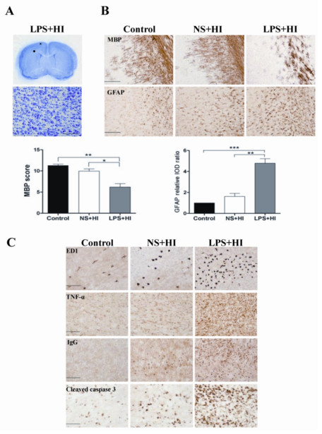
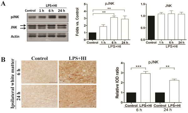
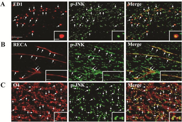
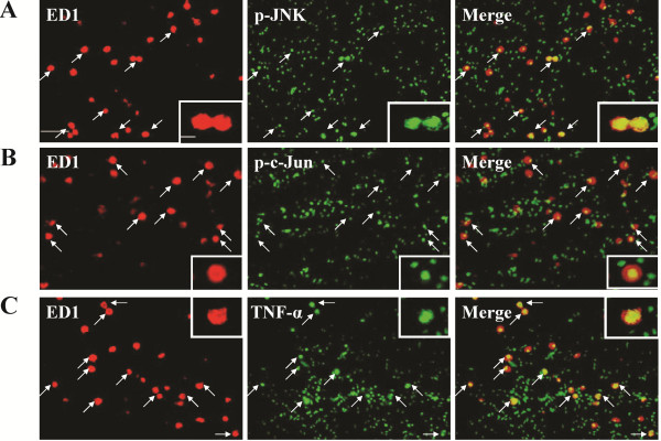
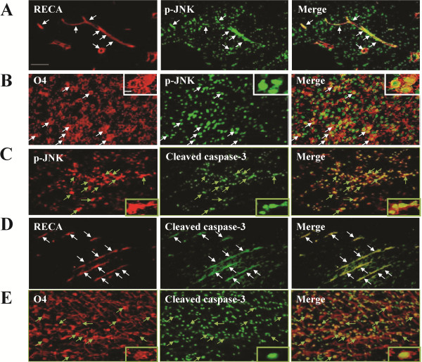
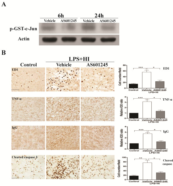
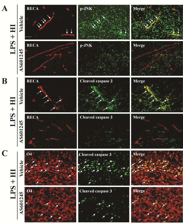
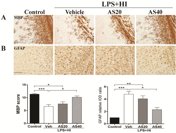
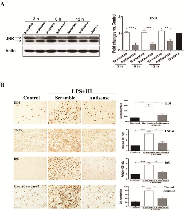
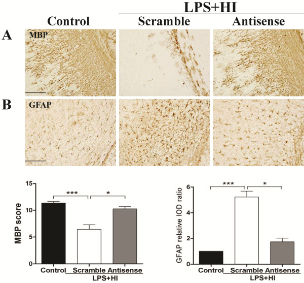
BACKGROUND: White matter injury is the major form of brain damage in very preterm infants. Selective white matter injury in the immature brain can be induced by lipopolysaccharide (LPS)-sensitized hypoxic-ischemia (HI) in the postpartum (P) day 2 rat pups whose brain maturation status is equivalent to that in preterm infants less than 30 weeks of gestation. Neuroinflammation, blood-brain barrier (BBB) damage and oligodendrocyte progenitor apoptosis may affect the susceptibility of LPS-sensitized HI in white matter injury. c-Jun N-terminal kinases (JNK) are important stress-responsive kinases in various forms of insults. We hypothesized that LPS-sensitized HI causes white matter injury through JNK activation-mediated neuroinflammation, BBB leakage and oligodendroglial apoptosis in the white matter of P2 rat pups. METHODS: P2 pups received LPS (0.05 mg/kg) or normal saline injection followed by 90-min HI. Immunohistochemistry and immunoblotting were used to determine microglia activation, TNF-α, BBB damage, cleaved caspase-3, JNK and phospho-JNK (p-JNK), myelin basic protein (MBP), and glial fibrillary acidic protein (GFAP) expression. Immunofluorescence was performed to determine the cellular distribution of p-JNK. Pharmacological and genetic approaches were used to inhibit JNK activity. RESULTS: P2 pups had selective white matter injury associated with upregulation of activated microglia, TNF-α, IgG extravasation and oligodendroglial progenitor apoptosis after LPS-sensitized HI. Immunohistochemical analyses showed early and sustained JNK activation in the white matter at 6 and 24 h post-insult. Immunofluorescence demonstrated upregulation of p-JNK in activated microglia, vascular endothelial cells and oligodendrocyte progenitors, and also showed perivascular aggregation of p-JNK-positive cells around the vessels 24 h post-insult. JNK inhibition by AS601245 or by antisense oligodeoxynucleotides (ODN) significantly reduced microglial activation, TNF-α immunoreactivity, IgG extravasation, and cleaved caspase-3 in the endothelial cells and oligodendrocyte progenitors, and also attenuated perivascular aggregation of p-JNK-positive cells 24 h post-insult. The AS601245 or JNK antisense ODN group had significantly increased MBP and decreased GFAP expression in the white matter on P11 than the vehicle or scrambled ODN group. CONCLUSIONS: LPS-sensitized HI causes white matter injury through JNK activation-mediated upregulation of neuroinflammation, BBB leakage and oligodendrocyte progenitor apoptosis in the immature brain.
Overview
The claims and statistics of this paper haven’t been checked yet.
Run the check — free, no account needed. It reads the open-access text and checks the sources it cites.
Integrity signals
Registry
Cites
Statistics
Figures
Verification
Claims the paper makes
The claims and statistics of this paper haven’t been checked yet.
Run the check — free, no account needed. It reads the open-access text and checks the sources it cites.
Claims other papers make about this one
Each is checked against this paper’s own text: does it say what the citing paper says it does?
In the citing paper
“Recently, the neuroinflammatory state was shown to activate the c-Jun N-terminal kinase (JNK) pathway not only in microglia but also in ECs, leading to microglial and endothelial activation with direct and microglial-induced BBB disruption, further cytokine production and perpetuation of the neuroinflammatory process (Tu et al., 2011; Wang et al., 2012).”
From The impact of microglial activation on blood-brain barrier in brain diseases (2014) · cites this paper as Wang et al., 2012
Self-check
Percentages, sums and reported differences re-computed from the paper’s own numbers.
The self-check needs the paper’s full text.
Citations
Cites
The rat brain in stereotaxic coordinates
Palkovits, M. (1983) · Neuropeptides · doi:10.1016/0143-4179(83)90049-5 · Cited by 28,959
The Rat Brain in Stereotaxic Coordinates
Paxinos, G., & Watson, C. (1983) · Elsevier eBooks · doi:10.1016/c2009-0-63235-9 · Cited by 28,585
AChE-stained horizontal sections of the rat brain in stereotaxic coordinates
Paxinos, G., WATSON, C. R. R., & Emson, P. C. (1980) · Journal of Neuroscience Methods · doi:10.1016/0165-0270(80)90021-7 · Cited by 4,514
Volpe, J. J. (1981) · PubMed · Cited by 2,859
Brain injury in premature infants: a complex amalgam of destructive and developmental disturbances
Volpe, J. J. (2008) · The Lancet Neurology · doi:10.1016/s1474-4422(08)70294-1 · Cited by 2,501
Kamata, H., Honda, S.-I., Maeda, S., Chang, L., Hirata, H., & Karin, M. (2005) · Cell · doi:10.1016/j.cell.2004.12.041 · Cited by 1,792
Signal transduction by tumor necrosis factor and its relatives.
Baud, V., & Karin, M. (2001) · Trends in Cell Biology · doi:10.1016/s0962-8924(01)02064-5 · Cited by 1,741
Stoll, B. J. (2004) · JAMA · doi:10.1001/jama.292.19.2357 · Cited by 1,535
Lehnardt, S., Massillon, L., Follett, P., Jensen, F. E., Ratan, R., Rosenberg, P. A., Volpe, J. J., & Vartanian, T. (2003) · Proceedings of the National Academy of Sciences · doi:10.1073/pnas.1432609100 · Cited by 1,052
Back, S. A., Luo, N. L., Borenstein, N. S., Levine, J. M., Volpe, J. J., & Kinney, H. C. (2001) · Journal of Neuroscience · doi:10.1523/jneurosci.21-04-01302.2001 · Cited by 936
Cited by
Sorted by citations
Items 101–119 of 119
Hammoodi, H. Z., & Al-Shawi, N. N. (2023) · Journal of Medicine and Life · doi:10.25122/jml-2023-0082 · Cited by 4
Xue, H., Liu, J., Shi, L., & Yang, H. (2020) · American Journal of Physiology-Cell Physiology · doi:10.1152/ajpcell.00576.2019 · Cited by 4
RetractedZhou, M., Li, H., Wan, M., Ji, Z., Liu, Z., Wan, Z., Cai, J., Shi, J., Qiu, X., & Zhang, X. (2026) · International Journal of Biological Macromolecules · doi:10.1016/j.ijbiomac.2026.151239 · Cited by 3
Huang, Z., Guo, X., Tan, P., Wang, M., Chen, H., Peng, Y., Xia, X., Tang, X., Li, Q., & Fu, W. (2021) · Journal of Food Biochemistry · doi:10.1111/jfbc.13905 · Cited by 3
Brown, S., Shallie, P. D., Sierra, C. A., Nayak, N., Odibo, A. O., Monaghan‐Nichols, P., & Nayak, N. R. (2025) · International Journal of Molecular Sciences · doi:10.3390/ijms26083862 · Cited by 3
Ball, B. K., Kuhn, M. K., Fleeman, R. M., Proctor, E. A., & Brubaker, D. K. (2025) · Brain Research · doi:10.1016/j.brainres.2025.149819 · Cited by 1
Чернышева, Г. А., Smol’yakova, V. I., Плотников, М. Б., Aliev, O. I., Ulyakhina, O. A., Osipenko, A. N., Kovrizhina, A. R., & Khlebnikov, А. I. (2025) · Bulletin of Experimental Biology and Medicine · doi:10.1007/s10517-025-06459-y · Cited by 1
Alam, M. S., Rallabandi, V. P. S., & Roy, P. K. (2016) · Springer eBooks · doi:10.1007/978-981-10-1711-7_9 · Cited by 1
Chen, R., Yuan, L., Cao, N., Li, P., Chen, H., Zhou, J., Hao, X., Liu, T., Yang, W., Cui, S., et al. (2019) · Journal of Cellular Biochemistry · doi:10.1002/jcb.28687 · Cited by 1
Cattani, D. (2013) · LA Referencia (Red Federada de Repositorios Institucionales de Publicaciones Científicas) · Cited by 1
Early-Life Feeding Exposure Impacts Brain Development in a Neonatal Piglet Model – a Pilot StudyPreprint
Nuñez, L., Fehrenkamp, B. D., McDonough, A., Piaskowski, J. L., & Chen, Y. (2024) · Preprints.org · doi:10.20944/preprints202405.1800.v1
Thioperamide treats neonatal hypoxic-ischemic encephalopathy by postsynaptic H1 receptors.
Jia, F., Du, L., Hao, Y., Liu, S., Li, N., & Jiang, H. (2013) · PubMed Central · doi:10.3969/j.issn.1673-5374.2013.19.009
Oxidative Stress and Inflammatory Response in Early Stage of Diffuse Axonal Injury in Rats
Ma, X., Song, J., & Zhao, Y. (2015) · Journal of Biosciences and Medicines · doi:10.4236/jbm.2015.310002
Glass, T. J. A. (2018) · TSpace (University of Toronto)
Dickerman, R. D., Bennett, M. T., Mathew, E., & East, J. W. (2025) · Expert Review of Neurotherapeutics · doi:10.1080/14737175.2025.2558195
Nomakuchi, T. T., Rippert, A. L., Leon, S. A. S. D., Gonzalez, E. M., Li, D., Angireddy, R., Finoti, L. S., Faletra, F., Musante, L., Rinne, T., et al. (2025) · medRxiv · doi:10.64898/2025.12.23.25342932
Zhang, Z. Y., Liang, F., Wen, D., Chen, H., Gu, N., Li, Z., Wang, L., Wang, Y., Pan, Q., Zhu, Y., et al. (2026) · Experimental Neurology · doi:10.1016/j.expneurol.2026.115667
Self-citationBernardo-Cervantes, A. G., & Castañeda‐Cabral, J. L. (2026) · Neurophysiology · doi:10.1007/s11062-026-10026-0
Nomakuchi, T. T., Rippert, A. L., Leon, S. A. S. D., Gonzalez, E. M., Li, D., Angireddy, R., Finoti, L. S., Faletra, F., Musante, L., Tuula, R., et al. (2026) · The American Journal of Human Genetics · doi:10.1016/j.ajhg.2026.09.014
Metadata
- Authors
- Wang, L.-W., Tu, Y.-F., Huang, C.-C., & Ho, C.-J.
- Affiliations
- National Cheng Kung University (TW)
- Corresponding author
- Huang, Chao-Ching
- Venue
- Journal of Neuroinflammation
- Published
- 2012-07-17
- Volume
- 9
- Issue
- 1
- Pages
- 175-175
- ISSN
- 1742-2094 (ISSN-L)
- PMC
- PMC3414763
- OpenAlex
- W1971748792
- Cited by
- 119
- References
- 72
- Citations by year
- 2026: 4 · 2025: 8 · 2024: 6 · 2023: 7 · 2022: 5 · 2021: 11 · 2020: 8 · 2019: 9 · 2018: 10 · 2017: 10 · 2016: 11 · 2015: 11 · 2014: 8 · 2013: 9 · 2012: 1
- FWCI
- 8.15
- Type
- journal article
- Language
- en
- Open access
- gold
- doi.org
- Locations
- Journal of Neuroinflammation (publishedVersion, cc-by)
- PubMed Central (publishedVersion, cc-by)
- PubMed (publishedVersion)
- DOAJ (DOAJ: Directory of Open Access Journals) (submittedVersion)
- Funders
- National Science Council
- National Health Research Institutes
- Chi Mei Medical Center
- Topics
- Health Sciences › Medicine › Pediatrics, Perinatology and Child Health › Neonatal and fetal brain pathology
- Life Sciences › Neuroscience › Neurology › Neuroinflammation and Neurodegeneration Mechanisms
- Health Sciences › Medicine › Clinical Neurology › Traumatic Brain Injury and Neurovascular Disturbances
- Keywords
- JNK signaling, white matter injury, neuroinflammation, blood-brain barrier disruption, hypoxia-ischemia, preterm infants, microglial activation, TNF-α, IgG extravasation, JNK inhibition, oligodeoxynucleotides
- MeSH terms
- Animals, Animals, Newborn, Blood-Brain Barrier, Brain, Inflammation, Nerve Fibers, Myelinated, Oligodendroglia, Rats, Sprague-Dawley, Apoptosis, Hypoxia-Ischemia, Brain, MAP Kinase Signaling System, Rats
About these checks
Registry, retraction, Cites and statistics results are computed, not judged, with one exception: a printed reference that no index lists is read once by a model before it counts as a citation error. Claims are extracted by a model and kept only when their quote appears verbatim in the text; each cited source is then checked, with a model’s help, for whether it says what the claim relies on. Those verdicts can be wrong — read the claim’s quote and the cited source before relying on one.
Statistics and arithmetic are recomputed, not judged.
Confidence bands are shown only for fields calibrated on our annotated test set. None is calibrated yet, so no band is shown.
What counts as a claim ›How we measure confidence ›How we check this ›
Are you an author, or do you see a problem with this page? Request a review
