

The DOI matches its Crossref record; 85 of the 86 references it cites were found in a registry or an index.
Cutaneous lesions of the external ear
Open accessAbstract
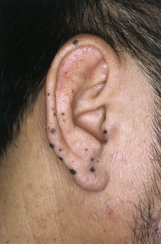
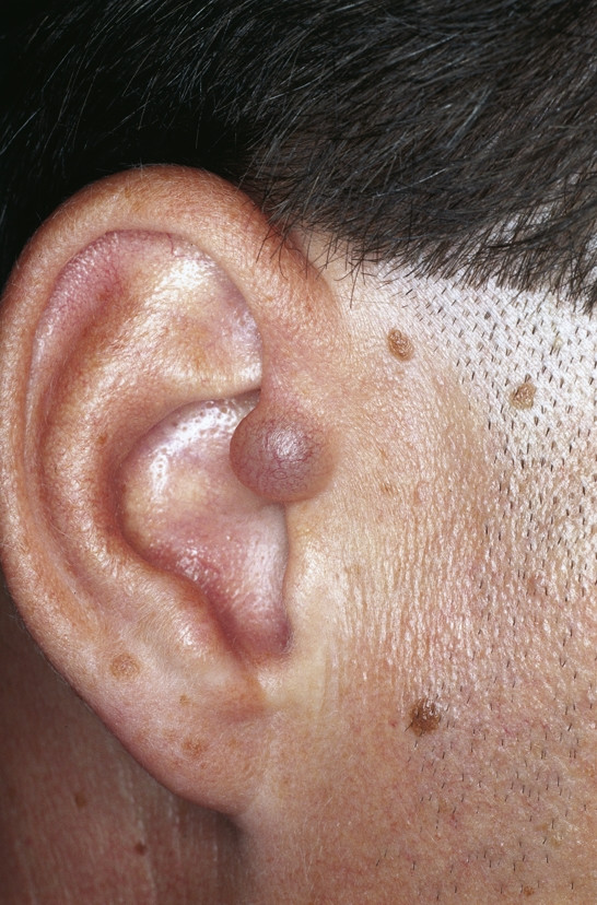
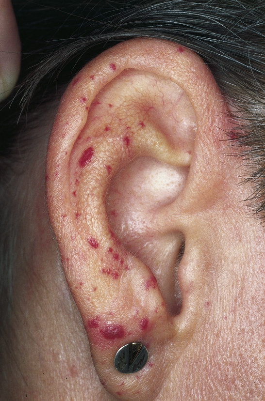
Skin diseases on the external aspect of the ear are seen in a variety of medical disciplines. Dermatologists, othorhinolaryngologists, general practitioners, general and plastic surgeons are regularly consulted regarding cutaneous lesions on the ear. This article will focus on those diseases wherefore surgery or laser therapy is considered as a possible treatment option or which are potentially subject to surgical evaluation.
Overview
The claims and statistics of this paper haven’t been checked yet.
Run the check — free, no account needed. It reads the open-access text and checks the sources it cites.
Integrity signals
Registry
Cites
Statistics
Figures
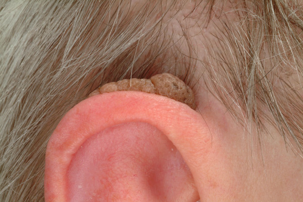
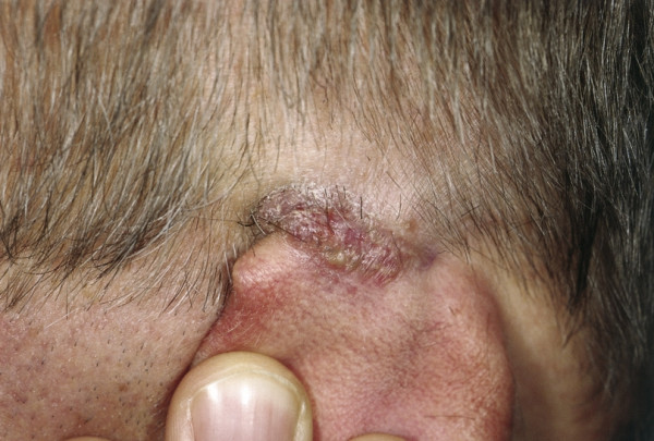
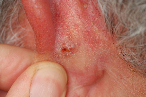
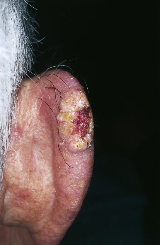
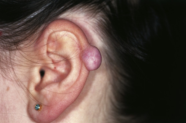
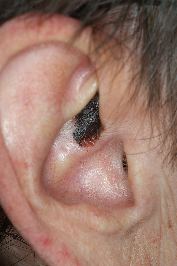
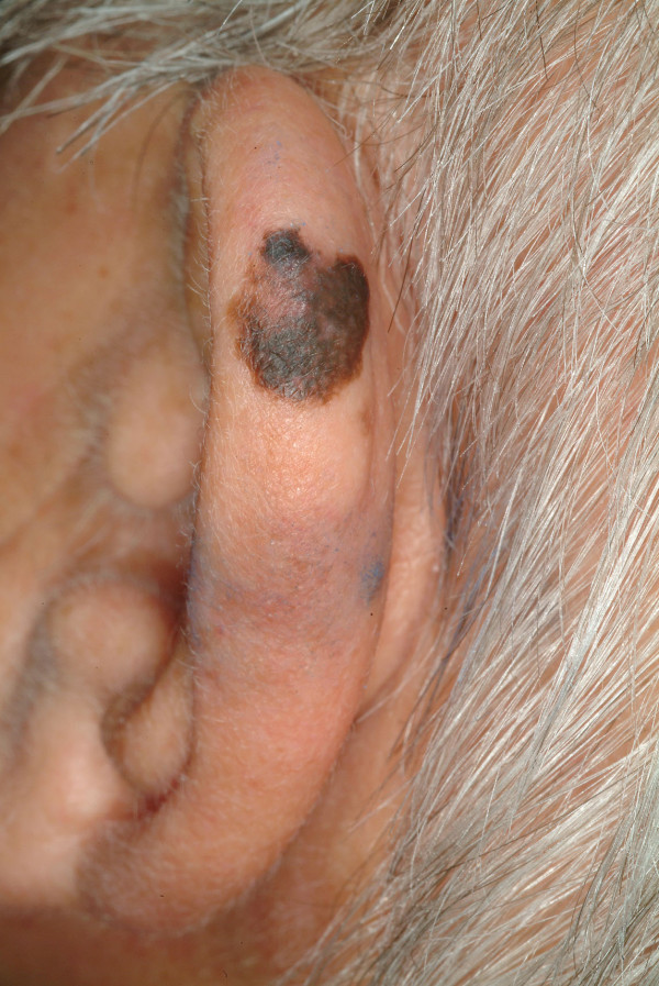
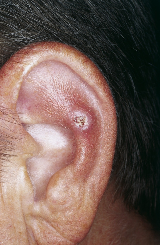
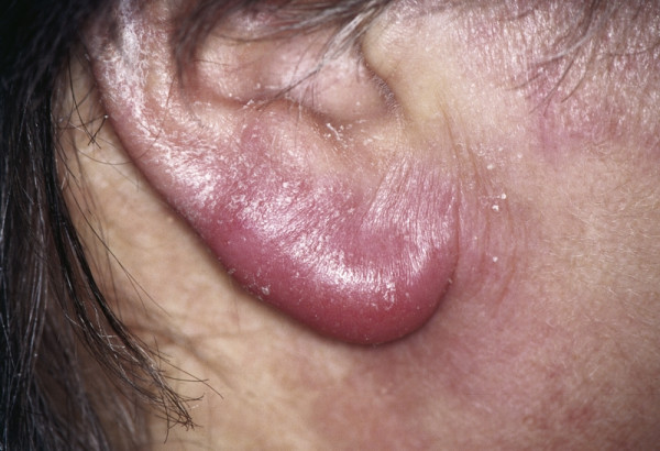
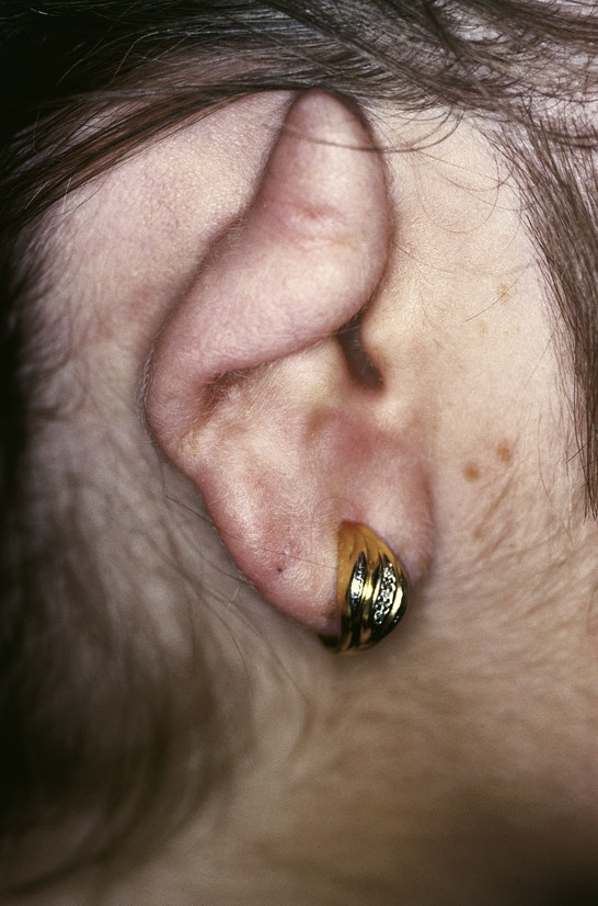
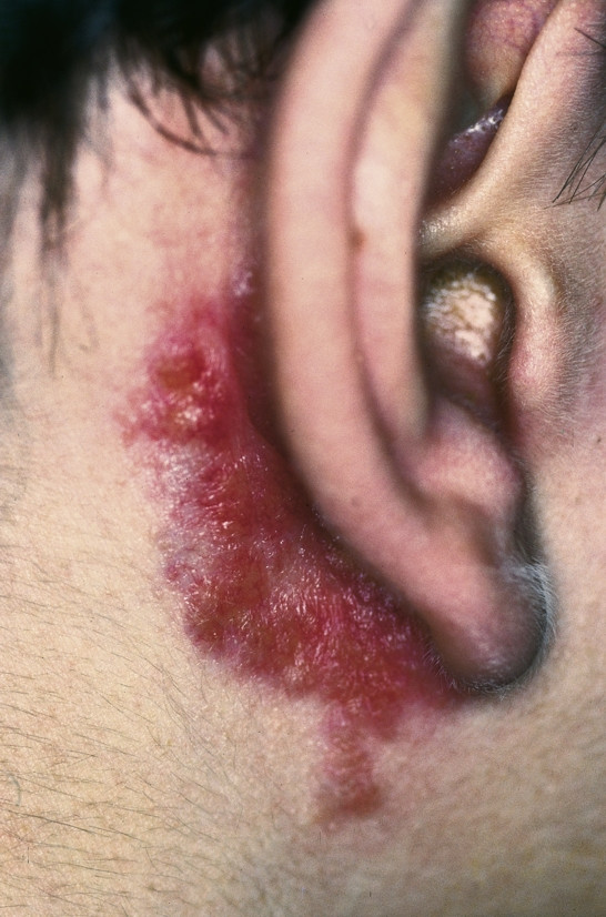
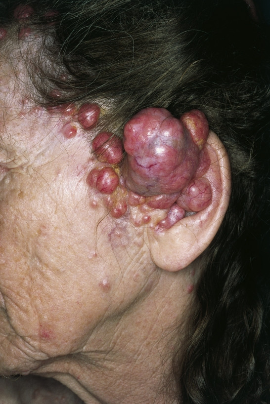
Verification
Claims the paper makes
The claims and statistics of this paper haven’t been checked yet.
Run the check — free, no account needed. It reads the open-access text and checks the sources it cites.
Self-check
Percentages, sums and reported differences re-computed from the paper’s own numbers.
The self-check needs the paper’s full text.
Citations
Cites
Firminger, H. I. (1968) · Armed Forces Institute of Pathology eBooks · Cited by 3,041
Lever, W. F., & Lippincott, J. B. (1950) · Plastic & Reconstructive Surgery · doi:10.1097/00006534-195005000-00012 · Cited by 2,666
ROWE, D. E., Carroll, R. J., & Day, C. L. (1992) · Journal of the American Academy of Dermatology · doi:10.1016/0190-9622(92)70144-5 · Cited by 1,517
ROWE, D. E., Carroll, R. J., & Day, C. L. (1989) · The Journal of Dermatologic Surgery and Oncology · doi:10.1111/j.1524-4725.1989.tb03166.x · Cited by 800
The Edwin Smith Surgical Papyrus
Breasted, J. H. (1931) · De Gruyter eBooks · doi:10.1515/9783112615263-011 · Cited by 725
Mohs Surgery Is the Treatment of Choice for Recurrent (Previously Treated) Basal Cell Carcinoma
ROWE, D. E., Carroll, R. J., & Day, C. L. (1989) · The Journal of Dermatologic Surgery and Oncology · doi:10.1111/j.1524-4725.1989.tb03249.x · Cited by 472
Shambaugh, G. E., & Glasscock, M. E. (1990) · Cited by 457
Cotton, D. W. K. (1992) · Journal of Clinical Pathology · doi:10.1136/jcp.45.9.842-b · Cited by 370
Prognostic Factors for Metastasis in Squamous Cell Carcinoma of the Skin
Cherpelis, B. S., Marcusen, C., & Lang, P. G. (2002) · Dermatologic Surgery · doi:10.1046/j.1524-4725.2002.01169.x · Cited by 309
Tumors of the Upper Respiratory Tract and Ear
(1988) · Annals of Internal Medicine · doi:10.7326/0003-4819-109-7-602_6 · Cited by 292
Research works printed but not linked: 4
- Mehregan AH: Cutaneous horn: a clinicopathological study. Dermatol Dig. 1965, 4: 45-54.
- MacKie RM: Epidermal skin tumours. Rook and Wilkinson and Ebling's Textbook of Dermatology. Edited by: Champion RH, Burton JL, Burns DA, Breathnach SM. 1998, Oxford: Blackwell Science, 1673-4. sixth
- Retz N: Traite des Maladies de la Peau et de celles de l'Esprit. 1790, Mequignon, Paris, 155-3
- Alibert JL: Note sur la chéloïde. Journal universel des sciences médicales. 1816, 2: 207-216.
Cited by
Sorted by citations
Sand, M. S., Sand, D., Thrandorf, C., Paech, V., Altmeyer, P. J., & Bechara, F. G. (2010) · Head & Face Medicine · doi:10.1186/1746-160x-6-7 · Cited by 43
Self-citationDermatologic diseases of the external ear
Öztürkcan, S., & Öztürkcan, S. (2013) · Clinics in Dermatology · doi:10.1016/j.clindermatol.2013.05.036 · Cited by 39
Pickrell, B., Hughes, C., & Maricevich, R. (2017) · Seminars in Plastic Surgery · doi:10.1055/s-0037-1603968 · Cited by 41
Reconstructive Surgery of Auricular Defects: An Overview
Ebrahimi, A., Kazemi, A., Rasouli, H. R., Kazemi, M., & Motamedi, M. H. K. (2015) · Trauma Monthly · doi:10.5812/traumamon.28202 · Cited by 39
Efficacy of triple therapy in auricular keloids
Sousa, R. F. D., Chakravarty, B., Sharma, A. K., Parwaz, M., & Malik, A. K. (2014) · Journal of Cutaneous and Aesthetic Surgery · doi:10.4103/0974-2077.138347 · Cited by 28
Gong, Y., Labh, S., Jin, Y., Diao, H., Li, X., Liu, Z.-Y., & Shi, Y. (2016) · Dermatologic Therapy · doi:10.1111/dth.12335 · Cited by 21
Imaging the External Ear: Practical Approach to Normal and Pathologic Conditions
Tsuno, N. S. G., Tsuno, M. Y., Neto, C., Noujaim, S. E., Decnop, M., Pacheco, F. T., Souza, S. A., Fonseca, A. P. A., & Garcia, M. R. T. (2022) · Radiographics · doi:10.1148/rg.210148 · Cited by 21
Cutaneous basal cell carcinoma arising within a keloid scar: a case report
Kornhaber, R. A., Goder, M., Bordoni, D., Winkler, E., Haik, J., & Tessone, A. (2016) · OncoTargets and Therapy · doi:10.2147/ott.s113724 · Cited by 19
3D Printing Smart Eyeglass Frames: A Review
Tsegay, F., Ghannam, R., Daniel, N., & Butt, H. (2023) · ACS Applied Engineering Materials · doi:10.1021/acsaenm.2c00265 · Cited by 18
Nanotools as potential approach in effective management of acne
Darvishi, M., Khan, M. S., Mohapatra, S., Singh, M., Ali, A., Rajabalizadeh, S., Rahman, H., Iqbal, Z., & Mirza, M. A. (2024) · Journal of Drug Delivery Science and Technology · doi:10.1016/j.jddst.2024.106380 · Cited by 16
Coating Medpor® Implant with Tissue-Engineered Elastic Cartilage
Lee, D. J., Kwon, J., Kim, Y., Kwon, Y. H., Min, S., & Shin, H. W. (2020) · Journal of Functional Biomaterials · doi:10.3390/jfb11020034 · Cited by 16
Assessment of Patients Who Underwent Nasal Reconstruction After Non-Melanoma Skin Cancer Excision
Uzun, H., Bitik, O., Kamburoğlu, H. O., Dadacı, M., Çalış, M., & Öcal, E. (2015) · Journal of Craniofacial Surgery · doi:10.1097/scs.0000000000001563 · Cited by 15
Medical Device-Related Pressure Injury to the Ear from a Mask
Levine, J. M., Ayello, E. A., Persaud, B., & Spinner, R. (2021) · Advances in Skin & Wound Care · doi:10.1097/01.asw.0000752712.31139.c0 · Cited by 14
Reconstruction of External Auditory Canal With a Laterocervical Twisted (Snail) Flap
Aksu, A. E., Uzun, H., Çalış, M., & Şafak, T. (2013) · Journal of Craniofacial Surgery · doi:10.1097/scs.0b013e318286994a · Cited by 10
The Efficacy of CT-Based Conformal Electron Beam Radiation Therapy After Keloid Excision
Hwang, N., Lee, N. K., Chae, J. H., Park, S. H., & Yoon, E. (2022) · Dermatologic Surgery · doi:10.1097/dss.0000000000003398 · Cited by 9
Acanthoma fissuratum: Lest we forget
Deshpande, N. S., Sen, A., Vasudevan, B., & Neema, S. (2017) · Indian Dermatology Online Journal · doi:10.4103/2229-5178.202267 · Cited by 9
Hypothesis: is yeast a clock model to study the onset of humans aging phenotypes?
Mazzoni, C., Mangiapelo, E., Palermo, V., & Falcone, C. (2012) · Frontiers in Oncology · doi:10.3389/fonc.2012.00203 · Cited by 8
Tammaro, A., Adebanjo, G. A. R., Chello, C., Parisella, F. R., Cantisani, C., Farnetani, F., & Pellacani, G. (2021) · Archives of Dermatological Research · doi:10.1007/s00403-021-02304-0 · Cited by 8
Valea, C.-V., Klein, M., Hallermann, C., Schulze, H.-J., Raguse, J.-D., & Wermker, K. (2025) · Frontiers in Oncology · doi:10.3389/fonc.2024.1438021 · Cited by 6
Mohs Micrographic Surgery for the Treatment of External Ear Melanoma: an Outcome Study
Puyana, C., Ham, P. J., & Tsoukas, M. M. (2019) · Dermatologic Surgery · doi:10.1097/dss.0000000000002229 · Cited by 6
Colao, B., & Khachemoune, A. (2024) · Archives of Dermatological Research · doi:10.1007/s00403-024-03127-5 · Cited by 6
Clinical and dermoscopic characterization of pediatric Spitz nevi of the ear
Vaccaro, M., Marafioti, I., Giuffrida, R., Borgia, F., & Zalaudek, I. (2021) · Pediatric Dermatology · doi:10.1111/pde.14616 · Cited by 6
Dmytrzak, A., Lewandowska, K., Boroń, A., Łoniewska, B., Grzesch, N., Brodkiewicz, A., Clark, J., Ciechanowicz, A., & Kostrzewa-Nowak, D. (2024) · International Journal of Molecular Sciences · doi:10.3390/ijms25105284 · Cited by 5
A Study of Skin Disease of the External Ear in Older Adults According to Anatomical Location
Kim, J. K., Bae, I. H., Kim, M. S., Choi, H., Na, C. H., & Shin, B. S. (2018) · Annals of Geriatric Medicine and Research · doi:10.4235/agmr.2018.22.2.88 · Cited by 5
De Rosa, A., Grieco, M. P., D’Antonio, S., Rullo, V., Castellaneta, F., & Fabrizio, T. (2025) · Plastic and Reconstructive Surgery - Global Open · doi:10.1097/gox.0000000000006531 · Cited by 4
Survival Outcomes of Patients with Mycosis Fungoides Involving the External Ear and Ear Canal
Wilkinson, A. J., Nader, M., Roberts, D. B., Duvic, M. A., Gunther, J. R., Dabaja, B. S., & Gidley, P. W. (2022) · The Laryngoscope · doi:10.1002/lary.30377 · Cited by 4
Ahmednaji, N. M., Wu, X., Wang, Y.-X., & Chen, X.-D. (2023) · Dermatologic Surgery · doi:10.1097/dss.0000000000003778 · Cited by 4
Dermatosurgical approach to the ear
Bilgen, A. S., Karadağ, A. S., Colcinar, A., Karakol, P., & Wollina, U. (2026) · Clinics in Dermatology · doi:10.1016/j.clindermatol.2026.05.016 · Cited by 2
A rare development of tumoral calcinosis of the ear auricle
Lee, S. Y., Lee, H. G., Kim, K. S., & Hwang, J. H. (2018) · Archives of Craniofacial Surgery · doi:10.7181/acfs.2018.02306 · Cited by 2
Cutaneous horn developing over a verrucous carcinoma: a rare entity with an unusual presentation
Gupta, R., Lavania, P., Bansal, V., Agarwal, N., & Singh, A. (2016) · International Surgery Journal · doi:10.18203/2349-2902.isj20161185 · Cited by 2
A retrospective study of demographic profile of keloid over the pinna in central Karnataka
Chaitanya, V. K., Kavitha, Y., P., B. K., & Joish, U. K. (2018) · International Journal of Otorhinolaryngology and Head and Neck Surgery · doi:10.18203/issn.2454-5929.ijohns20181860 · Cited by 2
The use of a tympanoplasty blade for tumor extirpation of the auricle
Richards, L. E., Samie, F. H., Troob, S. H., & Lewin, J. M. (2018) · Journal of the American Academy of Dermatology · doi:10.1016/j.jaad.2018.06.041 · Cited by 2
The Study of Ear Keloids by Surgical Excision with Fillet Flap in a Single Institution
Kim, C. J., Choi, H., Na, C. H., Shin, B. S., & Kim, M. S. (2020) · Korean Journal of Otorhinolaryngology - Head and Neck Surgery · doi:10.3342/kjorl-hns.2019.00675 · Cited by 2
Partial auricular reconstruction surgery for facial skin cancer
Rosi‐Schumacher, M., Shokri, T., & Bassichis, B. A. (2022) · Plastic and Aesthetic Research · doi:10.20517/2347-9264.2021.64 · Cited by 2
Squamous Cell Carcinoma of External Auditory Canal
Cortes, J. R. (2017) · Global Journal of Otolaryngology · doi:10.19080/gjo.2017.11.555805 · Cited by 2
A rare interesting case of seborrheic keratosis of pinna
Mundra, R. K., Agrawal, R., & Sinha, R. (2013) · Case Reports in Clinical Medicine · doi:10.4236/crcm.2013.28122 · Cited by 1
Dermatologic Conditions of the External Ear
Leibowitz, R., Koblinski, J. E., Hogue, L. M., Cheeley, J. T., & Blalock, T. W. (2023) · Otolaryngologic Clinics of North America · doi:10.1016/j.otc.2023.05.014 · Cited by 1
Benign and malignant cutaneous nasal lesions
Sarikhani, H., Zhao, K., Polacco, M. A., GROPPER, C. A., & Helman, S. N. (2024) · Eye & ENT Research · doi:10.1002/eer3.16 · Cited by 1
Epithelial and nonepithelial tumors of the external ear
Baykal, C., & Atcı, T. (2026) · Clinics in Dermatology · doi:10.1016/j.clindermatol.2026.05.012 · Cited by 1
Condritis auricular recidivante
Maldonado-García, C. A., Hernández, E. M., Ferra, D. S., Aranda, S. M., & Zavaleta, S. S. (2010) · Revista del Centro Dermatológico Pascua · Cited by 1
Applied Basic Science of the Auricular Cartilage
Abdalla, M. K. T. M. (2018) · InTech eBooks · doi:10.5772/intechopen.72479 · Cited by 1
Auricular and Periauricular Skin Cancers
Barton, B. M., & Moore, B. (2018) · Springer eBooks · doi:10.1007/978-3-319-74539-8_6 · Cited by 1
Cutaneous lesions of the auricular portion of external ear
Koyuncuer, A. (2020) · Annals of Medical Research · doi:10.5455/annalsmedres.2020.03.267 · Cited by 1
A one-stage method for external auditory canal reconstruction; the Wessex pre-auricular flap
Chessman, R., & Sipaul, F. (2021) · European Annals of Otorhinolaryngology Head and Neck Diseases · doi:10.1016/j.anorl.2021.03.014 · Cited by 1
Wound healing in the vulnerable: A novel case of postauricular wounds caused by mask ties
Yousef, A., Monte, O. L., Harmon, M. N., & Watson, D. J. (2023) · JEM Reports · doi:10.1016/j.jemrpt.2023.100038 · Cited by 1
Fabrizio, T. (2024) · Biomedical Journal of Scientific & Technical Research · doi:10.26717/bjstr.2024.55.008721 · Cited by 1
Gourgiotis, A., Isnard, H., Nonell, A., Aubert, M., Stadelmann, G., Dupont, E., Almahamid, I., Tiang, G., Rao, L., Lukens, W. W., et al. (2013) · Cited by 1
Ear Reconstruction following Mohs Micrographic Surgery
Mohan, V. C., Owens, W. R., Schmidt, J. L., Burns, H. R., & Buchanan, E. P. (2024) · Seminars in Plastic Surgery · doi:10.1055/s-0044-1791830
Principali patologie dermatologiche con riferimento alla topografia
Cannavò, S. P., & Trifirò, C. (2011) · Springer eBooks · doi:10.1007/978-88-470-2065-8_8
Induration, itch, redness, and scaling of the left Auricle
Propst, E., & Kopera, D. (2017) · Global Dermatology · doi:10.15761/god.1000207
Reconstrução da hélice: tripla transposição
Ferreira, B. D. F. M., Modesto, G. B. D. S., de Oliveira, C. S., do Carmo, M. C. L., & Silva, S. C. M. C. (2018) · Surgical & Cosmetic Dermatology · doi:10.5935/scd1984-8773.20181041099
Split plaque over postauricular area
Vinay, K., Guliani, A., Muhammed Razmi, T., & Aggarwal, D. (2019) · Indian Dermatology Online Journal · doi:10.4103/idoj.idoj_494_18
Nodular mass—A case of auricular schwannoma
Chuang, W.-C., Ueng, S., Chuang, W., & Chan, K. (2022) · Ear Nose & Throat Journal · doi:10.1177/01455613211068001
Chessman, R., & Sipaul, F. (2022) · Annales françaises d Oto-rhino-laryngologie et de Pathologie Cervico-faciale · doi:10.1016/j.aforl.2022.08.004
Jay (2022) · Cutis · doi:10.12788/cutis.0611
Mačiulaitis, T., Venciūtė-Stankevičė, R., & Jakutis, N. (2022) · Nigerian Journal of Clinical Practice · doi:10.4103/njcp.njcp_416_22
(2023) · International Research Journal of Modernization in Engineering Technology and Science · doi:10.56726/irjmets33817
Recurrent pseudolymphomatous reaction to ear piercing: 20-year history
Wilsher, M. J., & Marais, J. (2023) · Pathology · doi:10.1016/j.pathol.2023.03.013
Mane, B. S., & Gavali, R. M. (2023) · Indian Journal of Otolaryngology and Head & Neck Surgery · doi:10.1007/s12070-023-04132-2
"Black Mole Blocking the Ear Hole": Multiple Differentialscrossing the Mind
Basavannaiah, S. (2021) · Open Journal of Otolaryngology · doi:10.22259/2639-3603.0401001
Reconstruction of Partial Defect of Ear in a Tertiary Care Institute
Singh, K., Beniwal, M., Sharma, A., Singh, P., Kumar, A., & Anshul, A. (2023) · Indian Journal of Otolaryngology and Head & Neck Surgery · doi:10.1007/s12070-023-04420-x
Dermatolojik Hastalıklara Algoritmik Yaklaşım ve Tanısal İpuçları
Yavuz, C., Durdu, M., Yorulmaz, A., Günaydın, S. D., Çınar, S. L., Kızıltaç, K., Kocatürk, E., Hacınecipoğlu, F., Gönül, M., Çakmak, S. K., et al. (2022) · Akademisyen Kitabevi eBooks · doi:10.37609/akya.1231
Reconstruction of extensive defects involving the entire upper third of the auricle: A case report
Polverino, G., Russo, F., & D’Andrea, F. (2024) · Surgery Case Reports · doi:10.1016/j.sycrs.2024.100014
Clinical Analysis of Auricular Tumors
Park, J. M., Lee, G., Kim, K.-W., Lee, S. J., & Lee, J. M. (2024) · Korean Journal of Otorhinolaryngology - Head and Neck Surgery · doi:10.3342/kjorl-hns.2024.00199
Vorbereitung auf die Facharztprüfung HNO
Thölken, K. F. M., Plath, M., & Thölken, R. (2025) · HNO · doi:10.1007/s00106-025-01564-w
Santis, S. D., Galassi, S., Feci, L., Nosiglia, A., & Cambi, J. (2025) · European Archives of Oto-Rhino-Laryngology · doi:10.1007/s00405-025-09463-0
Fabrication of Implant-retained Auricular Prosthesis with Hader Bar Attachment
Alqahtani, N. M. (2025) · King Khalid University journal of health sciences. · doi:10.4103/kkujhs.kkujhs_3_25
Acquired Pathology of the External Ear
Casale, S., Piccin, E., Genovese, A. V., Caloro, E., Massullo, E., Scola, E., Conte, G., Gaini, L. M., Zanetti, D., & Triulzi, F. M. (2025) · Springer eBooks · doi:10.1007/978-3-032-08232-9_6
Kostopoulos, E., Avgerinos, N., Paraschakis, E., Deskoulidi, P. I., Papoulakis, T., Giannikis, G., & Gakis, C. (2026) · Journal of Craniofacial Surgery · doi:10.1097/scs.0000000000012638
Guida, S., ROSA, C. D., Bottino, V., & Rongioletti, F. (2026) · Clinics in Dermatology · doi:10.1016/j.clindermatol.2026.05.015
Metadata
- Authors
- Sand, M., Sand, D., Brors, D., Altmeyer, P., Mann, B., & Bechara, F. G.
- Affiliations
- University Hospitals of the Ruhr-University of Bochum (DE)
- University of California, Los Angeles (US)
- Ruhr University Bochum (DE)
- Corresponding author
- Sand, Michael
- Venue
- Head & Face Medicine
- Published
- 2008-02-08
- Volume
- 4
- Issue
- 1
- Pages
- 2-2
- ISSN
- 1746-160X (ISSN-L)
- PMC
- PMC2267455
- OpenAlex
- W2016651989
- Cited by
- 70
- References
- 86
- Citations by year
- 2026: 4 · 2025: 6 · 2024: 8 · 2023: 8 · 2022: 9 · 2021: 5 · 2020: 3 · 2019: 2 · 2018: 7 · 2017: 4 · 2016: 3 · 2015: 2 · 2014: 1 · 2013: 4 · 2012: 1
- FWCI
- 0.17
- Type
- journal article
- Language
- en
- Open access
- gold
- doi.org
- Licence
- Locations
- Head & Face Medicine (publishedVersion, cc-by)
- PubMed Central (publishedVersion, cc-by)
- PubMed (publishedVersion)
- DOAJ (DOAJ: Directory of Open Access Journals) (submittedVersion)
- Topics
- Health Sciences › Medicine › Surgery › Reconstructive Facial Surgery Techniques
- Health Sciences › Medicine › Dermatology › Nonmelanoma Skin Cancer Studies
- Health Sciences › Medicine › Oncology › Ear and Head Tumors
- Keywords
- skin lesions, external ear, surgical treatment, laser therapy, dermatologic surgery, otolaryngology consultation
- MeSH terms
- Ear Diseases, Head and Neck Neoplasms, Humans, Skin Diseases, Skin Neoplasms
About these checks
Registry, retraction, Cites and statistics results are computed, not judged, with one exception: a printed reference that no index lists is read once by a model before it counts as a citation error. Claims are extracted by a model and kept only when their quote appears verbatim in the text; each cited source is then checked, with a model’s help, for whether it says what the claim relies on. Those verdicts can be wrong — read the claim’s quote and the cited source before relying on one.
Statistics and arithmetic are recomputed, not judged.
Confidence bands are shown only for fields calibrated on our annotated test set. None is calibrated yet, so no band is shown.
What counts as a claim ›How we measure confidence ›How we check this ›
Are you an author, or do you see a problem with this page? Request a review
