

Multiple conserved regulatory domains promote Fezf2 expression in the developing cerebral cortex
Open accessThe DOI matches its Crossref record; 49 of the 50 references it cites were found in a registry or an index.
Abstract
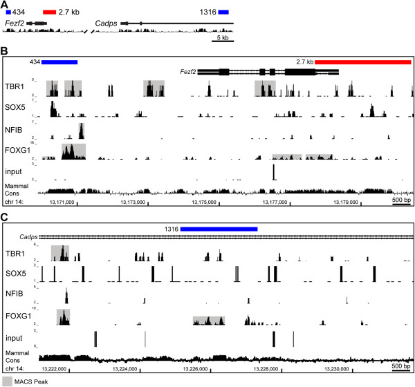
BACKGROUND: The genetic programs required for development of the cerebral cortex are under intense investigation. However, non-coding DNA elements that control the expression of developmentally important genes remain poorly defined. Here we investigate the regulation of Fezf2, a transcription factor that is necessary for the generation of deep-layer cortical projection neurons. RESULTS: Using a combination of chromatin immunoprecipitation followed by high throughput sequencing (ChIP-seq) we mapped the binding of four deep-layer-enriched transcription factors previously shown to be important for cortical development. Building upon this we characterized the activity of three regulatory regions around the Fezf2 locus at multiple stages throughout corticogenesis. We identified a promoter that was sufficient for expression in the cerebral cortex, and enhancers that drove reporter gene expression in distinct forebrain domains, including progenitor cells and cortical projection neurons. CONCLUSIONS: These results provide insight into the regulatory logic controlling Fezf2 expression and further the understanding of how multiple non-coding regulatory domains can collaborate to control gene expression in vivo.
Overview
The claims and statistics of this paper haven’t been checked yet.
Free: it reads the open-access text and checks the first 5 claims that cite a source we can check. Create a free account to check every claim.
Integrity signals
Registry
Cites
Statistics
Figures



Verification
Claims the paper makes
The claims and statistics of this paper haven’t been checked yet.
Free: it reads the open-access text and checks the first 5 claims that cite a source we can check. Create a free account to check every claim.
Claims other papers make about this one
Each is checked against this paper’s own text: does it say what the citing paper says it does?
In the citing paper
“For example, Lhx2-CreER and FezF2-CreER drivers captured embryonic day (E)12.5 RGs in the dorsal neuroepithelium, distributed along a medial-high and lateral-low gradient, consistent with their mRNA expression at this stage59,60.”
From A multimodal cell census and atlas of the mammalian primary motor cortex (2021) · cites this paper as [60]
Self-check
Percentages, sums and reported differences re-computed from the paper’s own numbers.
The self-check needs the paper’s full text.
Citations
Cites
Initial sequencing and analysis of the human genome
International Human Genome Sequencing Consortium, Whitehead Institute for Biomedical Research, C. F. G. R., Lander, E. S., Linton, L. M., Birren, B., Nusbaum, C., Zody, M. C., Baldwin, J., Devon, K., Dewar, K., et al. (2001) · Nature · doi:10.1038/35057062 · Cited by 24,216 · cited 1×
“Current estimates suggest that greater than half of evolutionarily conserved sequences in the human genome do not correspond to protein coding regions [1-4].”
BackgroundModel-based Analysis of ChIP-Seq (MACS)
Zhang, Y., Liu, T., Meyer, C. A., Eeckhoute, J., Johnson, D. S., Bernstein, B. E., Nusbaum, C., Myers, R. M., Brown, M., Li, W., et al. (2008) · Genome Biology · doi:10.1186/gb-2008-9-9-r137 · Cited by 18,386
The sequence of the human genome
Venter, J. C., Adams, M. D., Myers, E. W., Li, P. W., Mural, R. J., Sutton, G. G., Smith, H. O., Yandell, M., Evans, C. A., Holt, R. A., et al. (2001) · Science · doi:10.1126/science.1058040 · Cited by 13,909
A map of human genome variation from population-scale sequencing.
The 1000 Genomes Project Consortium (2010) · Nature · doi:10.1038/nature09534 · Cited by 7,877
Initial sequencing and comparative analysis of the mouse genome
Mouse Genome Sequencing Consortium (2002) · Nature · doi:10.1038/nature01262 · Cited by 7,187
Topological domains in mammalian genomes identified by analysis of chromatin interactions
Dixon, J. R., Selvaraj, S., Yue, F., Kim, A., Li, Y., Shen, Y., Hu, M., Liu, J. S., & Ren, B. (2012) · Nature · doi:10.1038/nature11082 · Cited by 6,829 · cited 1×
“Recently published high throughput chromosome conformation capture (Hi-C) suggests an interaction between enhancer 1316 and the Fezf2 promoter [38].”
BackgroundEvolutionarily conserved elements in vertebrate, insect, worm, and yeast genomes
Siepel, A., Bejerano, G., Pedersen, J. S., Hinrichs, A. S., Hou, M., Rosenbloom, K., Clawson, H., Spieth, J., Hillier, L. W., Richards, S., et al. (2005) · Genome Research · doi:10.1101/gr.3715005 · Cited by 3,971 · cited 1×
“Current estimates suggest that greater than half of evolutionarily conserved sequences in the human genome do not correspond to protein coding regions [1-4].”
BackgroundStrong association of de novo copy number mutations with autism
Sebat, J., Lakshmi, B., Malhotra, D., Troge, J., Lese-Martin, C., Walsh, T., Yamrom, B., Yoon, S., Krasnitz, A., Kendall, J., et al. (2007) · Science · doi:10.1126/science.1138659 · Cited by 2,795 · cited 1×
“Genome-wide association studies (GWAS) have identified sequence variations within putative enhancers that are associated with a wide range of neurological disorders such as autism, epilepsy, and schizophrenia [6-10].”
BackgroundA gene expression atlas of the central nervous system based on bacterial artificial chromosomes
Gong, S., Zheng, C., Doughty, M. L., Losos, K., Didkovsky, N., Schambra, U. B., Nowak, N. J., Joyner, A., Leblanc, G., Hatten, M. E., et al. (2003) · Nature · doi:10.1038/nature02033 · Cited by 2,108 · cited 1×
“Although enhancer 1316 is not contained within this BAC, GFP, or CRE expression in these transgenic mice recapitulates endogenous Fezf2 gene expression [36,39,40], suggesting that this enhancer is not essential for Fezf2 expression during”
BackgroundManipulating the Mouse Embryo: A Laboratory Manual
Behringer, R. R., Gertsenstein, M., Nagy, K. V., & Nagy, A. (2002) · Cited by 2,046
Cited by
A multimodal cell census and atlas of the mammalian primary motor cortex
BRAIN Initiative Cell Census Network (BICCN), BRAIN Initiative Cell Census Network (BICCN) Corresponding authors, Callaway, E. M., Dong, H.-W., Ecker, J. R., Hawrylycz, M. J., Huang, Z. J., Lein, E. S., Ngai, J., Osten, P., et al. (2021) · Nature · doi:10.1038/s41586-021-03950-0 · Cited by 522
Uzquiano, A., Kedaigle, A. J., Pigoni, M., Paulsen, B., Adiconis, X., Kim, K., Faits, T., Nagaraja, S., Antón-Bolaños, N., Gerhardinger, C., et al. (2022) · Cell · doi:10.1016/j.cell.2022.09.010 · Cited by 244
Genetic dissection of the glutamatergic neuron system in cerebral cortex
Matho, K. S., Huilgol, D., Galbavy, W., He, M., Kim, G., An, X., Lu, J., Wu, P., Di Bella, D. J., Shetty, A. S., et al. (2021) · Nature · doi:10.1038/s41586-021-03955-9 · Cited by 155
Toma, K., Kumamoto, T., & Hanashima, C. (2014) · Journal of Neuroscience · doi:10.1523/jneurosci.2334-14.2014 · Cited by 118
McKenna, W. L., Ortiz-Londono, C. F., Mathew, T. K., Hoang, K., Katzman, S., & Chen, B. (2015) · Proceedings of the National Academy of Sciences · doi:10.1073/pnas.1504144112 · Cited by 95
Self-citation
Metadata
- Authors
- Eckler, M. J., Larkin, K. A., McKenna, W. L., Katzman, S., Guo, C., Roque, R., Visel, A., Rubenstein, J. L. L., & Chen, B.
- Affiliations
- University of California, Santa Cruz (US)
- Lawrence Berkeley National Laboratory (US)
- Corresponding author
- Chen, Bin
- Venue
- Neural Development
- Published
- 2014-03-12
- Volume
- 9
- Issue
- 1
- Pages
- 6-6
- ISSN
- 1749-8104 (ISSN-L)
- PMC
- PMC4008173
- OpenAlex
- W2153518972
- Cited by
- 27
- References
- 50
- Citations by year
- 2025: 1 · 2024: 4 · 2023: 1 · 2022: 4 · 2021: 4 · 2020: 1 · 2019: 1 · 2017: 5 · 2016: 2 · 2015: 2 · 2014: 2
- FWCI
- 1.27
- Type
- journal article
- Language
- en
- Open access
- gold
- doi.org
- Licence
- cc-by
- Locations
- Neural Development (publishedVersion, cc-by)
- Europe PMC (PubMed Central) (publishedVersion, cc-by)
- PubMed Central (publishedVersion, cc-by)
- PubMed (publishedVersion)
- OSTI OAI (U.S. Department of Energy Office of Scientific and Technical Information) (submittedVersion)
- eScholarship (California Digital Library) (submittedVersion)
- eScholarship (California Digital Library) (submittedVersion, cc-by)
- Funders
- California Institute of Regenerative Medicine
- National Institute of Mental Health (R01MH094589)
- National Human Genome Research Institute (R01HG003988)
- National Institute of Neurological Disorders and Stroke (R01NS062859, R01 NS062859A, NS34661, R01NS034661, R01NS099099)
- Topics
- Life Sciences › Neuroscience › Developmental Neuroscience › Neurogenesis and neuroplasticity mechanisms
- Life Sciences › Biochemistry, Genetics and Molecular Biology › Molecular Biology › Genomics and Chromatin Dynamics
- Life Sciences › Biochemistry, Genetics and Molecular Biology › Molecular Biology › Developmental Biology and Gene Regulation
- Keywords
- Fezf2, cortical development, transcription factor binding sites, enhancers, promoter, non-coding regulatory elements, cortical progenitor cells, forebrain, gene expression regulation
- MeSH terms
- Animals, Cerebral Cortex, DNA-Binding Proteins, Enhancer Elements, Genetic, Mice, Transgenic, Nerve Tissue Proteins, Regulatory Elements, Transcriptional, Mice
About these checks
Registry, retraction, Cites and statistics results are computed, not judged, with one exception: a printed reference that no index lists is read once by a model before it counts as a citation error. Claims are extracted by a model and kept only when their quote appears verbatim in the text; each cited source is then checked, with a model’s help, for whether it says what the claim relies on. Those verdicts can be wrong — read the claim’s quote and the cited source before relying on one.
Statistics and arithmetic are recomputed, not judged.
Confidence bands are shown only for fields calibrated on our annotated test set. None is calibrated yet, so no band is shown.
What counts as a claim ›How we measure confidence ›How we check this ›
Are you an author, or do you see a problem with this page? Request a review
How to cite
Cite CoChat for the checks on this page, and the paper for its own findings.
- This page
- CoChat. (2026). Citation check: Multiple conserved regulatory domains promote Fezf2 expression in the developing cerebral cortex. https://app.cochat.ai/papers/10.1186/1749-8104-9-6
- The paper
- Eckler, M. J., Larkin, K. A., McKenna, W. L., Katzman, S., Guo, C., Roque, R., Visel, A., Rubenstein, J. L. L., & Chen, B. (2014). Multiple conserved regulatory domains promote Fezf2 expression in the developing cerebral cortex. Neural Development. https://doi.org/10.1186/1749-8104-9-6
Data sources
Bibliographic data: Semantic Scholar, OpenAlex, Crossref.
Contains information from the Semantic Scholar Academic Graph Datasets, made available under the ODC Attribution License (ODC-BY 1.0).
Semantic Scholar’s data is described in: Kinney, R., et al. (2023). The Semantic Scholar Open Data Platform. arXiv:2301.10140.
