Cited by 124Wnt/β-catenin signaling in development and cancerMuscle Physiology and DisordersDevelopmental Biology and Gene Regulation

The DOI matches its Crossref record; 39 of the 39 references it cites were found in a registry or an index.

Canonical Wnt signaling is involved in switching from cell proliferation to myogenic differentiation of mouse myoblast cells

Open access
Tanaka, Shingo, Terada, Kumiko, Nohno, Tsutomu·2011·Journal of Molecular Signaling

Abstract

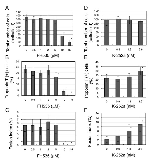
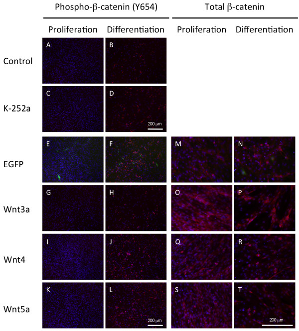
BACKGROUND: Wnt/β-catenin signaling is involved in various aspects of skeletal muscle development and regeneration. In addition, Wnt3a and β-catenin are required for muscle-specific gene transcription in embryonic carcinoma cells and satellite-cell proliferation during adult skeletal muscle regeneration. Downstream targets of canonical Wnt signaling are cyclin D1 and c-myc. However both target genes are suppressed during differentiation of mouse myoblast cells, C2C12. Underlying molecular mechanisms of β-catenin signaling during myogenic differentiation remain unknown. RESULTS: Using C2C12 cells, we examined intracellular signaling and gene transcription during myoblast proliferation and differentiation. We confirmed that several Wnt signaling components, including Wnt9a, Sfrp2 and porcupine, were consistently upregulated in differentiating C2C12 cells. Troponin T-positive myotubes were decreased by Wnt3a overexpression, but not Wnt4. TOP/FOP reporter assays revealed that co-expression with Wnt4 reduced Wnt3a-induced luciferase activity, suggesting that Wnt4 signaling counteracted Wnt3a signaling in myoblasts. FH535, a small-molecule inhibitor of β-catenin/Tcf complex formation, reduced basal β-catenin in the cytoplasm and decreased myoblast proliferation. K252a, a protein kinase inhibitor, increased both cytosolic and membrane-bound β-catenin and enhanced myoblast fusion. Treatments with K252a or Wnt4 resulted in increased cytoplasmic vesicles containing phosphorylated β-catenin (Tyr654) during myogenic differentiation. CONCLUSIONS: These results suggest that various Wnt ligands control subcellular β-catenin localization, which regulate myoblast proliferation and myotube formation. Wnt signaling via β-catenin likely acts as a molecular switch that regulates the transition from cell proliferation to myogenic differentiation.

Overview

The claims and statistics of this paper haven’t been checked yet.

Run the check — free, no account needed. It reads the open-access text and checks the sources it cites.

A few free checks a day · papers already verified open instantly · results are public

Integrity signals

Registry

Crossref · 10.1186/1750-2187-6-12

Cites

39 of 39 references verified0 citation errors · 0 not checkable

Statistics

Figures

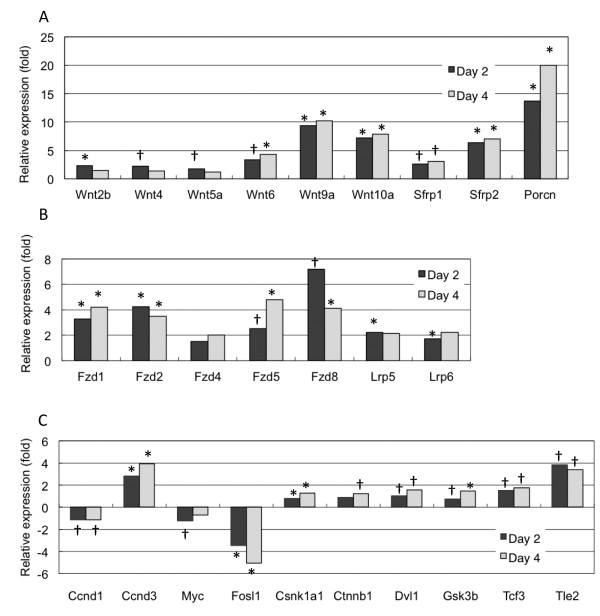
Figure 1 Endogenous Wnt signaling component expression during myogenic differentiation of C2C12 cells. Wnt signaling molecule expression was measured by quantitative RT-PCR. After 2 or 4 days culture in differentiation medium, cells were harvested, total RNA prepared and relative gene expression quantified by real-time PCR as follows: (A) wingless-related MMTV integration, Wnt; secreted frizzled-related protein, Sfrp; and porcupine, Porcn. (B) frizzled, Fzd; and low density lipoprotein receptor-related protein, Lrp. (C) cyclin D, Ccnd; myelocytomatosis oncogene, Myc; fos-like antigen 1, Fosl1; casein kinase 1, alpha 1, Csnk1a1; catenin, beta 1, Ctnnb1; dishevelled 1, Dvl1; glycogen synthase kinase 3 beta, Gsk3b; transcription factor 3, Tcf3; and transducin-like enhancer of split 2, Tle2. Day 2 and 4 time points were normalized to expression levels at day 0. Data are from three independent experiments, *P < 0.005 and † P < 0.05.
Figure 1 Endogenous Wnt signaling component expression during myogenic differentiation of C2C12 cells. Wnt signaling molecule expression was measured by quantitative RT-PCR. After 2 or 4 days culture in differentiation medium, cells were harvested, total RNA prepared and relative gene expression quantified by real-time PCR as follows: (A) wingless-related MMTV integration, Wnt; secreted frizzled-related protein, Sfrp; and porcupine, Porcn. (B) frizzled, Fzd; and low density lipoprotein receptor-related protein, Lrp. (C) cyclin D, Ccnd; myelocytomatosis oncogene, Myc; fos-like antigen 1, Fosl1; casein kinase 1, alpha 1, Csnk1a1; catenin, beta 1, Ctnnb1; dishevelled 1, Dvl1; glycogen synthase kinase 3 beta, Gsk3b; transcription factor 3, Tcf3; and transducin-like enhancer of split 2, Tle2. Day 2 and 4 time points were normalized to expression levels at day 0. Data are from three independent experiments, *P < 0.005 and † P < 0.05.CC BY · PubMed Central
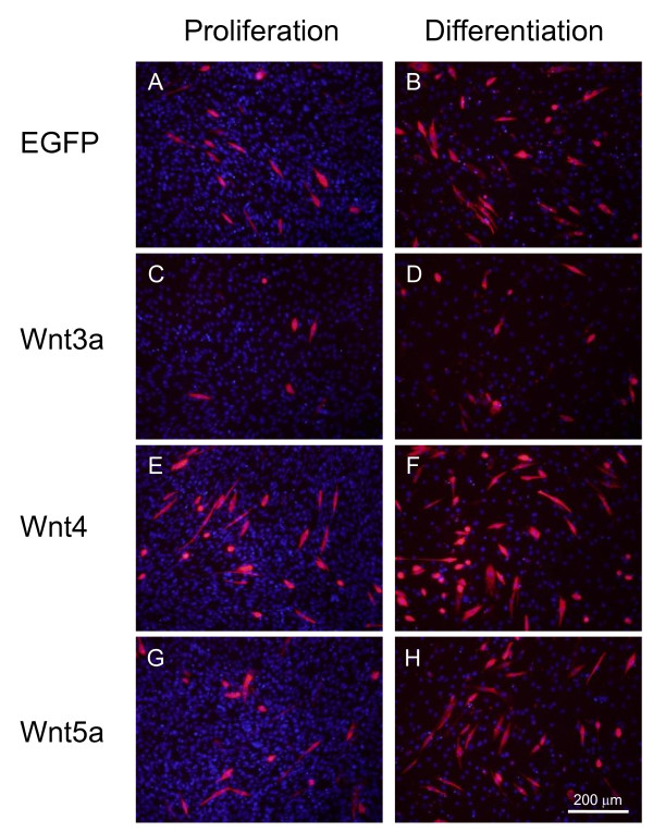
Figure 2 Wnt4 promotes myogenic differentiation of C2C12 cells. Expression constructs of Wnt family members in adenovirus vector were transfected into C2C12 cells and myogenic differentiation was determined by troponin T immunostaining after 2 days culture in differentiation medium, which commenced 24 h post-transfection.
Figure 2 Wnt4 promotes myogenic differentiation of C2C12 cells. Expression constructs of Wnt family members in adenovirus vector were transfected into C2C12 cells and myogenic differentiation was determined by troponin T immunostaining after 2 days culture in differentiation medium, which commenced 24 h post-transfection.CC BY · PubMed Central
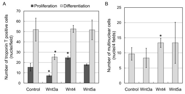
Figure 3 Wnt4 promotes myogenic differentiation and induces fusion of C2C12 cells. (A) Troponin T expression after 3 day culture in proliferation or differentiation medium with recombinant adenoviruses expressing Wnt3a, Wnt4, Wnt5a or eGFP (control). Cell numbers are the means and standard deviations of three independent experiments performed in triplicate, *P < 0.01 vs. control. (B) Differentiated myotubes were evaluated by counting nuclei within fused multinucleated cells expressing troponin T. Four fields in triplicate cultures were used for counting fused cells cultured under a differentiation condition, *P < 0.04 vs. control.
Figure 3 Wnt4 promotes myogenic differentiation and induces fusion of C2C12 cells. (A) Troponin T expression after 3 day culture in proliferation or differentiation medium with recombinant adenoviruses expressing Wnt3a, Wnt4, Wnt5a or eGFP (control). Cell numbers are the means and standard deviations of three independent experiments performed in triplicate, *P < 0.01 vs. control. (B) Differentiated myotubes were evaluated by counting nuclei within fused multinucleated cells expressing troponin T. Four fields in triplicate cultures were used for counting fused cells cultured under a differentiation condition, *P < 0.04 vs. control.CC BY · PubMed Central
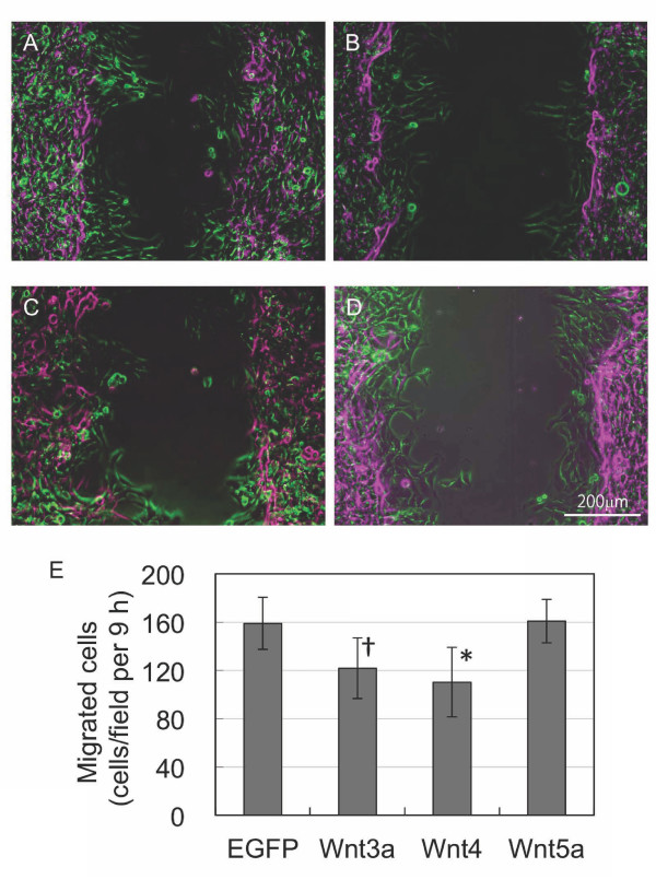
Figure 4 Effect of Wnt expression on cell migration. A scratch test was used to determine C2C12 cell proliferation and migration. Cell numbers were counted over 9 h, and mobility was monitored by time-lapse microscopy. Phase contrast images at 0 h (purple) and 9 h (green) were merged to show the migration of C2C12 cells expressing eGFP (A), Wnt3a (B), Wnt4(C) and Wnt5a (D). (E) Summary of cell numbers counted over 9 h (cells/field) with n = 3 for control, Wnt3a and Wnt4, and n = 4 for Wnt5a. *P < 0.01; † P < 0.02 vs. eGFP.
Figure 4 Effect of Wnt expression on cell migration. A scratch test was used to determine C2C12 cell proliferation and migration. Cell numbers were counted over 9 h, and mobility was monitored by time-lapse microscopy. Phase contrast images at 0 h (purple) and 9 h (green) were merged to show the migration of C2C12 cells expressing eGFP (A), Wnt3a (B), Wnt4(C) and Wnt5a (D). (E) Summary of cell numbers counted over 9 h (cells/field) with n = 3 for control, Wnt3a and Wnt4, and n = 4 for Wnt5a. *P < 0.01; † P < 0.02 vs. eGFP.CC BY · PubMed Central
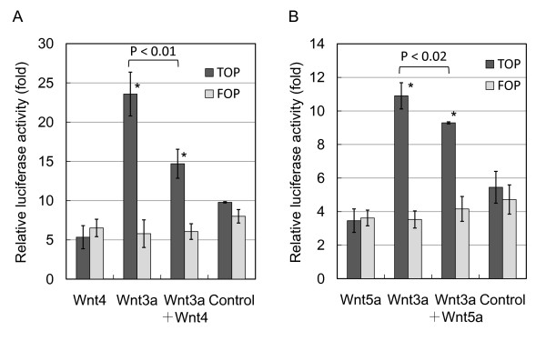
Figure 5 Wnt4 reduces Wnt-3a-induced transcriptional activity in myoblasts. Reporter assays were performed using TOP/FLASH and FOP/FLASH (luciferase assay) in C2C12 cells. Wnt3a significantly increases luciferase activity in TOP over FOP. (A) Wnt4 has no effect on luciferase activity, whereas Wnt4 inhibits Wnt3a activity. (B) Wnt5a poorly inhibits Wnt3a activity. Bars are the means of four independent experiments. *P < 0.01 between TOP and FOP; P < 0.01 between Wnt3a and Wnt3a + Wnt4 in TOP; P < 0.02 between Wnt3a and Wnt3a + Wnt5a in TOP.
Figure 5 Wnt4 reduces Wnt-3a-induced transcriptional activity in myoblasts. Reporter assays were performed using TOP/FLASH and FOP/FLASH (luciferase assay) in C2C12 cells. Wnt3a significantly increases luciferase activity in TOP over FOP. (A) Wnt4 has no effect on luciferase activity, whereas Wnt4 inhibits Wnt3a activity. (B) Wnt5a poorly inhibits Wnt3a activity. Bars are the means of four independent experiments. *P < 0.01 between TOP and FOP; P < 0.01 between Wnt3a and Wnt3a + Wnt4 in TOP; P < 0.02 between Wnt3a and Wnt3a + Wnt5a in TOP.CC BY · PubMed Central
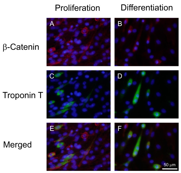
Figure 6 Localization of β-catenin and troponin T in proliferative and differentiating C2C12 cells. C2C12 cells cultured in proliferation and differentiation medium were double immunostained for β-catenin (A, B; red) and troponin T (C, D; green), and counterstained with DAPI (blue). Immunofluorescent images with various filters are merged (E, F).
Figure 6 Localization of β-catenin and troponin T in proliferative and differentiating C2C12 cells. C2C12 cells cultured in proliferation and differentiation medium were double immunostained for β-catenin (A, B; red) and troponin T (C, D; green), and counterstained with DAPI (blue). Immunofluorescent images with various filters are merged (E, F).CC BY · PubMed Central
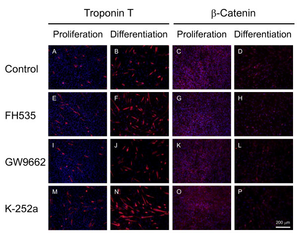
Figure 7 Effect of small-molecule inhibitors on C2C12 cell proliferation and differentiation. C2C12 cells were cultured without inhibitors (A-D) and with 1 μM FH535 (E-H), 1 μM GW9662 (I-L) and 3.6 nM K-252a (M-P), then immunostained with anti-troponin T (A, B, E, F, I, J, M, N) and anti-β-catenin (C, D, G, H, K, L, O, P) antibodies. FH535 interferes with β-catenin/Tcf complex formation, whereas GW9662 is structurally similar to FH535, but does not interfere with complex formation. K252a is a Trk kinase inhibitor, which phosphorylates β-catenin at tyrosine 654.
Figure 7 Effect of small-molecule inhibitors on C2C12 cell proliferation and differentiation. C2C12 cells were cultured without inhibitors (A-D) and with 1 μM FH535 (E-H), 1 μM GW9662 (I-L) and 3.6 nM K-252a (M-P), then immunostained with anti-troponin T (A, B, E, F, I, J, M, N) and anti-β-catenin (C, D, G, H, K, L, O, P) antibodies. FH535 interferes with β-catenin/Tcf complex formation, whereas GW9662 is structurally similar to FH535, but does not interfere with complex formation. K252a is a Trk kinase inhibitor, which phosphorylates β-catenin at tyrosine 654.CC BY · PubMed Central
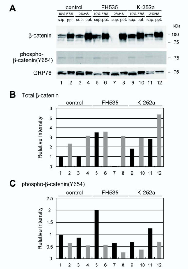
Figure 8 FH535 and K252a alter total and phospho-β-catenin (Y654) in C2C12 cells. (A) Western blots of cytosolic and membrane fractions from C2C12 cells treated with FH535 and K-252a using antibodies against total cellular-β-catenin (upper panel) and phosphorylated β-catenin (lower panel). GRP78 indicates loading control. (B, C) Relative levels of total and phosphorylated β-catenin in cytosolic (black) and membrane (grey) fractions were calculated as the β-catenin/GRP78 ratio. Band intensities are relative to the control cell values in the cytosolic fraction and defined as 1.0. Similar results were obtained in three independent experiments.
Figure 8 FH535 and K252a alter total and phospho-β-catenin (Y654) in C2C12 cells. (A) Western blots of cytosolic and membrane fractions from C2C12 cells treated with FH535 and K-252a using antibodies against total cellular-β-catenin (upper panel) and phosphorylated β-catenin (lower panel). GRP78 indicates loading control. (B, C) Relative levels of total and phosphorylated β-catenin in cytosolic (black) and membrane (grey) fractions were calculated as the β-catenin/GRP78 ratio. Band intensities are relative to the control cell values in the cytosolic fraction and defined as 1.0. Similar results were obtained in three independent experiments.CC BY · PubMed Central
Figure 9 Effect of FH535 and K252a on C2C12 cell proliferation and differentiation. C2C12 cells were cultured with or without various FH535 and K252a concentrations followed by troponin T immunostaining. Total cell numbers are the counted DAPI-stained nuclei and the fusion index is the percentage of multinucleated cells among troponin T-positive cells. *P < 0.001 vs. absence of inhibitors.
Figure 9 Effect of FH535 and K252a on C2C12 cell proliferation and differentiation. C2C12 cells were cultured with or without various FH535 and K252a concentrations followed by troponin T immunostaining. Total cell numbers are the counted DAPI-stained nuclei and the fusion index is the percentage of multinucleated cells among troponin T-positive cells. *P < 0.001 vs. absence of inhibitors.CC BY · PubMed Central
Figure 10 Wnt family members differentially regulate β-catenin phosphorylation and subcellular localization in C2C12 cells. (A-D) C2C12 cells were cultured with or without K252a, and then phospho-β-catenin (Y654) was immunostained. K252a caused an accumulation of vesicles containing phospho-β-catenin (Y654) in C2C12 cells. (E-L) Wnt3a, Wnt4 and Wnt5a are overexpressed in C2C12 cells by adenovirus-mediated gene transfer. After 2 day culture in differentiation medium commencing 24 h post-transfection, subcellular phospho-β-catenin (Y654) localization was determined by immunostaining. (M-T) C2C12 cells expressing eGFP, Wnt3a, Wnt4 and Wnt5a were immunostained with anti-pan-β-catenin antibodies. Cytoplasmic β-catenin is elevated with Wnt3a expression in proliferation medium and maintained at a higher level in differentiation medium, whereas nuclear and membrane-bound β-catenin is increased with Wnt4 expression.
Figure 10 Wnt family members differentially regulate β-catenin phosphorylation and subcellular localization in C2C12 cells. (A-D) C2C12 cells were cultured with or without K252a, and then phospho-β-catenin (Y654) was immunostained. K252a caused an accumulation of vesicles containing phospho-β-catenin (Y654) in C2C12 cells. (E-L) Wnt3a, Wnt4 and Wnt5a are overexpressed in C2C12 cells by adenovirus-mediated gene transfer. After 2 day culture in differentiation medium commencing 24 h post-transfection, subcellular phospho-β-catenin (Y654) localization was determined by immunostaining. (M-T) C2C12 cells expressing eGFP, Wnt3a, Wnt4 and Wnt5a were immunostained with anti-pan-β-catenin antibodies. Cytoplasmic β-catenin is elevated with Wnt3a expression in proliferation medium and maintained at a higher level in differentiation medium, whereas nuclear and membrane-bound β-catenin is increased with Wnt4 expression.CC BY · PubMed Central

Verification

Claims the paper makes

The claims and statistics of this paper haven’t been checked yet.

Run the check — free, no account needed. It reads the open-access text and checks the sources it cites.

A few free checks a day · papers already verified open instantly · results are public

Claims other papers make about this one

Each is checked against this paper’s own text: does it say what the citing paper says it does?

Self-check

Percentages, sums and reported differences re-computed from the paper’s own numbers.

The self-check needs the paper’s full text.

Citations

Cites

Show all 39 references

Cited by

Sorted by citations

See all 124

Metadata

Authors
Tanaka, S., Terada, K., & Nohno, T.
Affiliations
Kawasaki Medical School (JP)
Corresponding author
Nohno, Tsutomu
Venue
Journal of Molecular Signaling
Published
2011-10-05
Volume
6
Pages
12
ISSN
1750-2187 (ISSN-L)
OpenAlex
W2142673125
Cited by
124
References
39
Citations by year
2026: 7 · 2025: 9 · 2024: 5 · 2023: 6 · 2022: 7 · 2021: 5 · 2020: 5 · 2019: 7 · 2018: 13 · 2017: 12 · 2016: 16 · 2015: 6 · 2014: 9 · 2013: 10 · 2012: 7
FWCI
3.38
Type
journal article
Language
en
Open access
Locations
  • Journal of Molecular Signaling (publishedVersion, cc-by)
  • Europe PMC (PubMed Central) (publishedVersion, cc-by)
  • PubMed Central (publishedVersion, cc-by)
  • PubMed (publishedVersion)
  • Journal of Molecular Signaling (University of Oklahoma Health Sciences Center) (submittedVersion, cc-by)
Funders
  • Ministry of Health, Labour and Welfare
  • Kawasaki Medical School (23590227)
  • Japan Society for the Promotion of Science (21102, 23590227)
Topics
  • Life Sciences › Biochemistry, Genetics and Molecular Biology › Molecular Biology › Wnt/β-catenin signaling in development and cancer
  • Life Sciences › Biochemistry, Genetics and Molecular Biology › Molecular Biology › Muscle Physiology and Disorders
  • Life Sciences › Biochemistry, Genetics and Molecular Biology › Molecular Biology › Developmental Biology and Gene Regulation
Keywords
Wnt/β-catenin signaling, β-catenin, myogenesis, myoblast proliferation, C2C12 myoblasts, Wnt3a, Wnt4, Wnt9a, SFRP2, porcupines, β-catenin phosphorylation, myotube formation, myoblast fusion, FH535, K252a, skeletal muscle development

About these checks

Registry, retraction, Cites and statistics results are computed, not judged, with one exception: a printed reference that no index lists is read once by a model before it counts as a citation error. Claims are extracted by a model and kept only when their quote appears verbatim in the text; each cited source is then checked, with a model’s help, for whether it says what the claim relies on. Those verdicts can be wrong — read the claim’s quote and the cited source before relying on one.

Statistics and arithmetic are recomputed, not judged.

Confidence bands are shown only for fields calibrated on our annotated test set. None is calibrated yet, so no band is shown.

What counts as a claim ›How we measure confidence ›How we check this ›

Are you an author, or do you see a problem with this page? Request a review