

The DOI matches its Crossref record; 162 of the 163 references it cites were found in a registry or an index.
Placental contribution to the origins of sexual dimorphism in health and diseases: sex chromosomes and epigenetics
Open accessAbstract
Sex differences occur in most non-communicable diseases, including metabolic diseases, hypertension, cardiovascular disease, psychiatric and neurological disorders and cancer. In many cases, the susceptibility to these diseases begins early in development. The observed differences between the sexes may result from genetic and hormonal differences and from differences in responses to and interactions with environmental factors, including infection, diet, drugs and stress. The placenta plays a key role in fetal growth and development and, as such, affects the fetal programming underlying subsequent adult health and accounts, in part for the developmental origin of health and disease (DOHaD). There is accumulating evidence to demonstrate the sex-specific relationships between diverse environmental influences on placental functions and the risk of disease later in life. As one of the few tissues easily collectable in humans, this organ may therefore be seen as an ideal system for studying how male and female placenta sense nutritional and other stresses, such as endocrine disruptors. Sex-specific regulatory pathways controlling sexually dimorphic characteristics in the various organs and the consequences of lifelong differences in sex hormone expression largely account for such responses. However, sex-specific changes in epigenetic marks are generated early after fertilization, thus before adrenal and gonad differentiation in the absence of sex hormones and in response to environmental conditions. Given the abundance of X-linked genes involved in placentogenesis, and the early unequal gene expression by the sex chromosomes between males and females, the role of X- and Y-chromosome-linked genes, and especially those involved in the peculiar placenta-specific epigenetics processes, giving rise to the unusual placenta epigenetic landscapes deserve particular attention. However, even with recent developments in this field, we still know little about the mechanisms underlying the early sex-specific epigenetic marks resulting in sex-biased gene expression of pathways and networks. As a critical messenger between the maternal environment and the fetus, the placenta may play a key role not only in buffering environmental effects transmitted by the mother but also in expressing and modulating effects due to preconceptional exposure of both the mother and the father to stressful conditions.
Overview
The claims and statistics of this paper haven’t been checked yet.
Run the check — free, no account needed. It reads the open-access text and checks the sources it cites.
Integrity signals
Registry
Cites
Statistics
Figures
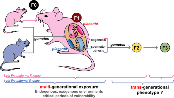
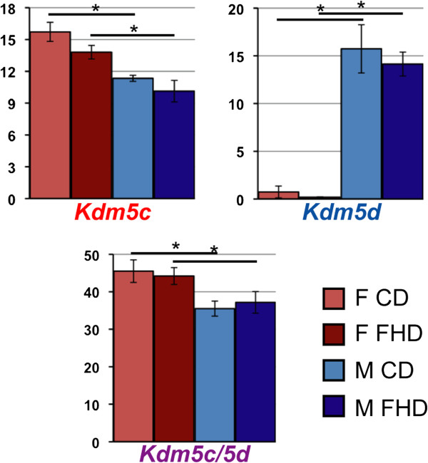
Verification
Claims the paper makes
The claims and statistics of this paper haven’t been checked yet.
Run the check — free, no account needed. It reads the open-access text and checks the sources it cites.
Claims other papers make about this one
Each is checked against this paper’s own text: does it say what the citing paper says it does?
In the citing paper
“Epigenetics is a rapidly expanding field, and there are now many reports of changes in placental DNA methylation and histone proteins in response to environmental cues that have been associated with developmental programming of the offspring (203, 417, 522).”
From Placental Origins of Chronic Disease (2016) · cites this paper as [203]
Self-check
Percentages, sums and reported differences re-computed from the paper’s own numbers.
The self-check needs the paper’s full text.
Citations
Cites
Eckel, R. H., Alberti, K., Grundy, S. M., & Zimmet, P. (2010) · The Lancet · doi:10.1016/s0140-6736(09)61794-3 · Cited by 6,317
The male-specific region of the human Y chromosome is a mosaic of discrete sequence classes
Skaletsky, H., Kuroda-Kawaguchi, T., Minx, P. J., Cordum, H. S., Hillier, L., Brown, L. G., Repping, S., Pyntikova, T., Ali, J., Bieri, T. A., et al. (2003) · Nature · doi:10.1038/nature01722 · Cited by 2,148
X-inactivation profile reveals extensive variability in X-linked gene expression in females
Carrel, L., & Willard, H. F. (2005) · Nature · doi:10.1038/nature03479 · Cited by 2,097
Spatio-temporal transcriptome of the human brain
Kang, H. J., Kawasawa, Y. I., Cheng, F., Zhu, Y., Xu, X., Li, M., Sousa, A. M. M., Pletikos, M., Meyer, K. A., Sedmak, G., et al. (2011) · Nature · doi:10.1038/nature10523 · Cited by 2,084
INTRAUTERINE GROWTH AS ESTIMATED FROM LIVEBORN BIRTH-WEIGHT DATA AT 24 TO 42 WEEKS OF GESTATION
Lubchenco, L. O., Hansman, C., Dressler, M., & Boyd, E. (1963) · PEDIATRICS · doi:10.1542/peds.32.5.793 · Cited by 1,865
Personal genomes: The case of the missing heritability
Maher, B. (2008) · Nature · doi:10.1038/456018a · Cited by 1,737
Chronic high-fat diet in fathers programs β-cell dysfunction in female rat offspring
Ng, S.-F., Lin, R. C. Y., Laybutt, D. R., Barres, R., Owens, J. A., & Morris, M. J. (2010) · Nature · doi:10.1038/nature09491 · Cited by 1,327
Dabelea, D., Hanson, R. L., Lindsay, R. S., Pettitt, D. J., Imperatore, G., Gabir, M. M., Roumain, J., Bennett, P. H., & Knowler, W. C. (2000) · Diabetes · doi:10.2337/diabetes.49.12.2208 · Cited by 1,277 · cited 1×
“Many of these factors have been shown to have the same range of defects and lead to the development of the metabolic syndrome [52-56], or mental health disorders in the offspring [57,58] with striking sexspecificity [15,59-63].”
Carone, B. R., Fauquier, L., Habib, N., Shea, J. M., Hart, C. E., Li, R., Bock, C., Li, C., Gu, H., Zamore, P. D., et al. (2010) · Cell · doi:10.1016/j.cell.2010.12.008 · Cited by 1,191
The Dutch Famine and Its Long-Term Consequences for Adult Health
Roseboom, T., de Rooij, S., & Painter, R. (2006) · Early Human Development · doi:10.1016/j.earlhumdev.2006.07.001 · Cited by 1,137
Self-citation
Research works printed but not linked: 1
- Eriksson JG, Kajantie E, Osmond C, Thornburg K, Barker DJ: Boys live dangerously in the womb. Am J Hum Biol 2009, 22: 330–335. · DOI · Paper page
Cited by
Sorted by citations
Items 101–200 of 345
Differential oxidative stress levels in mothers with preeclampsia delivering male and female babies
Roy, S., Dhobale, M. V., Dangat, K. D., Mehendale, S. S., Lalwani, S. K., & Joshi, S. (2014) · The Journal of Maternal-Fetal & Neonatal Medicine · doi:10.3109/14767058.2014.974537 · Cited by 34
Clark, J., Martin, E. M., Bulka, C. M., Smeester, L., Junior, H. P. D. O. S., O’Shea, T. M., & Fry, R. C. (2019) · Epigenetics · doi:10.1080/15592294.2019.1633865 · Cited by 35
Single-Step PCR-Based Genetic Sex Determination of Rat Tissues and Cells
Dhakal, P., & Soares, M. J. (2017) · BioTechniques · doi:10.2144/000114548 · Cited by 33
Preterm Birth is Associated With Increased Blood Pressure in Young Adult Women
Skudder‐Hill, L., Ahlsson, F., Lundgren, M., Cutfield, W. S., & Derraik, J. G. B. (2019) · Journal of the American Heart Association · doi:10.1161/jaha.119.012274 · Cited by 33
Prenatal exposure to polycyclic aromatic hydrocarbons and gestational age at birth
Freije, S. L., Enquobahrie, D. A., Day, D. B., Loftus, C. T., Szpiro, A. A., Karr, C. J., Trasande, L., Kahn, L. G., Barrett, E. S., Kannan, K., et al. (2022) · Environment International · doi:10.1016/j.envint.2022.107246 · Cited by 33
Carver, A. R., Tamayo, E. H., Perez-Polo, J. R., Saade, G. R., Hankins, G. D. V., & Costantine, M. M. (2013) · International Journal of Developmental Neuroscience · doi:10.1016/j.ijdevneu.2013.11.004 · Cited by 33
Parental Exposure to Dim Light at Night Prior to Mating Alters Offspring Adaptive Immunity
Cissé, Y. M., Russart, K. L. G., & Nelson, R. J. (2017) · Scientific Reports · doi:10.1038/srep45497 · Cited by 32
Gan, H., Xing, Y., Tong, J., Lu, M., Yan, S., Huang, K., Wu, X., Tao, S., Gao, H., Pan, Y., et al. (2024) · Environmental Science & Technology · doi:10.1021/acs.est.3c09611 · Cited by 32
Danielsen, I., Granström, C., Halldorsson, T. I., Rytter, D., Bech, B. H., Henriksen, T. B., Vaag, A. A., & Olsen, S. F. (2013) · PLoS ONE · doi:10.1371/journal.pone.0064887 · Cited by 32
Filis, P., Nagrath, N., Fraser, M., Hay, D. C., Iredale, J. P., O’Shaughnessy, P. J., & Fowler, P. (2015) · The Journal of Clinical Endocrinology & Metabolism · doi:10.1210/jc.2014-3941 · Cited by 32
Sex differences in umbilical artery Doppler indices: a longitudinal study
Widnes, C., Flo, K., Wilsgaard, T., Kiserud, T., & Acharya, G. (2018) · Biology of Sex Differences · doi:10.1186/s13293-018-0174-x · Cited by 31
O-GlcNAc: Regulator of Signaling and Epigenetics Linked to X-linked Intellectual Disability
Konzman, D., Abramowitz, L. K., Steenackers, A., Mukherjee, M., Na, H., & Hanover, J. A. (2020) · Frontiers in Genetics · doi:10.3389/fgene.2020.605263 · Cited by 31
Prenatal Exposure to BPA: The Effects on Hepatic Lipid Metabolism in Male and Female Rat Fetuses
Tonini, C., Segatto, M., Bertoli, S., Leone, A., Mazzoli, A., Cigliano, L., Barberio, L., Mandalà, M., & Pallottini, V. (2021) · Nutrients · doi:10.3390/nu13061970 · Cited by 31
The Placenta’s Role in Sexually Dimorphic Fetal Growth Strategies
Christians, J. K. (2021) · Reproductive Sciences · doi:10.1007/s43032-021-00780-3 · Cited by 31
Need for gender-specific pre-analytical testing: The dark side of the moon in laboratory testing
Franconi, F., Rosano, G. M. C., & Campesi, I. (2014) · International Journal of Cardiology · doi:10.1016/j.ijcard.2014.11.019 · Cited by 31
The placental imprinted DLK1-DIO3 domain: a new link to prenatal and postnatal growth in humans
Prats‐Puig, A., Carreras‐Badosa, G., Bassols, J., Cavelier, P., Magret, A., Sabench, C., de Zegher, F., Ibáñez, L., Feil, R., & López‐Bermejo, A. (2017) · American Journal of Obstetrics and Gynecology · doi:10.1016/j.ajog.2017.05.002 · Cited by 30
Phuthong, S., Reyes‐Hernández, C. G., Rodríguez‐Rodríguez, P., Ramiro‐Cortijo, D., Gil‐Ortega, M., González‐Blázquez, R., González, M. C., de Pablo, Á. L. L., & Arribas, S. M. (2020) · International Journal of Molecular Sciences · doi:10.3390/ijms22010237 · Cited by 30
Tang, P., He, W., Shao, Y., Liu, B., Huang, H., Liang, J., Liao, Q., Tang, Y., Mo, M., Zhou, Y., et al. (2022) · Environmental Pollution · doi:10.1016/j.envpol.2022.120451 · Cited by 30
Post-weaning diet determines metabolic risk in mice exposed to overnutrition in early life
King, V., Norman, J. E., Seckl, J. R., & Drake, A. J. (2014) · Reproductive Biology and Endocrinology · doi:10.1186/1477-7827-12-73 · Cited by 31
Widnes, C., Flo, K., & Acharya, G. (2016) · Placenta · doi:10.1016/j.placenta.2016.11.005 · Cited by 30
Sexual Differentiation and Sex Differences in Neural Development
Turano, A., Osborne, B. F., & Schwarz, J. M. (2018) · Current topics in behavioral neurosciences · doi:10.1007/7854_2018_56 · Cited by 33
Eaves, L. A., Phookphan, P., Rager, J. E., Bangma, J., Junior, H. P. D. O. S., Smeester, L., O’Shea, T. M., & Fry, R. C. (2020) · Epigenomics · doi:10.2217/epi-2020-0062 · Cited by 29
Bortolotto, C. C., Santos, I. S., Vaz, J. D. S., Matijasevich, A., Barros, A. J. D., Barros, F. C., Santos, L. P., & Munhoz, T. N. (2021) · BMC Public Health · doi:10.1186/s12889-021-10368-w · Cited by 30
Li, J., Lu, Y.-P., Tsuprykov, O., Hasan, A. A., Reichetzeder, C., Tian, M., Zhang, X. L., Zhang, Q., Sun, G., Guo, J., et al. (2018) · Diabetologia · doi:10.1007/s00125-018-4635-x · Cited by 30
Ram, S., Howland, M. A., Sandman, C. A., Davis, E. P., & Glynn, L. M. (2018) · Clinical Psychological Science · doi:10.1177/2167702618811079 · Cited by 28
Andræ, F., Abbott, D. H., Stridsklev, S., Schmedes, A., Odsæter, I. H., Vanky, E., & Salvesen, Ø. O. (2020) · The Journal of Clinical Endocrinology & Metabolism · doi:10.1210/clinem/dgaa605 · Cited by 28
Epigenetic Consequences of In Utero PFAS Exposure: Implications for Development and Long-Term Health
Abdulkadir, A., Kandel, S., Lewis, N., D’Auvergne, O., Rosby, R., & Hossain, E. (2025) · International Journal of Environmental Research and Public Health · doi:10.3390/ijerph22060917 · Cited by 29
Wei, Q., Zou, J., Ma, X., Xiao, X., Zhang, Y., & Shi, H. (2023) · Journal of Affective Disorders · doi:10.1016/j.jad.2023.01.103 · Cited by 29
Ryva, B. A., Pacyga, D. C., Anderson, K. Y., Calafat, A. M., Whalen, J., Aung, M. T., Gardiner, J. C., Braun, J. M., Schantz, S. L., & Strakovsky, R. S. (2024) · Environment International · doi:10.1016/j.envint.2024.108433 · Cited by 27
Responses of Bovine Early Embryos to S-adenosyl Methionine Supplementation in Culture
Saadi, H. A. S., Gagné, D., Fournier, É., Baldoceda, L., Sirard, M., & Robert, C. (2016) · Epigenomics · doi:10.2217/epi-2016-0022 · Cited by 27
Association Between Preterm Birth and Lower Adult Height in Women
Derraik, J. G. B., Lundgren, M., Cutfield, W. S., & Ahlsson, F. (2016) · American Journal of Epidemiology · doi:10.1093/aje/kww116 · Cited by 26
Prenatal factors associated with the neonatal line thickness in human deciduous incisors
Kurek, M., Żądzińska, E., Sitek, A., Borowska, B., Rosset, I., & Lorkiewicz, W. (2014) · HOMO · doi:10.1016/j.jchb.2014.11.001 · Cited by 26
Modelling foetal exposure to maternal smoking using hepatoblasts from pluripotent stem cells
Lucendo‐Villarin, B., Filis, P., Swortwood, M. J., Huestis, M. A., Meseguer-Ripolles, J., Cameron, K., Iredale, J. P., O’Shaughnessy, P. J., Fowler, P., & Hay, D. C. (2017) · Archives of Toxicology · doi:10.1007/s00204-017-1983-0 · Cited by 26
Maternal gut microbiota is associated with newborn anthropometrics in a sex-specific manner
Sato, Y., Sakurai, K., Tanabe, H., Kato, T., Nakanishi, Y., Ohno, H., & Mori, C. (2019) · Journal of Developmental Origins of Health and Disease · doi:10.1017/s2040174419000138 · Cited by 26
Freedman, A. N., Eaves, L. A., Rager, J. E., Gavino-Lopez, N., Smeester, L., Bangma, J., Junior, H. P. D. O. S., Joseph, R. M., Kuban, K., O’Shea, T. M., et al. (2022) · Epigenomics · doi:10.2217/epi-2022-0061 · Cited by 27
Cissé, Y. M., Chan, J. C., Nugent, B. M., Banducci, C., & Bale, T. L. (2020) · Placenta · doi:10.1016/j.placenta.2020.06.019 · Cited by 28
Intrinsic Sexual Dimorphism in the Placenta Determines the Differential Response to Benzene Exposure
Maxwell, A. J., Adzibolosu, N. K., Hu, A., You, Y., Stemmer, P. M., Ruden, D. M., Petriello, M. C., Sadagurski, M., Debarba, L. K., Koshko, L., et al. (2022) · iScience · doi:10.1016/j.isci.2023.106287 · Cited by 25
Derraik, J. G. B., Ahlsson, F., Lundgren, M., Jönsson, B., & Cutfield, W. S. (2015) · Journal of Epidemiology & Community Health · doi:10.1136/jech-2014-205368 · Cited by 24
Chiavaroli, V., Hopkins, S. A., Derraik, J. G. B., Biggs, J. B., Rodrigues, R. O., Brennan, C. H., Seneviratne, S. N., Higgins, C., Baldi, J. C., McCowan, L. M. E., et al. (2018) · Scientific Reports · doi:10.1038/s41598-018-30925-5 · Cited by 24
Widen, E. M., Nichols, A. R., Kahn, L. G., Factor-Litvak, P. R., Insel, B. J., Hoepner, L., Dube, S., Rauh, V., Perera, F. P., & Rundle, A. (2019) · BMC Pediatrics · doi:10.1186/s12887-019-1853-4 · Cited by 24
Bucher, M., Kadam, L., Ahuna, K., & Myatt, L. (2021) · International Journal of Molecular Sciences · doi:10.3390/ijms221910875 · Cited by 24
Ayano, G., Betts, K. S., Tait, R. J., Dachew, B. A., & Alati, R. (2021) · Journal of Affective Disorders · doi:10.1016/j.jad.2021.02.024 · Cited by 24
Winterbottom, E. F., Koestler, D. C., Fei, D. L., Wika, E., Capobianco, A. J., Marsit, C. J., Karagas, M. R., & Robbins, D. J. (2017) · Environmental Health · doi:10.1186/s12940-017-0267-8 · Cited by 23
Kautzky‐Willer, A. (2014) · Bundesgesundheitsblatt - Gesundheitsforschung - Gesundheitsschutz · doi:10.1007/s00103-014-2011-7 · Cited by 24
Chaparro, M. P., Koupil, I., & Byberg, L. (2017) · Public Health Nutrition · doi:10.1017/s1368980017002191 · Cited by 22
Maternal Diabetes and Postnatal High-Fat Diet on Pregnant Offspring
Sinzato, Y. K., Paula, V. G., Gallego, F. Q., Moraes-Souza, R. Q., Corrente, J. E., Volpato, G. T., & Damasceno, D. C. (2022) · Frontiers in Cell and Developmental Biology · doi:10.3389/fcell.2022.818621 · Cited by 22
Christians, J. K., & Chow, N. A. (2022) · Journal of Developmental Origins of Health and Disease · doi:10.1017/s2040174422000204 · Cited by 21
Wang, H., Wilson, G. J., Zhou, D., Lezmi, S., Chen, X., Layman, D. K., & Pan, Y. (2015) · British Journal Of Nutrition · doi:10.1017/s0007114515002172 · Cited by 21
Differential expression of lung adenocarcinoma transcriptome with signature of tobacco exposure
Hammouz, R. Y., Kostanek, J., Dudzisz, A., Witas, P. J., Orzechowska, M., & Bednarek, A. K. (2020) · Journal of Applied Genetics · doi:10.1007/s13353-020-00569-1 · Cited by 20
Clark, J., Eaves, L. A., Gaona, A. R., Junior, H. P. D. O. S., Smeester, L., Bangma, J., Rager, J. E., O’Shea, T. M., & Fry, R. C. (2021) · Scientific Reports · doi:10.1038/s41598-021-95051-1 · Cited by 20
Stenhouse, C., Bazer, F. W., & Ashworth, C. J. (2022) · Molecular Reproduction and Development · doi:10.1002/mrd.23573 · Cited by 20
Zhou, J., Teng, Y., Zhang, F., Ru, X., Li, P., Wang, J., Yan, S., Zhu, P., Tao, F., & Huang, K. (2022) · Brain Behavior and Immunity · doi:10.1016/j.bbi.2022.05.017 · Cited by 20
Utility of DNA methylation to assess placental health
Wilson, S. L., & Robinson, W. P. (2017) · Placenta · doi:10.1016/j.placenta.2017.12.013 · Cited by 24
Sun, Y., Tang, Y., Xu, X., Hu, K., Zhang, Z., Zhang, Y., Yi, Z., Zhu, Q., Xu, R., Zhang, Y., et al. (2020) · International Journal of Developmental Neuroscience · doi:10.1002/jdn.10067 · Cited by 20
Gan, H., Zhang, Y., Wang, Y.-F., Tao, F., & Gao, H. (2023) · Ecotoxicology and Environmental Safety · doi:10.1016/j.ecoenv.2023.114642 · Cited by 19
Ounjaijean, S., Wongthanee, A., Kulprachakarn, K., Rerkasem, A., Pruenglampoo, S., Mangklabruks, A., Rerkasem, K., & Derraik, J. G. B. (2021) · BMC Public Health · doi:10.1186/s12889-021-10678-z · Cited by 21
Pregnant women carrying female fetuses are at higher risk of placental malaria infection
Adam, I., Salih, M. M., Mohmmed, A. A., Rayis, D. A., & Elbashir, M. I. (2017) · PLoS ONE · doi:10.1371/journal.pone.0182394 · Cited by 19
Placental pathology associated with maternal age and maternal obesity in singleton pregnancy
Zhang, P., Haymar, T., Al-Sayyed, F., Dygulski, S., Dygulska, B., Devi, A., Lederman, S., Salafia, C. M., & Baergen, R. N. (2022) · The Journal of Maternal-Fetal & Neonatal Medicine · doi:10.1080/14767058.2022.2044777 · Cited by 18
A genome scale transcriptional regulatory model of the human placenta
Paquette, A. G., Ahuna, K., Hwang, Y. M., Pearl, J. R., Liao, H., Shannon, P. T., Kadam, L., Lapehn, S., Bucher, M., Roper, R. T., et al. (2024) · Science Advances · doi:10.1126/sciadv.adf3411 · Cited by 18
Pietrantonio, N. D., Sánchez-Ceinos, J., Shumliakivska, M., Rakow, A., Mandatori, D., Tomo, P. D., Formoso, G., Bonfini, T., BALDASSARRE, M. P. A., Sennström, M., et al. (2024) · European Heart Journal · doi:10.1093/eurheartj/ehae688 · Cited by 19
A transcriptomic comparison of in vitro models of the human placenta
Lapehn, S., Nair, S., Firsick, E. J., MacDonald, J. W., Thoreson, C., Litch, J. A., Bush, N. R., Kadam, L., Girard, S., Myatt, L., et al. (2024) · Placenta · doi:10.1016/j.placenta.2024.11.007 · Cited by 18
Body Mass Index, Overweight, and Obesity in Swedish Women Born Post‐term
Derraik, J. G. B., Lundgren, M., Cutfield, W. S., & Ahlsson, F. (2016) · Paediatric and Perinatal Epidemiology · doi:10.1111/ppe.12292 · Cited by 18
Cohen, C. C., Francis, E. C., Perng, W., Sauder, K. A., Scherzinger, A. L., Sundaram, S. S., Shankar, K., & Dabelea, D. (2022) · Pediatric Obesity · doi:10.1111/ijpo.12902 · Cited by 18
Novel relationships between porcine fetal size, sex, and endometrial angiogenesis†
Stenhouse, C., Hogg, C. O., & Ashworth, C. J. (2019) · Biology of Reproduction · doi:10.1093/biolre/ioz068 · Cited by 18
O-GlcNAcylation in Hyperglycemic Pregnancies: Impact on Placental Function
Ning, J., & Yang, H. (2021) · Frontiers in Endocrinology · doi:10.3389/fendo.2021.659733 · Cited by 17
Shireman, J. M., Ammanuel, S. G., Eickhoff, J. C., & Dey, M. (2022) · Neuro-Oncology Advances · doi:10.1093/noajnl/vdac082 · Cited by 17
Ebbing, C., Rasmussen, S., Kessler, J., & Moster, D. (2022) · Ultrasound in Obstetrics and Gynecology · doi:10.1002/uog.26047 · Cited by 17
Sánchez-Rubio, M., Abarzúa-Catalán, L., del Valle, A., Méndez-Ruette, M., Salazar, N., Sigala, J., Sandoval, S., Godoy, M. I., Luarte, A., Monteiro, L. J., et al. (2024) · Biological Research · doi:10.1186/s40659-024-00548-4 · Cited by 17
Nichols, A. R., Rundle, A., Factor-Litvak, P. R., Insel, B. J., Hoepner, L., Rauh, V., Perera, F. P., & Widen, E. M. (2019) · Journal of Developmental Origins of Health and Disease · doi:10.1017/s2040174419000412 · Cited by 17
Smith, A. L., Paul, E. N., McGee, D., Sinniah, R., Flom, E., Jackson‐Humbles, D. N., Harkema, J. R., & Racicot, K. E. (2020) · Frontiers in Immunology · doi:10.3389/fimmu.2019.03134 · Cited by 17
Endocrine disrupting chemicals (EDCs) and placental function: Impact on fetal brain development
Marinello, W. P., & Patisaul, H. B. (2021) · Advances in pharmacology · doi:10.1016/bs.apha.2021.04.003 · Cited by 17
Siwakoti, R. C., Park, S., Ferguson, K. K., Hao, W., Cantonwine, D. E., Mukherjee, B., McElrath, T. F., & Meeker, J. D. (2024) · Chemosphere · doi:10.1016/j.chemosphere.2024.142363 · Cited by 16
Sex-specific phenotypical, functional and metabolic profiles of human term placenta macrophages
Paparini, D., Grasso, E., Aguilera, F., Arslanian, M. A., Lella, V., Lara, B., Schafir, A., Gori, S., Merech, F., Hauk, V. C., et al. (2024) · Biology of Sex Differences · doi:10.1186/s13293-024-00652-w · Cited by 16
Épigénétique et réponses transgénérationnelles aux impacts de l’environnement
Junien, C., Panchenko, P. E., Fneich, S., Pirola, L., Chriett, S., Amarger, V., Kaeffer, B., Parnet, P., Torrisani, J., Bolaños, F. J., et al. (2016) · médecine/sciences · doi:10.1051/medsci/20163201007 · Cited by 18
Self-citationBernhardsen, G. P., Stensrud, T., Nystad, W., Dalene, K. E., Kolle, E., & Ekelund, U. (2019) · Scandinavian Journal of Medicine and Science in Sports · doi:10.1111/sms.13504 · Cited by 16
Wang, H., Elsaadawy, S. A., Wu, Z., & Bu, D. (2021) · Frontiers in Veterinary Science · doi:10.3389/fvets.2021.780731 · Cited by 16
Satokar, V. V., Derraik, J. G. B., Harwood, M. L. N., Okesene-Gafa, K. A. M., Beck, K. L., Cameron‐Smith, D., Garg, M. L., O'Sullivan, J. M., Sundborn, G., Pundir, S., et al. (2023) · American Journal of Clinical Nutrition · doi:10.1016/j.ajcnut.2023.02.007 · Cited by 16
Czamara, D., Dieckmann, L., Lahti‐Pulkkinen, M., Cruceanu, C., Henrich, W., Plagemann, A., Räikkönen, K., Braun, T., Binder, E. B., Lahti, J. M. T., et al. (2024) · Cellular and Molecular Life Sciences · doi:10.1007/s00018-024-05208-0 · Cited by 15
Breton, É., Gagné‐Ouellet, V., Thibeault, K., Guérin, R., Lieshout, R. V., Perron, P., Hivert, M., & Bouchard, L. (2019) · Epigenetics · doi:10.1080/15592294.2019.1666653 · Cited by 15
Kumar, S. N., Bastia, B., Borgohain, D., Agrawal, U., Raisuddin, S., & Jain, A. K. (2021) · Birth Defects Research · doi:10.1002/bdr2.1941 · Cited by 15
Zhang, X., Chiu, Y.-H. M., Kannan, S., Cowell, W., Deng, W., Coull, B. A., Wright, R. O., & Wright, R. J. (2022) · Environmental Research · doi:10.1016/j.envres.2022.112675 · Cited by 15
Panchenko, P. E., Lemaire, M., Fneich, S., Voisin, S. A. C., Jouin, M., Junien, C., & Gabory, A. (2015) · Biologie Aujourd hui · doi:10.1051/jbio/2015021 · Cited by 19
Self-citationZengeler, K. E., Shapiro, D. A., Bruch, K. R., Lammert, C. R., Ennerfelt, H. E., & Lukens, J. R. (2022) · Brain Behavior and Immunity · doi:10.1016/j.bbi.2022.10.024 · Cited by 15
Wang, H., Zhang, L., Wu, J., Wang, P., Li, Q., Sui, X., Xu, Y., Zhao, Y., Liu, Y., & Zhang, Y. (2023) · Eco-Environment & Health · doi:10.1016/j.eehl.2023.07.003 · Cited by 14
Zhao, M., Lei, J., Deng, F., Zhao, C., Xu, T., Ji, B., Fu, M., Wang, X., Sun, M., Zhang, M., et al. (2024) · Journal of the American Heart Association · doi:10.1161/jaha.123.032079 · Cited by 15
Sex and gender differences in health and disease. Part II. Clinical and medical-social
Drapkina, O. M., & Kim, O. T. (2024) · CARDIOVASCULAR THERAPY AND PREVENTION · doi:10.15829/10.15829/1728-8800-2023-3831 · Cited by 14
Olmos‐Ortiz, A., García‐Quiroz, J., Halhali, A., Avila, E., Zaga‐Clavellina, V., Chavira‐Ramírez, R., Garcı́a-Becerra, R., Caldiño‐Soto, F., Larrea, F., & Dı́az, L. (2018) · The Journal of Steroid Biochemistry and Molecular Biology · doi:10.1016/j.jsbmb.2018.10.009 · Cited by 14
Pregnancy and placental development in horses: an update
Chavatte‐Palmer, P., Derisoud, E., & Robles, M. (2021) · Domestic Animal Endocrinology · doi:10.1016/j.domaniend.2021.106692 · Cited by 14
Fesser, E. A., Gianatiempo, O., Berardino, B. G., Alberca, C. D., Urrutia, L., Falasco, G., Sonzogni, S. V., Chertoff, M., & Cánepa, E. T. (2021) · Experimental Neurology · doi:10.1016/j.expneurol.2021.113911 · Cited by 14
Anelli, G. M., Parisi, F., Sarno, L., Fornaciari, O., Carlea, A., Coco, C., Porta, M. G. D., Mollo, N., Villa, P. M., Guida, M., et al. (2022) · Nutrients · doi:10.3390/nu14173631 · Cited by 14
Periconceptional Seafood Intake and Fetal Growth
Mohanty, A. F., Thompson, M. L., Burbacher, T. M., Siscovick, D. S., Williams, M. A., & Enquobahrie, D. A. (2015) · Paediatric and Perinatal Epidemiology · doi:10.1111/ppe.12205 · Cited by 14
Boone‐Heinonen, J., Markwardt, S., Fortmann, S. P., & Thornburg, K. L. R. (2015) · Pediatric Obesity · doi:10.1111/ijpo.12040 · Cited by 13
Barros, M. P., Bachi, A. L. L., Santos, J. M. B., Lambertucci, R. H., Figueirôa, R. I., Polotow, T. G., Caldo‐Silva, A., Valente, P. A., Hogervorst, E., & Furtado, G. E. (2021) · Experimental Gerontology · doi:10.1016/j.exger.2021.111562 · Cited by 13
Griffith, J. A., Garner, K. L., Bowdridge, E. C., DeVallance, E., Schafner, K. J., Engles, K. J., Batchelor, T., Goldsmith, W. T., Wix, K., Hussain, S., et al. (2022) · Toxicological Sciences · doi:10.1093/toxsci/kfac055 · Cited by 13
Gender differences in the relationship between risk of hypertension and fruit intake
Song, H. J., Paek, Y., Choi, M. K., & Lee, H. (2014) · Preventive Medicine · doi:10.1016/j.ypmed.2014.07.016 · Cited by 12
van Duijn, L., Rousian, M., Laven, J. S. E., & Steegers‐Theunissen, R. P. M. (2021) · International Journal of Obesity · doi:10.1038/s41366-021-00901-7 · Cited by 12
Baker, B., Lui, S., Lorne, I., Heazell, A. E. P., Forbes, K. A., & Jones, R. L. (2021) · Biology of Sex Differences · doi:10.1186/s13293-021-00405-z · Cited by 12
Prenatal exposure to glycol ethers and sex steroid hormones at birth
Warembourg, C., Binter, A.-C., Giton, F., Fiet, J., Labat, L., Monfort, C., Chevrier, C., Multigner, L., Cordier, S., & Garlantézec, R. (2018) · Environment International · doi:10.1016/j.envint.2018.01.013 · Cited by 12
Qiu, F. X., Zhang, H., Cui, Y., Zhang, L., Zhou, W., Huang, M., Xia, W., Xu, S., & Li, Y. (2023) · Environmental Pollution · doi:10.1016/j.envpol.2023.123163 · Cited by 11
Chevalley, T., Dübi, M., Fumeaux, L., Merli, M. S., Sarre, A., Schaer, N., Siméoni, U., & Yzydorczyk, C. (2025) · Cells · doi:10.3390/cells14060467 · Cited by 13
Metadata
- Authors
- Gabory, A., Roseboom, T. J., Moore, T., Moore, L. G., & Junien, C.
- Affiliations
- Biologie de la Reproduction, Environnement, Epigénétique et Développement (FR)
- Amsterdam UMC Location University of Amsterdam (NL)
- University College Cork (IE)
- University of Colorado Denver (US)
- Université de Versailles Saint-Quentin-en-Yvelines (FR)
- Corresponding author
- Junien, Claudine
- Venue
- Biology of Sex Differences
- Published
- 2013-03-21
- Volume
- 4
- Issue
- 1
- Pages
- 5-5
- ISSN
- 2042-6410 (ISSN-L)
- PMC
- PMC3618244
- OpenAlex
- W2150272037
- Cited by
- 344
- References
- 163
- Citations by year
- 2026: 17 · 2025: 24 · 2024: 26 · 2023: 19 · 2022: 26 · 2021: 37 · 2020: 31 · 2019: 23 · 2018: 32 · 2017: 24 · 2016: 23 · 2015: 32 · 2014: 21 · 2013: 9
- FWCI
- 26.45
- Type
- journal article
- Language
- en
- Open access
- gold
- doi.org
- Licence
- cc-by
- Locations
- Biology of Sex Differences (publishedVersion, cc-by)
- Europe PMC (PubMed Central) (publishedVersion, cc-by)
- PubMed Central (publishedVersion, cc-by)
- PubMed (publishedVersion)
- Pure Amsterdam UMC (publishedVersion)
- Arrow@dit (Dublin Institute of Technology) (acceptedVersion)
- Data Archiving and Networked Services (DANS) (submittedVersion)
- HAL (submittedVersion, other-oa)
- Cork Open Research Archive (University College Cork, Ireland) (submittedVersion, cc-by)
- Lenus, The Irish Health Repository (Dr Steevens Hospital Library) (submittedVersion)
- Funders
- Science Foundation Ireland
- Agence Nationale de la Recherche
- National Institutes of Health (079647)
- Topics
- Health Sciences › Medicine › Pediatrics, Perinatology and Child Health › Birth, Development, and Health
- Life Sciences › Biochemistry, Genetics and Molecular Biology › Genetics › Genetic and Clinical Aspects of Sex Determination and Chromosomal Abnormalities
- Life Sciences › Biochemistry, Genetics and Molecular Biology › Genetics › Genetic Syndromes and Imprinting
- Keywords
- placenta, sexual dimorphism, sex differences in disease, sex chromosomes, X-linked genes, Y chromosome, epigenetics, developmental origins of health and disease, fetal programming, sex-specific gene expression, endocrine disruptors, sex hormones, non-communicable diseases, maternal environment
About these checks
Registry, retraction, Cites and statistics results are computed, not judged, with one exception: a printed reference that no index lists is read once by a model before it counts as a citation error. Claims are extracted by a model and kept only when their quote appears verbatim in the text; each cited source is then checked, with a model’s help, for whether it says what the claim relies on. Those verdicts can be wrong — read the claim’s quote and the cited source before relying on one.
Statistics and arithmetic are recomputed, not judged.
Confidence bands are shown only for fields calibrated on our annotated test set. None is calibrated yet, so no band is shown.
What counts as a claim ›How we measure confidence ›How we check this ›
Are you an author, or do you see a problem with this page? Request a review
