

The DOI matches its Crossref record; 61 of the 61 references it cites were found in a registry or an index.
Differential response of skeletal muscles to mTORC1 signaling during atrophy and hypertrophy
Open accessAbstract
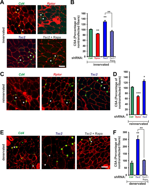
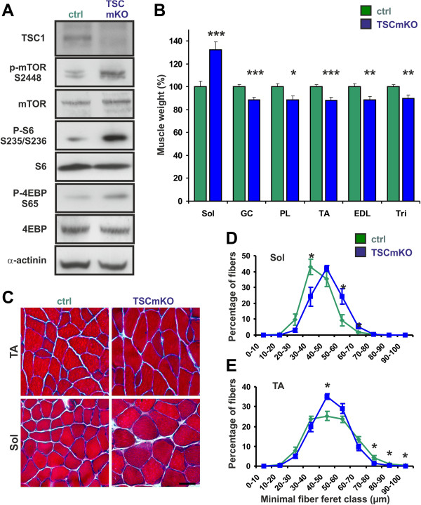
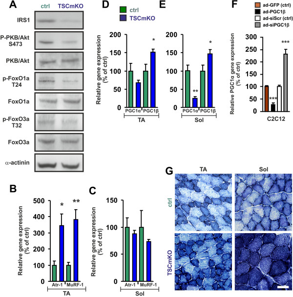
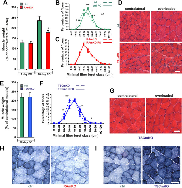
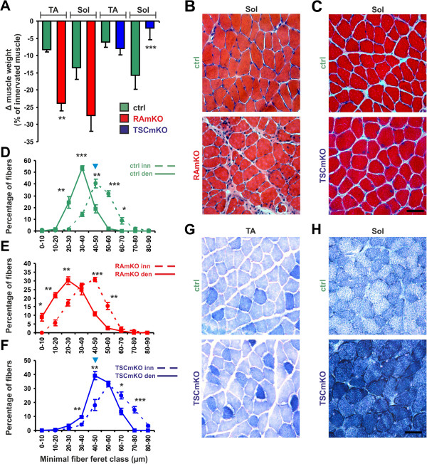
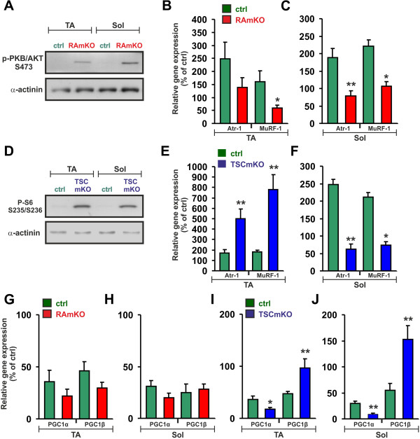
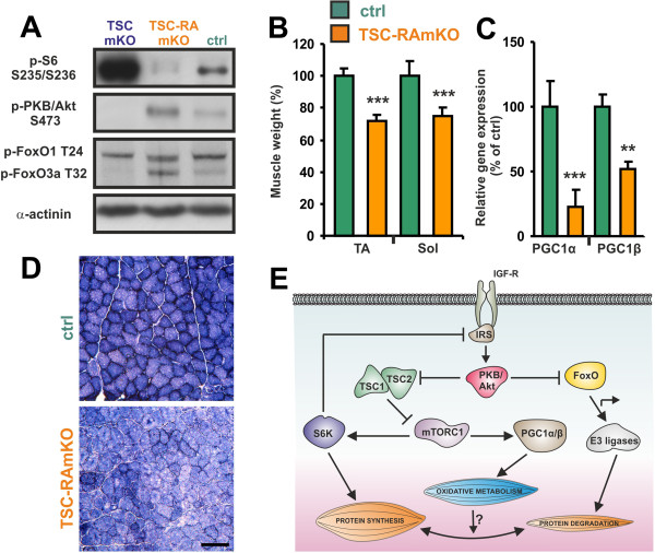
BACKGROUND: Skeletal muscle mass is determined by the balance between protein synthesis and degradation. Mammalian target of rapamycin complex 1 (mTORC1) is a master regulator of protein translation and has been implicated in the control of muscle mass. Inactivation of mTORC1 by skeletal muscle-specific deletion of its obligatory component raptor results in smaller muscles and a lethal dystrophy. Moreover, raptor-deficient muscles are less oxidative through changes in the expression PGC-1α, a critical determinant of mitochondrial biogenesis. These results suggest that activation of mTORC1 might be beneficial to skeletal muscle by providing resistance to muscle atrophy and increasing oxidative function. Here, we tested this hypothesis by deletion of the mTORC1 inhibitor tuberous sclerosis complex (TSC) in muscle fibers. METHOD: Skeletal muscles of mice with an acute or a permanent deletion of raptor or TSC1 were examined using histological, biochemical and molecular biological methods. Response of the muscles to changes in mechanical load and nerve input was investigated by ablation of synergistic muscles or by denervation . RESULTS: Genetic deletion or knockdown of raptor, causing inactivation of mTORC1, was sufficient to prevent muscle growth and enhance muscle atrophy. Conversely, short-term activation of mTORC1 by knockdown of TSC induced muscle fiber hypertrophy and atrophy-resistance upon denervation, in both fast tibialis anterior (TA) and slow soleus muscles. Surprisingly, however, sustained activation of mTORC1 by genetic deletion of Tsc1 caused muscle atrophy in all but soleus muscles. In contrast, oxidative capacity was increased in all muscles examined. Consistently, TSC1-deficient soleus muscle was atrophy-resistant whereas TA underwent normal atrophy upon denervation. Moreover, upon overloading, plantaris muscle did not display enhanced hypertrophy compared to controls. Biochemical analysis indicated that the atrophy response of muscles was based on the suppressed phosphorylation of PKB/Akt via feedback inhibition by mTORC1 and subsequent increased expression of the E3 ubiquitin ligases MuRF1 and atrogin-1/MAFbx. In contrast, expression of both E3 ligases was not increased in soleus muscle suggesting the presence of compensatory mechanisms in this muscle. CONCLUSIONS: Our study shows that the mTORC1- and the PKB/Akt-FoxO pathways are tightly interconnected and differentially regulated depending on the muscle type. These results indicate that long-term activation of the mTORC1 signaling axis is not a therapeutic option to promote muscle growth because of its strong feedback induction of the E3 ubiquitin ligases involved in protein degradation.
Overview
The claims and statistics of this paper haven’t been checked yet.
Run the check — free, no account needed. It reads the open-access text and checks the sources it cites.
Integrity signals
Registry
Cites
Statistics
Figures
Verification
Claims the paper makes
The claims and statistics of this paper haven’t been checked yet.
Run the check — free, no account needed. It reads the open-access text and checks the sources it cites.
Self-check
Percentages, sums and reported differences re-computed from the paper’s own numbers.
The self-check needs the paper’s full text.
Citations
Cites
mTOR Signaling in Growth Control and Disease
Laplante, M., & Sabatini, D. M. (2012) · Cell · doi:10.1016/j.cell.2012.03.017 · Cited by 8,509
TOR signaling in growth and metabolism
Wullschleger, S., Loewith, R., & Hall, M. N. (2006) · Cell · doi:10.1016/j.cell.2006.01.016 · Cited by 5,787
Sandri, M., Sandri, C., Gilbert, A., Skurk, C., Calabria, E., Picard, A., Walsh, K., Schiaffino, S., Lecker, S. H., & Goldberg, A. L. (2004) · Cell · doi:10.1016/s0092-8674(04)00400-3 · Cited by 3,012
Bodine, S. C., Stitt, T. N., Gonzalez, M., Kline, W. O., Stover, G. L., Bauerlein, R., Zlotchenko, E., Scrimgeour, A., Lawrence, J. C., Glass, D. J., et al. (2001) · Nature Cell Biology · doi:10.1038/ncb1101-1014 · Cited by 2,752
Sarbassov, D. D., Ali, S. M., Kim, D.-H., Guertin, D. A., Latek, R. R., Erdjument-Bromage, H., Tempst, P., & Sabatini, D. M. (2004) · Current Biology · doi:10.1016/j.cub.2004.06.054 · Cited by 2,665
Transcriptional co-activator PGC-1α drives the formation of slow-twitch muscle fibres
Lin, J., Wu, H., Tarr, P. T., Zhang, C.-Y., Wu, Z., Boss, O., Michael, L. F., Puigserver, P., Isotani, E., Olson, E. N., et al. (2002) · Nature · doi:10.1038/nature00904 · Cited by 2,546
Mammalian TOR complex 2 controls the actin cytoskeleton and is rapamycin insensitive
Jacinto, E., Loewith, R., Schmidt, A., Lin, S., Rüegg, M. A., Hall, A., & Hall, M. N. (2004) · Nature Cell Biology · doi:10.1038/ncb1183 · Cited by 2,097
Puigserver, P., & Spiegelman, B. M. (2003) · Endocrine Reviews · doi:10.1210/er.2002-0012 · Cited by 2,076
Stitt, T. N., Drujan, D., Clarke, B. A., Panaro, F., Timofeyva, Y., Kline, W. O., Gonzalez, M., Yancopoulos, G. D., & Glass, D. J. (2004) · Molecular Cell · doi:10.1016/s1097-2765(04)00211-4 · Cited by 1,964
FoxO3 Controls Autophagy in Skeletal Muscle In Vivo
Mammucari, C., Milan, G., Romanello, V., Masiero, E., Rudolf, R., Del Piccolo, P., Burden, S. J., Di Lisi, R., Sandri, C., Zhao, J., et al. (2007) · Cell Metabolism · doi:10.1016/j.cmet.2007.11.001 · Cited by 1,953
Cited by
Sorted by citations
Satellite Cells and Skeletal Muscle Regeneration
Dumont, N. A., Bentzinger, C. F., Sincennes, M., & Rudnicki, M. A. (2015) · Comprehensive Physiology · doi:10.1002/cphy.c140068 · Cited by 719
Self-citationProtein breakdown in muscle wasting: Role of autophagy-lysosome and ubiquitin-proteasome
Sandri, M. (2013) · The International Journal of Biochemistry & Cell Biology · doi:10.1016/j.biocel.2013.04.023 · Cited by 689
Signaling pathways controlling skeletal muscle mass
Egerman, M. A., & Glass, D. J. (2014) · Critical Reviews in Biochemistry and Molecular Biology · doi:10.3109/10409238.2013.857291 · Cited by 555
Mechanisms Regulating Neuromuscular Junction Development and Function and Causes of Muscle Wasting
Tintignac, L. A. J., Brenner, H.-R., & Rüegg, M. A. (2015) · Physiological Reviews · doi:10.1152/physrev.00033.2014 · Cited by 449
Leal, L. G., Lopes, M. A., & Batista, M. L. (2018) · Frontiers in Physiology · doi:10.3389/fphys.2018.01307 · Cited by 382
“Skeletal Muscle as an Immunogenic Organ The Concept of Myokines The SkM is a highly organized tissue at the micro and macroscopic level and is the body’s main protein reserve (Hornberger, 2011; Bentzinger et al., 2013).”
Background
Metadata
- Authors
- Bentzinger, C. F., Lin, S., Romanino, K., Castets, P., Guridi, M., Summermatter, S., Handschin, C., Tintignac, L. A., Hall, M. N., & Rüegg, M. A.
- Affiliations
- University of Basel (CH)
- Corresponding author
- Rüegg, Markus A
- ORCID
- Venue
- Skeletal Muscle
- Published
- 2013-03-06
- Volume
- 3
- Issue
- 1
- Pages
- 6-6
- ISSN
- 2044-5040 (ISSN-L)
- PMC
- PMC3622636
- OpenAlex
- W2149451793
- Cited by
- 153
- References
- 61
- Citations by year
- 2026: 4 · 2025: 13 · 2024: 14 · 2023: 8 · 2022: 10 · 2021: 14 · 2020: 11 · 2019: 16 · 2018: 11 · 2017: 8 · 2016: 11 · 2015: 10 · 2014: 14 · 2013: 8
- FWCI
- 6.42
- Type
- journal article
- Language
- en
- Open access
- gold
- doi.org
- Licence
- cc-by
- Locations
- Skeletal Muscle (publishedVersion, cc-by)
- Europe PMC (PubMed Central) (publishedVersion, cc-by)
- PubMed Central (publishedVersion, cc-by)
- PubMed (publishedVersion)
- edoc (University of Basel) (acceptedVersion)
- Archive ouverte UNIGE (University of Geneva) (submittedVersion, cc-by)
- HAL (submittedVersion, cc-by)
- edoc (University of Basel)
- Funders
- National Science Foundation
- Schweizerischer Nationalfonds zur Förderung der Wissenschaftlichen Forschung
- Topics
- Life Sciences › Biochemistry, Genetics and Molecular Biology › Molecular Biology › Muscle Physiology and Disorders
- Health Sciences › Nursing › Nutrition and Dietetics › Muscle metabolism and nutrition
- Life Sciences › Biochemistry, Genetics and Molecular Biology › Molecular Biology › PI3K/AKT/mTOR signaling in cancer
- Keywords
- mTORC1 signaling, skeletal muscle atrophy, muscle hypertrophy, raptors, tuberous sclerosis complex 1, denervation, muscle overload, oxidative capacity, PGC-1α, mitochondrial biogenesis, Akt signaling, FOXO, MuRF1, atrogin-1/MAFbx, E3 ubiquitin ligase, muscle fiber type, soleus muscle
About these checks
Registry, retraction, Cites and statistics results are computed, not judged, with one exception: a printed reference that no index lists is read once by a model before it counts as a citation error. Claims are extracted by a model and kept only when their quote appears verbatim in the text; each cited source is then checked, with a model’s help, for whether it says what the claim relies on. Those verdicts can be wrong — read the claim’s quote and the cited source before relying on one.
Statistics and arithmetic are recomputed, not judged.
Confidence bands are shown only for fields calibrated on our annotated test set. None is calibrated yet, so no band is shown.
What counts as a claim ›How we measure confidence ›How we check this ›
Are you an author, or do you see a problem with this page? Request a review
