

The DOI matches its Crossref record; 131 of the 131 references it cites were found in a registry or an index.
Sequence and structural analysis of BTB domain proteins
Open accessAbstract
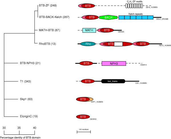
Background: The BTB domain (also known as the POZ domain) is a versatile protein-protein interaction motif that participates in a wide range of cellular functions, including transcriptional regulation, cytoskeleton dynamics, ion channel assembly and gating, and targeting proteins for ubiquitination. Several BTB domain structures have been experimentally determined, revealing a highly conserved core structure. Results: We surveyed the protein architecture, genomic distribution and sequence conservation of BTB domain proteins in 17 fully sequenced eukaryotes. The BTB domain is typically found as a single copy in proteins that contain only one or two other types of domain, and this defines the BTB-zinc finger (BTB-ZF), BTB-BACK-kelch (BBK), voltage-gated potassium channel T1 (T1-Kv), MATH-BTB, BTB-NPH3 and BTB-BACK-PHR (BBP) families of proteins, among others. In contrast, the Skp1 and ElonginC proteins consist almost exclusively of the core BTB fold. There are numerous lineage-specific expansions of BTB proteins, as seen by the relatively large number of BTB-ZF and BBK proteins in vertebrates, MATH-BTB proteins in Caenorhabditis elegans , and BTB-NPH3 proteins in Arabidopsis thaliana . Using the structural homology between Skp1 and the PLZF BTB homodimer, we present a model of a BTB-Cul3 SCF-like E3 ubiquitin ligase complex that shows that the BTB dimer or the T1 tetramer is compatible in this complex. Conclusion: Despite widely divergent sequences, the BTB fold is structurally well conserved. The fold has adapted to several different modes of self-association and interactions with non-BTB proteins.
Overview
The claims and statistics of this paper haven’t been checked yet.
Free: it reads the open-access text and checks the first 5 claims that cite a source we can check. Create a free account to check every claim.
Integrity signals
Registry
Cites
Statistics
Figures



Verification
Claims the paper makes
The claims and statistics of this paper haven’t been checked yet.
Free: it reads the open-access text and checks the first 5 claims that cite a source we can check. Create a free account to check every claim.
Self-check
Percentages, sums and reported differences re-computed from the paper’s own numbers.
The self-check needs the paper’s full text.
Citations
Cites
Gapped BLAST and PSI-BLAST: a new generation of protein database search programs
Altschul, S. (1997) · Nucleic Acids Research · doi:10.1093/nar/25.17.3389 · Cited by 73,302
Berman, H. M. (2000) · Nucleic Acids Research · doi:10.1093/nar/28.1.235 · Cited by 40,196
The PyMOL Molecular Graphics System
DeLano, W. L. (2002) · Medical Entomology and Zoology · Cited by 18,147
PHYLIP-Phylogeny inference package (Version 3.2)
Felsenstein, J. (1989) · Cladistics · Cited by 17,682
The Pfam Protein Families Database
Bateman, A. (2002) · Nucleic Acids Research · doi:10.1093/nar/30.1.276 · Cited by 14,000
SWISS‐MODEL and the Swiss‐Pdb Viewer: An environment for comparative protein modeling
Guex, N., & Peitsch, M. C. (1997) · ELECTROPHORESIS · doi:10.1002/elps.1150181505 · Cited by 11,241
Murzin, A. G., Brenner, S. E., Hubbard, T., & Chothia, C. (1995) · Journal of Molecular Biology · doi:10.1016/s0022-2836(05)80134-2 · Cited by 6,445
MEGA2: molecular evolutionary genetics analysis software
Kumar, S., Tamura, K., Jakobsen, I. B., & Nei, M. (2001) · Bioinformatics · doi:10.1093/bioinformatics/17.12.1244 · Cited by 6,097
Multiple sequence alignment with the Clustal series of programs
Chenna, R. (2003) · Nucleic Acids Research · doi:10.1093/nar/gkg500 · Cited by 4,875
The PSIPRED protein structure prediction server
McGuffin, L. J., Bryson, K., & Jones, D. T. (2000) · Bioinformatics · doi:10.1093/bioinformatics/16.4.404 · Cited by 3,838
Research works printed but not linked: 1
- Swofford D: PAUP. 1998, Sunderland, MA: Sinauer Associates
Cited by
The Molecular Mechanisms Regulating the KEAP1-NRF2 Pathway
Baird, L., & Yamamoto, M. (2020) · Molecular and Cellular Biology · doi:10.1128/mcb.00099-20 · Cited by 1,542
Sturgeon, C. M., Duffy, M. J., Stenman, U.-H., Lilja, H., Brünner, N., Chan, D. W., Babaian, R., Bast, R. C., Dowell, B., Esteva, F. J., et al. (2008) · Clinical Chemistry · doi:10.1373/clinchem.2008.105601 · Cited by 1,129
Li, W., Bengtson, M. H., Ulbrich, A., Matsuda, A., Reddy, V. A., Orth, A., Chanda, S. K., Batalov, S., & Joazeiro, C. A. P. (2008) · PLoS ONE · doi:10.1371/journal.pone.0001487 · Cited by 838
“. . . . . . . . . . . . . . . . . . . . . . . . . . . . . . . . . . . . . . . . . . . . . . . . . . . . . . . . . . . . . . . . . . . . . . Protein family Gene name Gene symbol Ubox PRP19 YLL036C UFD2 YDL190C HECT TOM1 YDR457W HUL4 YJR036C HUL5 YGL141W UFD4 YKL010C RSP5 YER125W F BOX COS111 YBR203W SAF1 YBR280C MFB1 Y…”
BackgroundInfluentialTong, K. I., Katoh, Y., Kusunoki, H., Itoh, K., Tanaka, T., & Yamamoto, M. (2006) · Molecular and Cellular Biology · doi:10.1128/mcb.26.8.2887-2900.2006 · Cited by 759
“Except for the DDB1, core BTB folds are found in SKP1, Elongin C and other BTB proteins (54).”
BackgroundArgout, X., Salse, J., Aury, J.-M., Guiltinan, M. J., Droc, G., Gouzy, J., Allegre, M., Chaparro, C., Legavre, T., Maximova, S. N., et al. (2011) · Nature Genetics · doi:10.1038/ng.736 · Cited by 746
Metadata
- Authors
- Stogios, P. J., Downs, G. S., Jauhal, J. J., Nandra, S. K., & Privé, G. G.
- Affiliations
- University of Toronto (CA)
- Seneca Polytechnic (CA)
- Corresponding author
- Privé, Gilbert G
- Venue
- Genome Biology
- Published
- 2005-09-15
- Volume
- 6
- Issue
- 10
- Pages
- R82-R82
- ISSN
- 1465-6906 (ISSN-L)
- 1474-760X
- PMC
- PMC1257465
- OpenAlex
- W2159956236
- Cited by
- 694
- References
- 131
- Citations by year
- 2026: 21 · 2025: 31 · 2024: 48 · 2023: 24 · 2022: 45 · 2021: 27 · 2020: 37 · 2019: 48 · 2018: 37 · 2017: 25 · 2016: 39 · 2015: 25 · 2014: 35 · 2013: 37 · 2012: 42
- FWCI
- 9.82
- Type
- journal article
- Language
- en
- Open access
- gold
- doi.org
- Licence
- cc-by
- creativecommons.org/licenses/by/2.0/ (tdm)
- creativecommons.org/licenses/by/2.0/ (vor)
- Locations
- Genome biology (publishedVersion, cc-by)
- Europe PMC (PubMed Central) (publishedVersion)
- PubMed Central (publishedVersion)
- PubMed (publishedVersion)
- TSpace (University of Toronto) (submittedVersion)
- Topics
- Life Sciences › Biochemistry, Genetics and Molecular Biology › Molecular Biology › Ubiquitin and proteasome pathways
- Life Sciences › Biochemistry, Genetics and Molecular Biology › Molecular Biology › Protein Structure and Dynamics
- Life Sciences › Biochemistry, Genetics and Molecular Biology › Molecular Biology › Genomics and Chromatin Dynamics
- Keywords
- BTB domain, POZ domain, protein-protein interaction motifs, Skp1, SCF ubiquitin ligase, protein domain architecture, sequence conservation, structural conservation
- MeSH terms
- Amino Acid Sequence, Animals, DNA-Binding Proteins, Models, Molecular, Molecular Sequence Data, Phylogeny, Protein Binding, Zinc Fingers, Genome, Sequence Analysis, DNA, Protein Structure, Tertiary, Protein Folding, Sequence Analysis, Protein
About these checks
Registry, retraction, Cites and statistics results are computed, not judged, with one exception: a printed reference that no index lists is read once by a model before it counts as a citation error. Claims are extracted by a model and kept only when their quote appears verbatim in the text; each cited source is then checked, with a model’s help, for whether it says what the claim relies on. Those verdicts can be wrong — read the claim’s quote and the cited source before relying on one.
Statistics and arithmetic are recomputed, not judged.
Confidence bands are shown only for fields calibrated on our annotated test set. None is calibrated yet, so no band is shown.
What counts as a claim ›How we measure confidence ›How we check this ›
Are you an author, or do you see a problem with this page? Request a review
How to cite
Cite CoChat for the checks on this page, and the paper for its own findings.
- This page
- CoChat. (2026). Citation check: Sequence and structural analysis of BTB domain proteins. https://app.cochat.ai/papers/10.1186/gb-2005-6-10-r82
- The paper
- Stogios, P. J., Downs, G. S., Jauhal, J. J., Nandra, S. K., & Privé, G. G. (2005). Sequence and structural analysis of BTB domain proteins. Genome Biology. https://doi.org/10.1186/gb-2005-6-10-r82
Data sources
Bibliographic data: Semantic Scholar, OpenAlex, Crossref.
Contains information from the Semantic Scholar Academic Graph Datasets, made available under the ODC Attribution License (ODC-BY 1.0).
Semantic Scholar’s data is described in: Kinney, R., et al. (2023). The Semantic Scholar Open Data Platform. arXiv:2301.10140.
