

The DOI matches its Crossref record; 145 of the 145 references it cites were found in a registry or an index.
The ubiquitin–proteasome system in regulation of the skeletal muscle homeostasis and atrophy: from basic science to disorders
Open accessAbstract
Skeletal muscle is one of the most abundant and highly plastic tissues. The ubiquitin-proteasome system (UPS) is recognised as a major intracellular protein degradation system, and its function is important for muscle homeostasis and health. Although UPS plays an essential role in protein degradation during muscle atrophy, leading to the loss of muscle mass and strength, its deficit negatively impacts muscle homeostasis and leads to the occurrence of several pathological phenotypes. A growing number of studies have linked UPS impairment not only to matured muscle fibre degeneration and weakness, but also to muscle stem cells and deficiency in regeneration. Emerging evidence suggests possible links between abnormal UPS regulation and several types of muscle diseases. Therefore, understanding of the role of UPS in skeletal muscle may provide novel therapeutic insights to counteract muscle wasting, and various muscle diseases. In this review, we focussed on the role of proteasomes in skeletal muscle and its regeneration, including a brief explanation of the structure of proteasomes. In addition, we summarised the recent findings on several diseases and elaborated on how the UPS is related to their pathological states.
Overview
The claims and statistics of this paper haven’t been checked yet.
Run the check — free, no account needed. It reads the open-access text and checks the sources it cites.
Integrity signals
Registry
Cites
Statistics
Figures
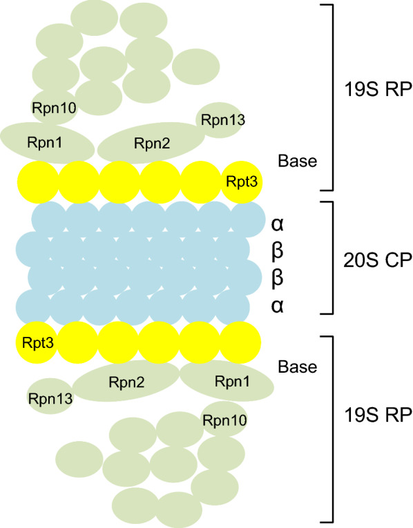
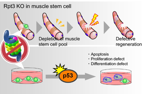
Verification
Claims the paper makes
The claims and statistics of this paper haven’t been checked yet.
Run the check — free, no account needed. It reads the open-access text and checks the sources it cites.
Claims other papers make about this one
Each is checked against this paper’s own text: does it say what the citing paper says it does?
The cited source says what the claim attributes to it.
In the citing paper
“The ubiquitin–proteasome system plays a major role in protein content turnover by degrading misfolded or defective proteins [163,164].”
From Main Pathogenic Mechanisms and Recent Advances in COPD Peripheral Skeletal Muscle Wasting (2023) · cites this paper as [163]
Self-check
Percentages, sums and reported differences re-computed from the paper’s own numbers.
The self-check needs the paper’s full text.
Citations
Cites
2 retracted
Azakir, B., Fulvio, S. D., Kinter, J., & Sinnreich, M. (2012) · Journal of Biological Chemistry · doi:10.1074/jbc.m111.329078 · Cited by 36
RetractedAzakir, B., Fulvio, S. D., Kinter, J., & Sinnreich, M. (2017) · Journal of Biological Chemistry · doi:10.1074/jbc.a111.329078 · Cited by 1
RetractedRepertoires of Autophagy in the Pathogenesis of Ocular Diseases
Li, Y., Jiang, Q. L., Cao, G.-F., Yao, J., & Yan, B. (2015) · Cellular Physiology and Biochemistry · doi:10.1159/000373980 · Cited by 16,351
Cui, C., Cui, J., Jin, F., Cui, Y., Li, R., Jiang, X., Tian, Y., Wang, R. C., Jiang, P., & Gao, J. (2017) · Cellular Physiology and Biochemistry · doi:10.1159/000479571 · Cited by 5,214
Pankiv, S., Clausen, T. H., Lamark, T., Brech, A., Bruun, J.-A., Outzen, H., Øvervatn, A., Bjørkøy, G., & Johansen, T. (2007) · Journal of Biological Chemistry · doi:10.1074/jbc.m702824200 · Cited by 4,578
The Ubiquitin-Proteasome Proteolytic Pathway: Destruction for the Sake of Construction
Glickman, M. H., & Ciechanover, A. J. (2002) · Physiological Reviews · doi:10.1152/physrev.00027.2001 · Cited by 4,304
Autophagy regulates lipid metabolism
Singh, R., Kaushik, S., Wang, Y., Xiang, Y., Novak, I., Komatsu, M., Tanaka, K., Cuervo, A. M., & Czaja, M. J. (2009) · Nature · doi:10.1038/nature07976 · Cited by 3,975
Identification of ubiquitin ligases required for skeletal muscle atrophy.
Bodine, S. C., Latres, E., Baumhueter, S., Lai, V. K.-M., Nunez, L., Clarke, B. A., Poueymirou, W. T., Panaro, F. J., Na, E., Dharmarajan, K., et al. (2001) · Science · doi:10.1126/science.1065874 · Cited by 3,521
Sandri, M., Sandri, C., Gilbert, A., Skurk, C., Calabria, E., Picard, A., Walsh, K., Schiaffino, S., Lecker, S. H., & Goldberg, A. L. (2004) · Cell · doi:10.1016/s0092-8674(04)00400-3 · Cited by 3,012
The role of autophagy during the early neonatal starvation period
Kuma, A., Hatano, M., Matsui, M., Yamamoto, A., Nakaya, H., Yoshimori, T., Ohsumi, Y., Tokuhisa, T., & Mizushima, N. (2004) · Nature · doi:10.1038/nature03029 · Cited by 2,865
Cited by
Sorted by citations
Mitochondrial dysfunction: roles in skeletal muscle atrophy
Chen, X., Ji, Y., Liu, R., Zhu, X., Wang, K., Yang, X., Liu, B., Gao, Z., Huang, Y. H., Shen, Y., et al. (2023) · Journal of Translational Medicine · doi:10.1186/s12967-023-04369-z · Cited by 362
Cancer cachexia: molecular mechanisms and treatment strategies
Setiawan, T., Sari, I. N., Wijaya, Y. T., Julianto, N. M., Muhammad, J. A., Lee, H., Chae, J. H., & Kwon, H. Y. (2023) · Journal of Hematology & Oncology · doi:10.1186/s13045-023-01454-0 · Cited by 342
Inflammation: Roles in Skeletal Muscle Atrophy
Ji, Y., Li, M., Chang, M., Liu, R., Qiu, J., Wang, K., Deng, C., Shen, Y., Zhu, J., Wang, W., et al. (2022) · Antioxidants · doi:10.3390/antiox11091686 · Cited by 329
Voiriot, G., Oualha, M., Pierre, A., Salmon-Gandonnière, C., Gaudet, A., Jouan, Y., Kallel, H., Radermacher, P., Vodovar, D., Sarton, B., et al. (2022) · Annals of Intensive Care · doi:10.1186/s13613-022-01038-0 · Cited by 229
Mitochondrial dysfunction and skeletal muscle atrophy: Causes, mechanisms, and treatment strategies
Kubat, G. B., Bouhamida, E., Ülger, Ö., Türkel, İ., Pedriali, G., Ramaccini, D., Ekinci, Ö., Özerkliğ, B., Atalay, Ö., Patergnani, S., et al. (2023) · Mitochondrion · doi:10.1016/j.mito.2023.07.003 · Cited by 134
Metadata
- Authors
- Kitajima, Y., Yoshioka, K., & Suzuki, N.
- Affiliations
- Institute of Molecular Embryology and Genetics, Kumamoto University (JP)
- Tohoku University (JP)
- Corresponding author
- Kitajima, Yasuo
- Suzuki, Naoki
- Venue
- The Journal of Physiological Sciences
- Publisher
- Elsevier BV
- Published
- 2020
- Volume
- 70
- Issue
- 1
- Pages
- 40
- ISSN
- 1880-6546 (ISSN-L)
- PMC
- PMC10717345
- OpenAlex
- W3085918938
- Cited by
- 171
- References
- 145
- Citations by year
- 2026: 28 · 2025: 43 · 2024: 33 · 2023: 25 · 2022: 30 · 2021: 12
- FWCI
- 1.60
- Type
- journal article
- Language
- en
- Open access
- gold
- doi.org
- Licence
- Locations
- The Journal of Physiological Sciences (publishedVersion, cc-by)
- PubMed Central (publishedVersion, cc-by)
- PubMed (publishedVersion)
- Funders
- Takeda Science Foundation
- Grant-in-Aid for Scientific Research KAKENHI (15H05667, 16H05318, 18K07519, 18K17857, 20H04078)
- Suzuken Memorial Foundation (YK)
- Topics
- Life Sciences › Biochemistry, Genetics and Molecular Biology › Molecular Biology › Muscle Physiology and Disorders
- Life Sciences › Biochemistry, Genetics and Molecular Biology › Molecular Biology › Ubiquitin and proteasome pathways
- Health Sciences › Nursing › Nutrition and Dietetics › Muscle metabolism and nutrition
- Keywords
- ubiquitin-proteasome system, skeletal muscle homeostasis, muscle atrophy, proteasome, muscle regeneration, muscle stem cells, muscle fiber degeneration, myopathy, protein degradation
- MeSH terms
- Animals, Homeostasis, Humans, Muscle Proteins, Muscular Atrophy, Regeneration, Muscle, Skeletal, Muscle Development, Ubiquitin, Satellite Cells, Skeletal Muscle, Proteasome Endopeptidase Complex, Ubiquitination, Proteolysis
About these checks
Registry, retraction, Cites and statistics results are computed, not judged, with one exception: a printed reference that no index lists is read once by a model before it counts as a citation error. Claims are extracted by a model and kept only when their quote appears verbatim in the text; each cited source is then checked, with a model’s help, for whether it says what the claim relies on. Those verdicts can be wrong — read the claim’s quote and the cited source before relying on one.
Statistics and arithmetic are recomputed, not judged.
Confidence bands are shown only for fields calibrated on our annotated test set. None is calibrated yet, so no band is shown.
What counts as a claim ›How we measure confidence ›How we check this ›
Are you an author, or do you see a problem with this page? Request a review
