

The DOI matches its Crossref record; 4 of the 4 references it cites were found in a registry or an index.
Integrative multi-omics analysis reveals the contribution of neoVTX genes to venom diversity of Synanceia verrucosa
Open accessPublished version of a preprint
Abstract
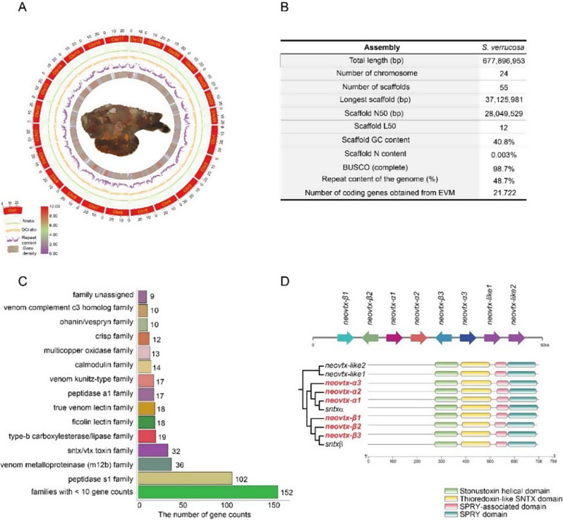
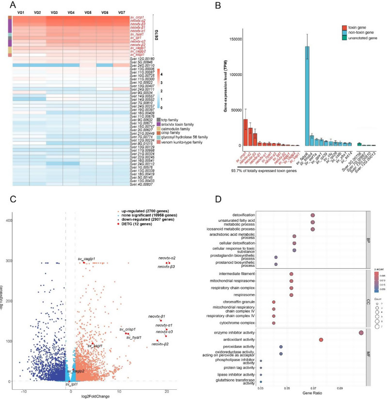
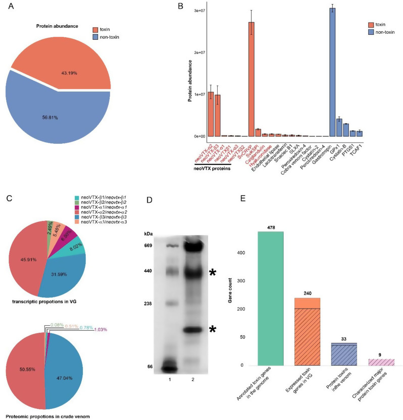
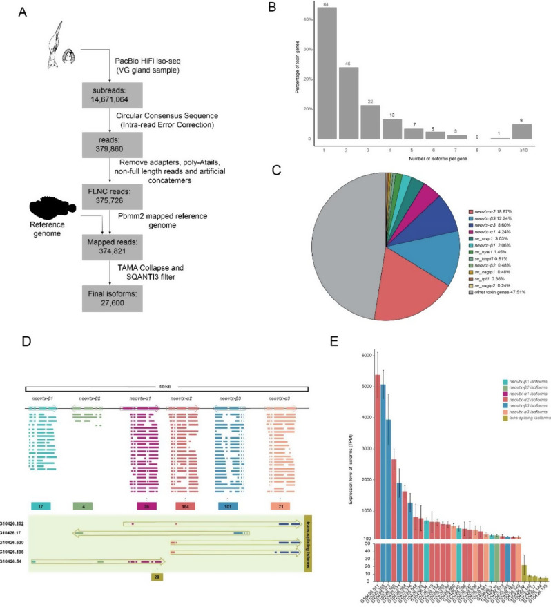
BACKGROUND: Animal venom systems are considered as valuable model for investigating the molecular mechanisms underlying phenotypic evolution. Stonefish are the most venomous and dangerous fish because of severe human envenomation and occasionally fatalities, whereas the genomic background of their venom has not been fully explored compared with that in other venomous animals. RESULTS: In this study, we followed modern venomic pipelines to decode the Synanceia verrucosa venom components. A catalog of 478 toxin genes was annotated based on our assembled chromosome-level genome. Integrative analysis of the high-quality genome, the transcriptome of the venom gland, and the proteome of crude venom revealed mechanisms underlying the venom complexity in S. verrucosa. Six tandem-duplicated neoVTX subunit genes were identified as the major source for the neoVTX protein production. Further isoform sequencing revealed massive alternative splicing events with a total of 411 isoforms demonstrated by the six genes, which further contributed to the venom diversity. We then characterized 12 dominantly expressed toxin genes in the venom gland, and 11 of which were evidenced to produce the venom protein components, with the neoVTX proteins as the most abundant. Other major venom proteins included a presumed CRVP, Kuntiz-type serine protease inhibitor, calglandulin protein, and hyaluronidase. Besides, a few of highly abundant non-toxin proteins were also characterized and they were hypothesized to function in housekeeping or hemostasis maintaining roles in the venom gland. Notably, gastrotropin like non-toxin proteins were the second highest abundant proteins in the venom, which have not been reported in other venomous animals and contribute to the unique venom properties of S. verrucosa. CONCLUSIONS: The results identified the major venom composition of S. verrucosa, and highlighted the contribution of neoVTX genes to the diversity of venom composition through tandem-duplication and alternative splicing. The diverse neoVTX proteins in the venom as lethal particles are important for understanding the adaptive evolution of S. verrucosa. Further functional studies are encouraged to exploit the venom components of S. verrucosa for pharmaceutical innovation.
Overview
The claims and statistics of this paper haven’t been checked yet.
Run the check — free, no account needed. It reads the open-access text and checks the sources it cites.
Integrity signals
Registry
Cites
Statistics
Figures
Verification
Claims the paper makes
The claims and statistics of this paper haven’t been checked yet.
Run the check — free, no account needed. It reads the open-access text and checks the sources it cites.
Self-check
Percentages, sums and reported differences re-computed from the paper’s own numbers.
The self-check needs the paper’s full text.
Citations
Cites
Global quantification of mammalian gene expression control
Schwanhäusser, B., Busse, D., Li, N., Dittmar, G., Schuchhardt, J., Wolf, J., Chen, W., & Selbach, M. (2011) · Nature · doi:10.1038/nature10098 · Cited by 6,566
RepeatModeler2 for automated genomic discovery of transposable element families
Flynn, J. M., Hubley, R., Goubert, C., Rosen, J., Clark, A. G., Feschotte, C., & Smit, A. F. (2020) · Proceedings of the National Academy of Sciences · doi:10.1073/pnas.1921046117 · Cited by 3,848
Large-scale ruminant genome sequencing provides insights into their evolution and distinct traits
Chen, L., Qiu, Q., Jiang, Y., Wang, K., Lin, Z., Li, Z., Bibi, F., Yang, Y., Wang, J., Nie, W., et al. (2019) · Science · doi:10.1126/science.aav6202 · Cited by 441
Self-citationBittenbinder, M. A., van Thiel, J., Cardoso, F. C., Casewell, N. R., Gutiérrez, J.-M., Kool, J., & Vonk, F. J. (2024) · Communications Biology · doi:10.1038/s42003-024-06019-6 · Cited by 132
Research works printed but not linked: 87
- Zancolli G, Casewell NR. Venom systems as models for studying the origin and regulation of evolutionary novelties. Mol Biol Evol. 2020;37(10):2777–90. · DOI
- Bordon KCF, Cologna CT, Fornari-Baldo EC, Pinheiro-Júnior EL, Cerni FA, Amorim FG, Anjolette FAP, Cordeiro FA, Wiezel GA, Cardoso IA, et al. From animal poisons and venoms to medicines: achievements, challenges and perspectives in drug discovery. Front Pharmacol. 2020;11:1132. · DOI
- von Reumont BM, Anderluh G, Antunes A, Ayvazyan N, Beis D, Caliskan F, Crnković A, Damm M, Dutertre S, Ellgaard L et al. Modern venomics-Current insights, novel methods, and future perspectives in biological and applied animal venom research. GigaScience 2022, 11. · DOI
- Suranse V, Srikanthan A, Sunagar K. Animal Venoms: Origin, Diversity and Evolution. In: eLS. John Wiley & Sons, Ltd; 2018: 1–20. · DOI
- Suryamohan K, Krishnankutty SP, Guillory J, Jevit M, Schröder MS, Wu M, Kuriakose B, Mathew OK, Perumal RC, Koludarov I, et al. The Indian cobra reference genome and transcriptome enables comprehensive identification of venom toxins. Nat Genet. 2020;52(1):106–17. · DOI
- Ludington AJ, Hammond JM, Breen J, Deveson IW, Sanders KL. New chromosome-scale genomes provide insights into marine adaptations of sea snakes (Hydrophis: Elapidae). BMC Biol 2023, 21(1). · DOI
- Margres MJ, Rautsaw RM, Strickland JL, Mason AJ, Schramer TD, Hofmann EP, Stiers E, Ellsworth SA, Nystrom GS, Hogan MP et al. The Tiger Rattlesnake genome reveals a complex genotype underlying a simple venom phenotype. Proc Natl Acad Sci U S A 2021, 118(4). · DOI
- Zhu B, Jin P, Hou Z, Li J, Wei S, Li S. Chromosomal-level genome of a sheet-web spider provides insight into the composition and evolution of venom. Mol Ecol Resour. 2022;22(6):2333–48. · DOI
- You Y, Tang Y, Yin W, Liu X, Gao P, Zhang C, Tembrock LR, Zhao Y, Yang Z. From genome to proteome: Comprehensive identification of venom toxins from the Chinese funnel-web spider (Macrothelidae: Macrothele yani). Int J Biol Macromol. 2024;268(Pt 2). · DOI
- Herráez-Pérez A, Pardos-Blas JR, Afonso CML, Tenorio MJ, Zardoya R. Chromosome-level genome of the venomous snail Kalloconus canariensis: a valuable model for venomics and comparative genomics. Gigascience. 2022;12. · DOI
- Peng C, Huang Y, Bian C, Li J, Liu J, Zhang K, You X, Lin Z, He Y, Chen J, et al. The first Conus genome assembly reveals a primary genetic central dogma of conopeptides in C. betulinus. Cell Discov. 2021;7(1):11. · DOI
- Farhat S, Modica MV, Puillandre N. Whole genome duplication and gene evolution in the hyperdiverse venomous gastropods. Mol Biol Evol. 2023;40(8):msad171. · DOI
- Wei S, Zhou W, Fan H, Zhang Z, Guo W, Peng Z, Wei F. Chromosome-level genome assembly of the yellow boxfish (Ostracion cubicus) provides insights into the evolution of bone plates and ostracitoxin secretion. Front Mar Sci. 2023;10:1170704. · DOI
- Martinson EO, Mrinalini, Kelkar YD, Chang C-H, Werren JH. The evolution of venom by co-option of single-copy genes. Curr Biol. 2017;27(13):2007–13. e2008. · DOI
- Zhang Z-Y, Lv Y, Wu W, Yan C, Tang C-Y, Peng C, Li J-T. The structural and functional divergence of a neglected three-finger toxin subfamily in lethal elapids. Cell Rep. 2022;40(2):111079. · DOI
- Almeida DD, Viala VL, Nachtigall PG, Broe M, Gibbs HL, Serrano SMT, Moura-da-Silva AM, Ho PL, Nishiyama-Jr MY, Junqueira-de-Azevedo ILM. Tracking the recruitment and evolution of snake toxins using the evolutionary context provided by the Bothrops jararaca genome. Proc Natl Acad Sci U S A. 2021;118(20):e2015159118. · DOI
- Koludarov I, Senoner T, Jackson TNW, Dashevsky D, Heinzinger M, Aird SD, Rost B. Domain loss enabled evolution of novel functions in the snake three-finger toxin gene superfamily. Nat Commun. 2023;14(1):4861. · DOI
- Zancolli G, Reijnders M, Waterhouse RM, Robinson-Rechavi M. Convergent evolution of venom gland transcriptomes across Metazoa. Proc Natl Acad Sci U S A. 2022;119(1):e2111392119. · DOI
- Ye X, He C, Yang Y, Sun YH, Xiong S, Chan KC, Si Y, Xiao S, Zhao X, Lin H, et al. Comprehensive isoform-level analysis reveals the contribution of alternative isoforms to venom evolution and repertoire diversity. Genome Res. 2023;33(9):1554–67. · DOI
- Harris RJ, Jenner RA. Evolutionary ecology of fish venom: Adaptations and consequences of evolving a venom system. Toxins. 2019;11(2):60. · DOI
- Maillaud C, Hoang-Oppermann T, Hoang-Oppermann V, Rigot H, Girardot S, Nour M. Is stonefish Synanceia verrucosa envenomation potentially lethal? Toxicon. 2020;184:78–82. · DOI
- Lopez CAJ, Magee CAJ, Belyea CCM, Gumboc L. Finger flexor tenosynovitis from Stonefish envenomation injury. J Am Acad Orthop Surg Glob Res Rev. 2019;3(5):e024.
- Khoo HE. Bioactive proteins from stonefish venom. Clin Exp Pharmacol Physiol. 2002;29(9):802–6. · DOI
- Poh CH, Yuen R, Khoo HE, Chung M, Gwee M, Gopalakrishnakone P. Purification and partial characterization of stonustoxin (lethal factor) from Synanceja horrida venom. Comp Biochem Physiol B. 1991;99(4):793–8. · DOI
- Ziegman R, Undheim EAB, Baillie G, Jones A, Alewood PF. Investigation of the estuarine stonefish (Synanceia horrida) venom composition. J Proteom. 2019;201:12–26. · DOI
- Ellisdon AM, Reboul CF, Panjikar S, Huynh K, Oellig CA, Winter KL, Dunstone MA, Hodgson WC, Seymour J, Dearden PK, et al. Stonefish toxin defines an ancient branch of the perforin-like superfamily. Proc Natl Acad Sci U S A. 2015;112(50):15360–5. · DOI
- Saggiomo SL, Firth C, Wilson DT, Seymour J, Miles JJ, Wong Y. The geographic distribution, venom components, pathology and treatments of stonefish (Synanceia spp.) venom. Mar Drugs. 2021;19(6):302. · DOI
- Tang T, Huang Y, Peng C, Liao Y, Lv Y, Shi Q, Gao B. A chromosome-level genome assembly of the reef stonefish (Synanceia verrucosa) provides novel insights into Stonustoxin (sntx) Genes. Mol Biol Evol. 2023;40(10):msad215. · DOI
- Garnier P, Sauviat M-p, Goudey-perriere F, Perriere C. Cardiotoxicity of verrucotoxin, a protein isolated from the venom of Synanceia verrucosa. Toxicon. 1997;35(1):47–55. · DOI
- Garnier P, Goudey-Perrière F, Breton P, Dewulf C, Petek F, Perrière C. Enzymatic properties of the stonefish (Synanceia verrucosa Bloch and Schneider, 1801) venom and purification of a lethal, hypotensive and cytolytic factor. Toxicon. 1995;33(2):143–55. · DOI
- Garnier P, Ducancel F, Ogawa T, Boulain J-C, Goudey-Perrière F, Perrière C, Ménez A. Complete amino-acid sequence of the β-subunit of VTX from venom of the stonefish (Synanceia verrucosa) as identified from cDNA cloning experiments. Biochim Biophys Acta. 1997;1337(1):1–5. · DOI
- Harris RJ, Youngman NJ, Chan W, Bosmans F, Cheney KL, Fry BG. Getting stoned: Characterisation of the coagulotoxic and neurotoxic effects of reef stonefish (Synanceia verrucosa) venom. Toxicol Lett. 2021;346:16–22. · DOI
- Campos FV, Fiorotti HB, Coitinho JB, Figueiredo SG. Fish Cytolysins in All Their Complexity. Toxins. 2021;13(12):877. · DOI
- Madokoro M, Ueda A, Kiriake A, Shiomi K. Properties and cDNA cloning of a hyaluronidase from the stonefish Synanceia verrucosa venom. Toxicon. 2011;58(4):285–92. · DOI
- Kato K, Nakagawa H, Shinohara M, Ohura K. Purification of a novel lectin from the dorsal spines of the stonefish, Synanceia verrucosa. J Osaka Dent Univ. 2016;50(2):55–61.
- Cheng H, Concepcion GT, Feng X, Zhang H, Li H. Haplotype-resolved de novo assembly using phased assembly graphs with hifiasm. Nat Methods. 2021;18(2):170–5. · DOI · Paper page
- Guan D, McCarthy SA, Wood J, Howe K, Wang Y, Durbin R. Identifying and removing haplotypic duplication in primary genome assemblies. Bioinformatics. 2020;36(9):2896–8. · DOI
- Zhang H, Song L, Wang X, Cheng H, Wang C, Meyer CA, Liu T, Tang M, Aluru S, Yue F, et al. Fast alignment and preprocessing of chromatin profiles with Chromap. Nat Commun. 2021;12(1):6566. · DOI
- Zhou C, McCarthy SA, Durbin R. YaHS: yet another Hi-C scaffolding tool. Bioinformatics 2023, 39(1). · DOI · Paper page
- Manni M, Berkeley MR, Seppey M, Simão FA, Zdobnov EM. BUSCO update: novel and streamlined workflows along with broader and deeper phylogenetic coverage for scoring of eukaryotic, prokaryotic, and viral genomes. Mol Biol Evol. 2021;38(10):4647–54. · DOI · Paper page
- Bao W, Kojima KK, Kohany O. Repbase Update, a database of repetitive elements in eukaryotic genomes. Mob DNA. 2015;6:11. · DOI
- Tarailo-Graovac M, Chen N. Using RepeatMasker to identify repetitive elements in genomic sequences. Curr Protoc Bioinf. 2009;Chap. 4:Unit 4.10. · DOI
- Benson G. Tandem repeats finder: a program to analyze DNA sequences. Nucleic Acids Res. 1999;27(2):573–80. · DOI · Paper page
- Haas BJ, Salzberg SL, Zhu W, Pertea M, Allen JE, Orvis J, White O, Buell CR, Wortman JR. Automated eukaryotic gene structure annotation using EVidenceModeler and the program to assemble spliced alignments. Genome Biol. 2008;9(1):R7. · DOI
- Kim D, Paggi JM, Park C, Bennett C, Salzberg SL. Graph-based genome alignment and genotyping with HISAT2 and HISAT-genotype. Nat Biotechnol. 2019;37(8):907–15. · DOI · Paper page
- Pertea M, Pertea GM, Antonescu CM, Chang TC, Mendell JT, Salzberg SL. StringTie enables improved reconstruction of a transcriptome from RNA-seq reads. Nat Biotechnol. 2015;33(3):290–5. · DOI
- Stanke M, Diekhans M, Baertsch R, Haussler D. Using native and syntenically mapped cDNA alignments to improve de novo gene finding. Bioinformatics. 2008;24(5):637–44. · DOI · Paper page
- Burge CB, Karlin S. Finding the genes in genomic DNA. Curr Opin Struct Biol. 1998;8(3):346–54. · DOI
- Majoros WH, Salzberg SL. An empirical analysis of training protocols for probabilistic gene finders. BMC Bioinformatics. 2004;5:206. · DOI
- Kuo RI, Cheng Y, Zhang R, Brown JWS, Smith J, Archibald AL, Burt DW. Illuminating the dark side of the human transcriptome with long read transcript sequencing. BMC Genomics. 2020;21(1):751. · DOI
- Tardaguila M, de la Fuente L, Marti C, Pereira C, Pardo-Palacios FJ, Del Risco H, Ferrell M, Mellado M, Macchietto M, Verheggen K, et al. SQANTI: extensive characterization of long-read transcript sequences for quality control in full-length transcriptome identification and quantification. Genome Res. 2018;28(3):396–411. · DOI
- Liao Y, Smyth GK, Shi W. featureCounts: an efficient general purpose program for assigning sequence reads to genomic features. Bioinformatics. 2014;30(7):923–30. · DOI · Paper page
- Love MI, Huber W, Anders S. Moderated estimation of fold change and dispersion for RNA-seq data with DESeq2. Genome Biol. 2014;15(12):550. · DOI · Paper page
- Margres MJ, Rautsaw RM, Strickland JL, Mason AJ, Schramer TD, Hofmann EP, Stiers E, Ellsworth SA, Nystrom GS, Hogan MP, et al. The Tiger Rattlesnake genome reveals a complex genotype underlying a simple venom phenotype. Proc Natl Acad Sci U S A. 2021;118(4):e2014634118. · DOI
- Mistry J, Chuguransky S, Williams L, Qureshi M, Salazar Gustavo A, Sonnhammer ELL, Tosatto SCE, Paladin L, Raj S, Richardson LJ, et al. Pfam: The protein families database in 2021. Nucleic Acids Res. 2020;49(D1):D412–9. · DOI · Paper page
- Tasoulis T, Isbister GK. A current perspective on snake venom composition and constituent protein families. Arch Toxicol. 2023;97(1):133–53. · DOI
- Morgenstern D, Rohde BH, King GF, Tal T, Sher D, Zlotkin E. The tale of a resting gland: transcriptome of a replete venom gland from the scorpion Hottentotta judaicus. Toxicon. 2011;57(5):695–703. · DOI
- Almeida DD, Scortecci KC, Kobashi LS, Agnez-Lima LF, Medeiros SR, Silva-Junior AA, Junqueira-de-Azevedo Ide L. Fernandes-Pedrosa Mde F: Profiling the resting venom gland of the scorpion Tityus stigmurus through a transcriptomic survey. BMC Genomics. 2012;13:362. · DOI
- Liu ZC, Zhang R, Zhao F, Chen ZM, Liu HW, Wang YJ, Jiang P, Zhang Y, Wu Y, Ding JP, et al. Venomic and transcriptomic analysis of centipede Scolopendra subspinipes dehaani. J Proteome Res. 2012;11(12):6197–212. · DOI
- Benton JT, Bayly-Jones C. Challenges and approaches to studying pore-forming proteins. Biochem Soc Trans. 2021;49(6):2749–65. · DOI
- Wijanarko A, Firdianna A, Ginting MJ, Lischer K, Sahlan M, Hermansyah H. Isolation and anticancer activity assay of stonefish (Synanceia horrida) bioactive stonustoxin. AIP Conference Proceedings 2022, 2537(1). · DOI
- Angus JA, Wright CE, Xi Q. Targetting voltage-gated calcium channels in cardiovascular therapy. Lancet. 2000;356(9238):1287–9. · DOI
- Ueda A, Suzuki M, Honma T, Nagai H, Nagashima Y, Shiomi K. Purification, properties and cDNA cloning of neoverrucotoxin (neoVTX), a hemolytic lethal factor from the stonefish Synanceia verrucosa venom. Biochim Biophys Acta. 2006;1760(11):1713–22. · DOI
- Wong ESW, Belov K. Venom evolution through gene duplications. Gene. 2012;496(1):1–7. · DOI
- Haney RA, Matte T, Forsyth FS, Garb JE. Alternative transcription at venom genes and its role as a complementary mechanism for the generation of venom complexity in the common house spider. Front Ecol Evol. 2019;7:85. · DOI
- Ogawa T, Oda-Ueda N, Hisata K, Nakamura H, Chijiwa T, Hattori S, Isomoto A, Yugeta H, Yamasaki S, Fukumaki Y, et al. Alternative mRNA splicing in three venom families underlying a possible production of divergent venom proteins of the habu snake, Protobothrops flavoviridis. Toxins (Basel). 2019;11(10):581. · DOI
- Mochca-Morales J, Martin BM, Possani LD. Isolation and characterization of Helothermine, a novel toxin from Heloderma horridum horridum (Mexican beaded lizard) venom. Toxicon. 1990;28(3):299–309. · DOI
- Van Petegem F. Ryanodine receptors: structure and function. J Biol Chem. 2012;287(38):31624–32. · DOI
- Morrissette J, Krätzschmar J, Haendler B, el-Hayek R, Mochca-Morales J, Martin BM, Patel JR, Moss RL, Schleuning WD, Coronado R, et al. Primary structure and properties of helothermine, a peptide toxin that blocks ryanodine receptors. Biophys J. 1995;68(6):2280–8. · DOI
- Inagaki H. Snake Venom Protease Inhibitors: Enhanced Identification, Expanding Biological Function, and Promising Future. In: Snake Venoms. Edited by Inagaki H, Vogel C-W, Mukherjee AK, Rahmy TR, Gopalakrishnakone P. Dordrecht: Springer Netherlands; 2017: 161–186. · DOI
- Ma H, Xiao-Peng T, Yang S-L, Lu Q-M, Lai R. Protease inhibitor in scorpion (Mesobuthus eupeus) venom prolongs the biological activities of the crude venom. Chin J Nat Med. 2016;14(8):607–14.
- Thakur R, Mukherjee AK. Pathophysiological significance and therapeutic applications of snake venom protease inhibitors. Toxicon. 2017;131:37–47. · DOI
- Junqueira-de-Azevedo ILM, Pertinhez T, Spisni A, Carreño FR, Farah CS, Ho PL. Cloning and expression of calglandulin, a new EF-hand protein from the venom glands of Bothrops insularis snake in E. coli. Biochim Biophys Acta. 2003;1648(1):90–8. · DOI
- Pierre LS, Woods R, Earl S, Masci PP, Lavin MF. Identification and analysis of venom gland-specific genes from the coastal taipan (Oxyuranus scutellatus) and related species. Cell Mol Life Sci. 2005;62(22):2679–93. · DOI
- Cai L, Yang F, Wang Y, Yang J, Zhu Y, Ma X, Höfer J, Wang Y, Ma Y, Xiao L. A combined protein toxin screening based on the transcriptome and proteome of Solenopsis invicta. Proteome Sci. 2022;20(1):15. · DOI
- Kirchhoff KN, Billion A, Voolstra CR, Kremb S, Wilke T, Vilcinskas A. Stingray venom proteins: Mechanisms of action revealed using a novel network pharmacology approach. Mar Drugs. 2021;20(1):27. · DOI
- Bala E, Hazarika R, Singh P, Yasir M, Shrivastava R. A biological overview of Hyaluronidase: A venom enzyme and its inhibition with plants materials. Materials Today 2018, 5(2, Part 1):6406–6412. · DOI
- Silva de França F, Tambourgi DV. Hyaluronan breakdown by snake venom hyaluronidases: From toxins delivery to immunopathology. Front Immunol. 2023;14:1125899. · DOI
- Boia-Ferreira M, Moreno KG, Basílio ABC, da Silva LP, Vuitika L, Soley B, Wille ACM, Donatti L, Barbaro KC, Chaim OM, et al. TCTP from Loxosceles Intermedia (Brown Spider) venom contributes to the allergic and inflammatory response of cutaneous loxoscelism. Cells. 2019;8(12):1489. · DOI
- Senff-Ribeiro A. Translationally Controlled Tumor Protein (TCTP/HRF) in Animal Venoms. In: TCTP/tpt1 - Remodeling Signaling from Stem Cell to Disease. Edited by Telerman A, Amson R. Cham: Springer International Publishing; 2017: 193–200. · DOI
- Xie B, Yu H, Kerkkamp H, Wang M, Richardson M, Shi Q. Comparative transcriptome analyses of venom glands from three scorpionfishes. Genomics. 2019;111(3):231–41. · DOI
- Rokyta DR, Lemmon AR, Margres MJ, Aronow K. The venom-gland transcriptome of the eastern diamondback rattlesnake (Crotalus adamanteus). BMC Genomics. 2012;13(1):312. · DOI
- Francischetti IMB, Assumpção TCF, Ma D, Li Y, Vicente EC, Uieda W, Ribeiro JMC. The Vampirome: Transcriptome and proteome analysis of the principal and accessory submaxillary glands of the vampire bat Desmodus rotundus, a vector of human rabies. J Proteom. 2013;82:288–319. · DOI
- Calvete JJ, Fasoli E, Sanz L, Boschetti E, Righetti PG. Exploring the venom proteome of the western diamondback rattlesnake, Crotalus atrox, via snake venomics and combinatorial peptide ligand library approaches. J Proteome Res. 2009;8(6):3055–67. · DOI
- Jebali J, Bazaa A, Sarray S, Benhaj K, Karboul A, El Ayeb M, Marrakchi N, Gargouri A. C-type lectin protein isoforms of Macrovipera lebetina: cDNA cloning and genetic diversity. Toxicon. 2009;53(2):228–37. · DOI
- Fritzinger DC, Bredehorst R, Vogel CW. Molecular cloning and derived primary structure of cobra venom factor. Proc Natl Acad Sci U S A. 1994;91(26):12775–9. · DOI
- McKillop IH, Girardi CA, Thompson KJ. Role of fatty acid binding proteins (FABPs) in cancer development and progression. Cell Signal. 2019;62:109336. · DOI
Metadata
- Authors
- Zhang, Z., Li, Q., Li, H., Wei, S., Yu, W., Peng, Z., Wei, F., & Zhou, W.
- Affiliations
- Southern Marine Science and Engineering Guangdong Laboratory (Guangzhou) (CN)
- Guangzhou University (CN)
- Chinese Academy of Sciences (CN)
- Corresponding author
- Zhou, Wenliang
- Venue
- BMC Genomics
- Published
- 2024-12-18
- Volume
- 25
- Issue
- 1
- Pages
- 1210-1210
- ISSN
- 1471-2164 (ISSN-L)
- PMC
- PMC11657881
- OpenAlex
- W4405514974
- Cited by
- 6
- References
- 4
- Citations by year
- 2026: 4 · 2025: 2
- FWCI
- 1.59
- Type
- journal article
- Language
- en
- Open access
- gold
- doi.org
- Licence
- Locations
- BMC Genomics (publishedVersion, cc-by-nc-nd)
- Research Square (acceptedVersion, cc-by)
- PubMed Central (publishedVersion, cc-by-nc-nd)
- PubMed (publishedVersion)
- DOAJ (DOAJ: Directory of Open Access Journals) (submittedVersion)
- Funders
- Ministry of Science and Technology of the People’s Republic of China (2023YFF1304900)
- Science and Technology Department of Guangdong Province (2024A1515013196)
- Guangdong Forestry Administration (SLYJ2023B4004)
- PI Project of Southern Marine Science and Engineering Guangdong Laboratory (GML2020GD0804, GML2022GD0804)
- National Natural Science Foundation of China (32222014)
- Ministry of Science and Technology of China (2021YFF0502804)
- Topics
- Life Sciences › Pharmacology, Toxicology and Pharmaceutics › Toxicology › Venomous Animal Envenomation and Studies
- Life Sciences › Agricultural and Biological Sciences › Animal Science and Zoology › Marine Invertebrate Physiology and Ecology
- Physical Sciences › Environmental Science › Environmental Chemistry › Marine Toxins and Detection Methods
- Keywords
- Synanceia verrucosa, stonefish venom, venom diversity, venom toxins, tandem duplication, alternative splicing, venom gland transcriptome, venom proteome, serine protease inhibitors, hyaluronidase, venom evolution, chromosome-level genome assembly
- MeSH terms
- Multiomics, Animals, Fish Venoms, Fishes, Phylogeny, Alternative Splicing, Proteome, Gene Expression Profiling, Genomics, Proteomics, Transcriptome
About these checks
Registry, retraction, Cites and statistics results are computed, not judged, with one exception: a printed reference that no index lists is read once by a model before it counts as a citation error. Claims are extracted by a model and kept only when their quote appears verbatim in the text; each cited source is then checked, with a model’s help, for whether it says what the claim relies on. Those verdicts can be wrong — read the claim’s quote and the cited source before relying on one.
Statistics and arithmetic are recomputed, not judged.
Confidence bands are shown only for fields calibrated on our annotated test set. None is calibrated yet, so no band is shown.
What counts as a claim ›How we measure confidence ›How we check this ›
Are you an author, or do you see a problem with this page? Request a review
