

The DOI matches its Crossref record; 3 of the 3 references it cites were found in a registry or an index.
PRIME-BSPre: a genome-wide protein-RNA binding sites prediction method based on templates
Open accessPublished version of a preprint
Abstract
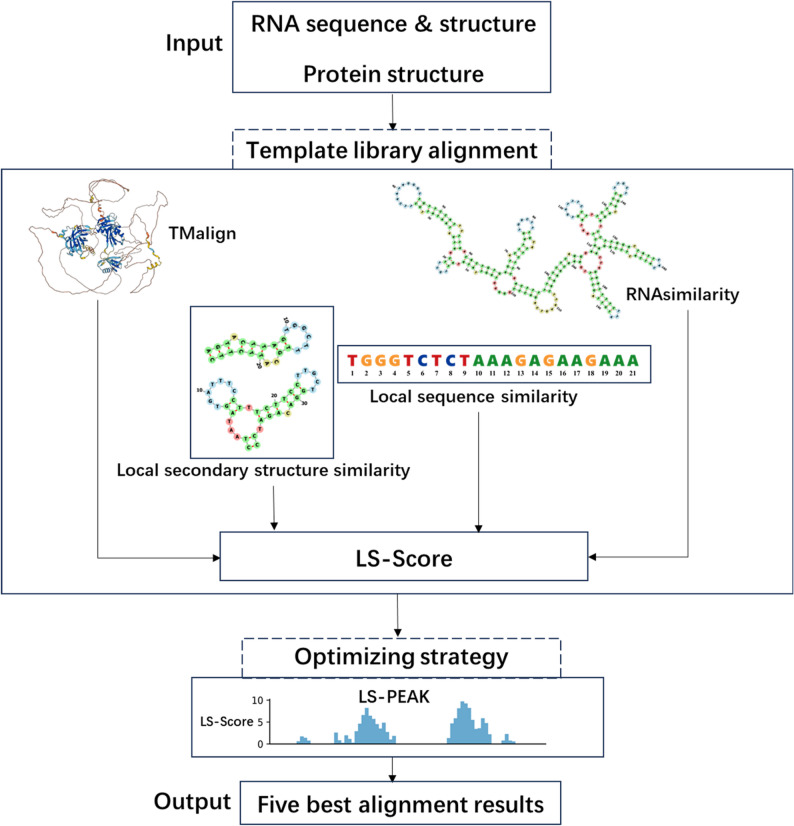
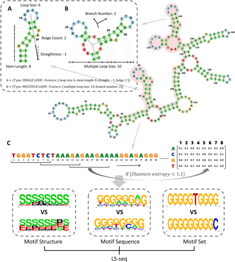
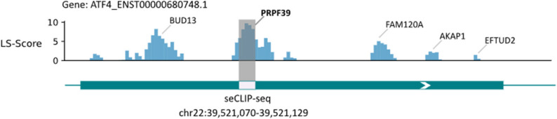
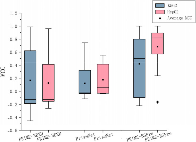
In this paper, we present PRIME-BSPre, a template-based genome-wide method for predicting protein-RNA binding sites that incorporates the RNA sequence and secondary structure as well as the tertiary structure of corresponding RBPs. We are pioneers in introducing low Shannon entropy algorithm in PRIME-BSPre to describe the binding preferences of RBPs on RNA motifs. The LS-PEAK derived from LS-Scores in PRIME-BSPre is utilized to optimize the alignments screening. PRIME-BSPre has been successfully benchmarked on the human genome, demonstrating its excellent prediction performance on independent RBP datasets and its robustness across different cell lines.
Overview
The claims and statistics of this paper haven’t been checked yet.
Run the check — free, no account needed. It reads the open-access text and checks the sources it cites.
Integrity signals
Registry
Cites
Statistics
Figures
Verification
Claims the paper makes
The claims and statistics of this paper haven’t been checked yet.
Run the check — free, no account needed. It reads the open-access text and checks the sources it cites.
Self-check
Percentages, sums and reported differences re-computed from the paper’s own numbers.
The self-check needs the paper’s full text.
Citations
Cites
Highly accurate protein structure prediction with AlphaFold
Jumper, J., Evans, R., Pritzel, A., Green, T., Figurnov, M., Ronneberger, O., Tunyasuvunakool, K., Bates, R., Žídek, A., Potapenko, A., et al. (2021) · Nature · doi:10.1038/s41586-021-03819-2 · Cited by 43,764
Context-dependent control of alternative splicing by RNA-binding proteins
Fu, X.-D., & Ares, M. (2014) · Nature Reviews Genetics · doi:10.1038/nrg3778 · Cited by 1,044
RNA sequence‐ and shape‐dependent recognition by proteins in the ribonucleoprotein particle
Stefl, R., Skrisovska, L., & Allain, F. H. ‐. (2005) · The EMBO Reports · doi:10.1038/sj.embor.7400325 · Cited by 175
Research works printed but not linked: 43
- England WE, et al. An atlas of posttranslational modifications on RNA binding proteins. Nucleic Acids Res. 2022;50:4329–39. · DOI
- Spitale RC, et al. Structural imprints in vivo Decode RNA regulatory mechanisms. Nature. 2015;519:486–90. · DOI
- Van Nostrand EL, et al. A large-scale binding and functional map of human RNA-binding proteins. Nature. 2020;583:711–9. · DOI · Paper page
- Van Nostrand EL, et al. Robust transcriptome-wide discovery of RNA-binding protein binding sites with enhanced CLIP (eCLIP). Nat Methods. 2016;13:508–14. · DOI · Paper page
- Gerstberger S, Hafner M, Tuschl T. A census of human RNA-binding proteins. Nat Rev Genet. 2014;15:829–45. · DOI · Paper page
- Kuret, et al. Positional motif analysis reveals the extent of specificity of protein-RNA interactions observed by CLIP. Genome Biol. 2022;23:191. · DOI
- Xu Y, et al. PrismNet: predicting protein-RNA interaction using in vivo RNA structural information. Nucleic Acids Res. 2023;51:W468–77. · DOI
- Laverty KU, et al. PRIESSTESS: interpretable, high-performing models of the sequence and structure preferences of RNA-binding proteins. Nucleic Acids Res. 2022;50:e111. · DOI
- Konig J, et al. iCLIP reveals the function of HnRNP particles in splicing at individual nucleotide resolution. Nat Struct Mol Biol. 2010;17:909–15. · DOI · Paper page
- Hafner M, et al. Transcriptome-wide identification of RNA-binding protein and MicroRNA target sites by PAR-CLIP. Cell. 2010;141:129–41. · DOI · Paper page
- Cook KB, Hughes TR, Morris QD. High-throughput characterization of protein-RNA interactions. Brief Funct Genomics. 2015;14:74–89. · DOI
- Cook KB, et al. RNAcompete-S: combined RNA sequence/structure preferences for RNA binding proteins derived from a single-step in vitro selection. Methods. 2017;126:18–28. · DOI
- Lambert N, et al. RNA Bind-n-Seq: quantitative assessment of the sequence and structural binding specificity of RNA binding proteins. Mol Cell. 2014;54:887–900. · DOI
- Zheng J, Hong X, Xie J, Tong X, Liu S. P3DOCK: a protein–RNA Docking webserver based on template-based and template-free Docking. Bioinformatics. 2020;36(1):96–103. · DOI
- Zheng J, Kundrotas PJ, Vakser IA, Liu S. Template-Based modeling of Protein-RNA interactions. PLoS Comput Biol. 2016;12(9):e1005120. · DOI
- Orenstein Y, Wang Y, Berger B. RCK: accurate and efficient inference of sequence- and structure-based protein-RNA binding models from RNAcompete data. Bioinformatics. 2016;32:i351–9. · DOI
- Maticzka D, Lange SJ, Costa F, Backofen R. GraphProt: modeling binding preferences of RNA-binding proteins. Genome Biol. 2014;15:R17. · DOI
- Kazan H, Ray D, Chan ET, Hughes TR, Morris Q. RNAcontext: a new method for learning the sequence and structure binding preferences of RNA-binding proteins. PLoS Comput Biol. 2010;6:e1000832. · DOI
- Ding Y, Lawrence CE. A statistical sampling algorithm for RNA secondary structure prediction. Nucleic Acids Res. 2003;31:7280–301. · DOI
- Spitale R, Flynn R, Zhang Q, et al. Structural imprints in vivo Decode RNA regulatory mechanisms. Nature. 2015;519:486–90. · DOI
- Alipanahi B, Delong A, Weirauch MT, Frey BJ. Predicting the sequence specificities of DNA- and RNA-binding proteins by deep learning. Nat Biotechnol. 2015;33:831–8. · DOI · Paper page
- Hiller M, Pudimat R, Busch A, Backofen R. Using RNA secondary structures to guide sequence motif finding towards single-stranded regions. Nucleic Acids Res. 2006;34:e117. · DOI
- Dominguez D, et al. Sequence, Structure, and context preferences of human RNA binding proteins. Mol Cell. 2018;70:854–e867859. · DOI · Paper page
- Ray D, et al. A compendium of RNA-binding motifs for decoding gene regulation. Nature. 2013;499:172–7. · DOI · Paper page
- Xie J, Zheng J, Hong X, Tong X, Liu S. PRIME-3D2D is a 3D2D model to predict binding sites of protein-RNA interaction. Commun Biol. 2020;3:384. · DOI
- Luo Y, et al. New developments on the encyclopedia of DNA elements (ENCODE) data portal. Nucleic Acids Res. 2020;48:D882–9. · DOI
- Cunningham F, et al. Ensembl 2022. Nucleic Acids Res. 2022;50:D988–95. · DOI · Paper page
- Danecek P, et al. Twelve years of samtools and BCFtools. Gigascience. 2021;10:giab008. · DOI
- Lander ES, et al. Initial sequencing and analysis of the human genome. Nature. 2001;409:860–921. · DOI · Paper page
- Reuter JS, Mathews DH. RNAstructure: software for RNA secondary structure prediction and analysis. BMC Bioinformatics. 2010;11:129. · DOI
- Varadi M, et al. AlphaFold protein structure database: massively expanding the structural coverage of protein-sequence space with high-accuracy models. Nucleic Acids Res. 2022;50:D439–44. · DOI · Paper page
- Baltz AG, et al. The mRNA-bound proteome and its global occupancy profile on protein-coding transcripts. Mol Cell. 2012;46:674–90. · DOI
- Lodde V, et al. Systematic identification of NF90 target RNAs by iCLIP analysis. Sci Rep. 2022;12:364. · DOI
- Blue SM, et al. Transcriptome-wide identification of RNA-binding protein binding sites using seCLIP-seq. Nat Protoc. 2022;17:1223–65. · DOI
- Li Q, et al. Measuring reproducibility of high-throughput experiments. Annals Appl Stat. 2011;5:1752–79. · DOI
- Xinzhou Ge. et al. Clipper: p-value-free FDR control on high-throughput data from two conditions. Genome Biol. 2021;22:288. · DOI
- Zhang Y, Skolnick J. TM-align: a protein structure alignment algorithm based on the TM-score. Nucleic Acids Res. 2005;33:2302–9. · DOI · Paper page
- Will S, Joshi T, Hofacker IL, Stadler PF, Backofen R. LocARNA-P: accurate boundary prediction and improved detection of structural RNAs. RNA. 2012;18:900–14. · DOI
- Gemmell NJ, Repetitive DNA. Genomic dark matter matters. Nat Rev Genet. 2021;22:342. · DOI
- de Koning AP, Gu W, Castoe TA, Batzer MA, Pollock DD. Repetitive elements May comprise over two-thirds of the human genome. PLoS Genet. 2011;7:e1002384. · DOI
- Zhu H, et al. Dynamic characterization and interpretation for protein-RNA interactions across diverse cellular conditions using HDRNet. Nat Commun. 2023;14:6824. · DOI
- Shen X, et al. A deep learning model for characterizing protein-RNA interactions from sequences at single-base resolution. Patterns (N Y). 2025;6:101150. · DOI
- Sun L, et al. Predicting dynamic cellular protein-RNA interactions by deep learning using in vivo RNA structures. Cell Res. 2021;31:495–516. · DOI
Metadata
- Authors
- Wei, X., Duan, Y., Li, D., Liu, X., Xie, J., & Liu, S.
- Affiliations
- Huazhong University of Science and Technology (CN)
- Corresponding author
- Liu, Shiyong
- Venue
- BMC Genomics
- Published
- 2026-02-21
- Volume
- 27
- Issue
- 1
- ISSN
- 1471-2164 (ISSN-L)
- PMC
- PMC13032426
- OpenAlex
- W7130849569
- Cited by
- 0
- References
- 3
- FWCI
- 0.00
- Type
- journal article
- Language
- en
- Open access
- gold
- doi.org
- Licence
- Locations
- BMC Genomics (publishedVersion, cc-by)
- bioRxiv (Cold Spring Harbor Laboratory) (acceptedVersion, cc-by-nc-nd)
- PubMed Central (publishedVersion, cc-by)
- PubMed (publishedVersion)
- DOAJ (DOAJ: Directory of Open Access Journals) (submittedVersion, cc-by-sa)
- Funders
- National Natural Science Foundation of China (32271267)
- Topics
- Life Sciences › Biochemistry, Genetics and Molecular Biology › Molecular Biology › RNA Research and Splicing
- Life Sciences › Biochemistry, Genetics and Molecular Biology › Molecular Biology › Genomics and Phylogenetic Studies
- Life Sciences › Biochemistry, Genetics and Molecular Biology › Molecular Biology › RNA and protein synthesis mechanisms
- Keywords
- RNA-binding proteins, template-based modeling, RNA secondary structure, RNA tertiary structure, RNA motifs, genomic prediction, human genome
- MeSH terms
- Prediction Algorithms, Algorithms, Binding Sites, Humans, Nucleic Acid Conformation, Protein Binding, RNA, Genome, Human, RNA-Binding Proteins, Computational Biology, Genomics
About these checks
Registry, retraction, Cites and statistics results are computed, not judged, with one exception: a printed reference that no index lists is read once by a model before it counts as a citation error. Claims are extracted by a model and kept only when their quote appears verbatim in the text; each cited source is then checked, with a model’s help, for whether it says what the claim relies on. Those verdicts can be wrong — read the claim’s quote and the cited source before relying on one.
Statistics and arithmetic are recomputed, not judged.
Confidence bands are shown only for fields calibrated on our annotated test set. None is calibrated yet, so no band is shown.
What counts as a claim ›How we measure confidence ›How we check this ›
Are you an author, or do you see a problem with this page? Request a review
