

The DOI matches its Crossref record; the references it cites are not checked yet.
Global landscape of antibiotic resistance genes in the human gut microbiome metagenome-assembled genomes
Open accessPublished version of a preprint
Abstract
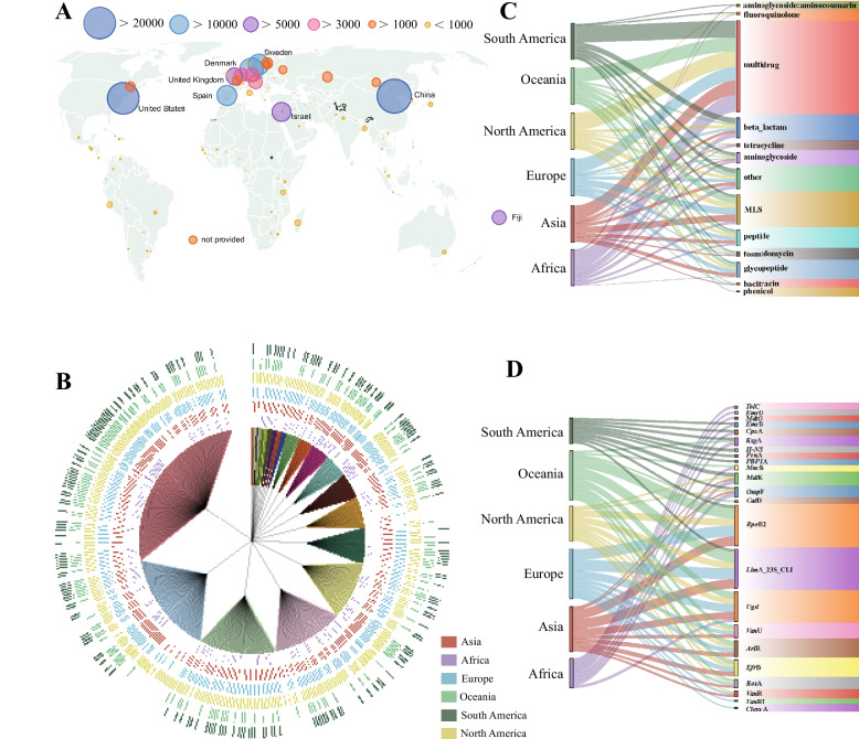
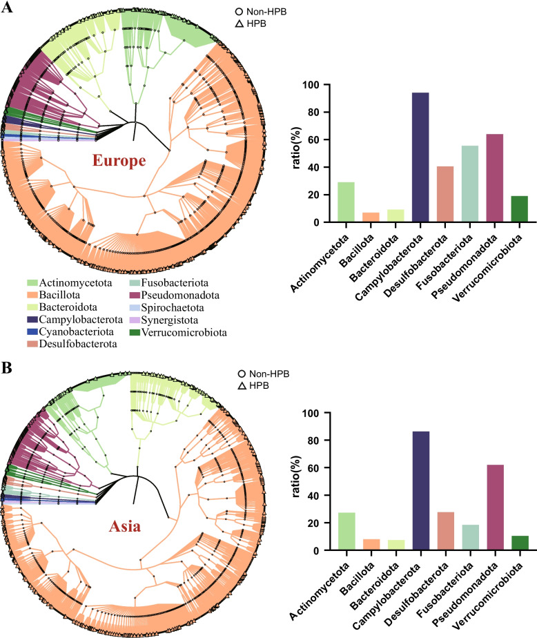
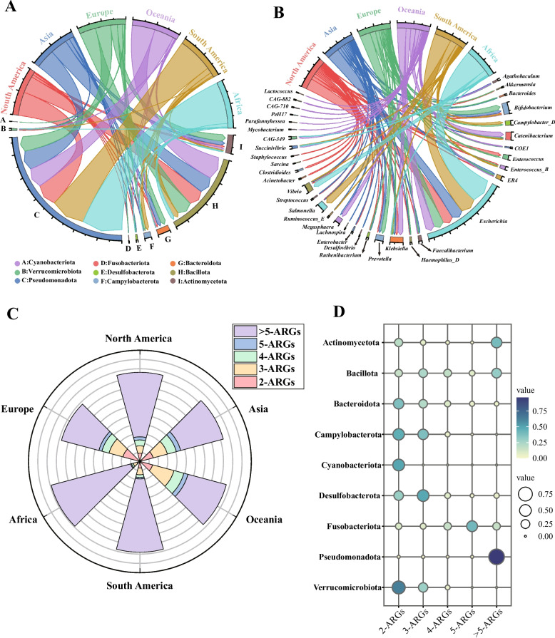
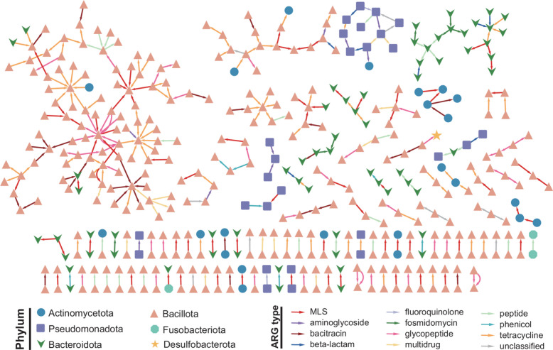
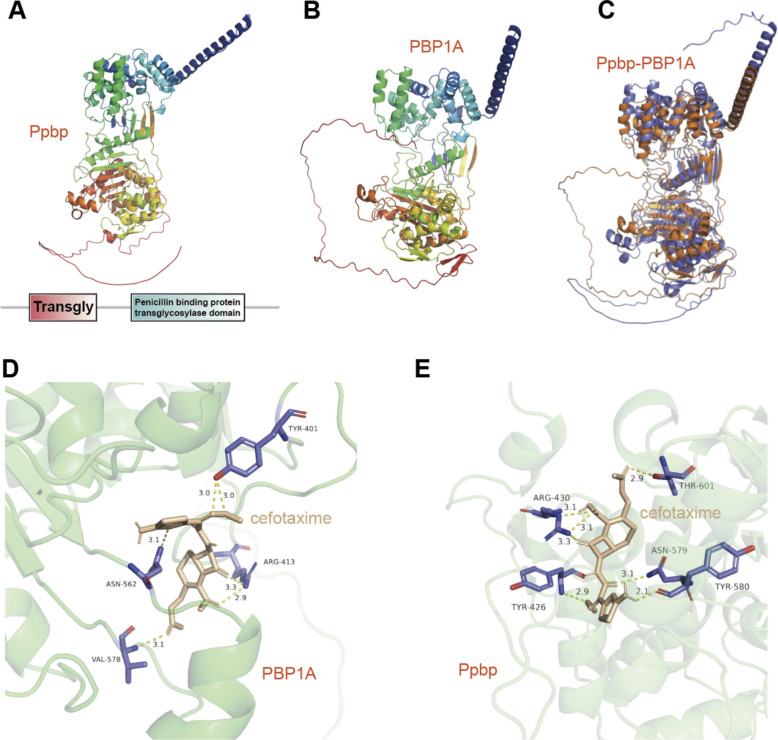
Antibiotic resistance poses a significant threat to human health, and the human gut microbiota serves as a major reservoir of antibiotic resistance genes (ARGs). In this study, we analyzed 149,515 metagenome-assembled genomes (MAGs) from human gut microbiomes and revealed marked geographic variations in the global distribution of gut-associated ARGs. Asia exhibits the highest diversity of ARGs. At the phylum level, Pseudomonadota was identified as the predominant ARG host among pathogenic bacteria, with its pathogenic strains frequently exhibiting high levels of multidrug resistant strains harboring ≥ 5 ARGs accounting for up to 88.5% and 79.1% in Africa and South America, respectively. Campylobacterota was also recognized as a potential high-risk ARG host phylum. Horizontal gene transfer (HGT) analysis revealed that ARG transmission predominantly occurred within the same phylum, with Bacillota being the most active donor, which was likely influenced by antibiotic selection pressure. Actinomycetota and Bacteroidota were identified as major recipients of interphylum HGT, indicating their greater capacity to acquire exogenous ARGs. Through the integration of deep learning and structural calculation, we also identified a potentially novel class of β-lactam resistance genes. This study provides a comprehensive global landscape of gut-associated resistomes, underscores the critical roles of public health infrastructure, antibiotic misuse, and HGT in shaping antimicrobial resistance (AMR), and offers methodological insights for the discovery of novel ARGs. Our findings highlight urgent challenges and provide a scientific basis for developing global AMR mitigation strategies.
Overview
The claims and statistics of this paper haven’t been checked yet.
Run the check — free, no account needed. It reads the open-access text and checks the sources it cites.
Integrity signals
Registry
Cites
Statistics
Figures
Verification
Claims the paper makes
The claims and statistics of this paper haven’t been checked yet.
Run the check — free, no account needed. It reads the open-access text and checks the sources it cites.
Self-check
Percentages, sums and reported differences re-computed from the paper’s own numbers.
The self-check needs the paper’s full text.
Citations
The citation lists haven’t been fetched yet
References 56 · Cited by 6
Research works printed but not linked: 53
- Murray CJL, Ikuta KS, Sharara F, Swetschinski L, Robles Aguilar G, Gray A, et al. Global burden of bacterial antimicrobial resistance in 2019: a systematic analysis. Lancet. 2022;399:629–55. https://doi.org/10.1016/S0140-6736(21)02724-0. · DOI · Paper page
- De Kraker MEA, Stewardson AJ, Harbarth S. Will 10 million people die a year due to antimicrobial resistance by 2050? PLoS Med. 2016;13:e1002184. https://doi.org/10.1371/journal.pmed.1002184. · DOI
- Stokes JM, Yang K, Swanson K, Jin W, Cubillos-Ruiz A, Donghia NM, et al. A deep learning approach to antibiotic discovery. Cell. 2020;180:688-702.e13. https://doi.org/10.1016/j.cell.2020.01.021. · DOI · Paper page
- Leviatan S, Shoer S, Rothschild D, Gorodetski M, Segal E. An expanded reference map of the human gut microbiome reveals hundreds of previously unknown species. Nat Commun. 2022;13:3863. https://doi.org/10.1038/s41467-022-31502-1. · DOI
- Sender R, Fuchs S, Milo R. Revised estimates for the number of human and bacteria cells in the body. PLoS Biol. 2016;14:e1002533. https://doi.org/10.1371/journal.pbio.1002533. · DOI · Paper page
- Sommer F, Bäckhed F. The gut microbiota — masters of host development and physiology. Nat Rev Microbiol. 2013;11:227–38. https://doi.org/10.1038/nrmicro2974. · DOI · Paper page
- Sampson TR, Debelius JW, Thron T, Janssen S, Shastri GG, Ilhan ZE, et al. Gut microbiota regulate motor deficits and neuroinflammation in a model of Parkinson’s disease. Cell. 2016;167:1469-1480.e12. https://doi.org/10.1016/j.cell.2016.11.018. · DOI · Paper page
- De Vos WM, Tilg H, Van Hul M, Cani PD. Gut microbiome and health: mechanistic insights. Gut. 2022;71:1020–32. https://doi.org/10.1136/gutjnl-2021-326789. · DOI · Paper page
- Sommer MOA, Dantas G, Church GM. Functional characterization of the antibiotic resistance reservoir in the human microflora. Science. 2009;325:1128–31. https://doi.org/10.1126/science.1176950. · DOI · Paper page
- Kim D-W, Cha C-J. Antibiotic resistome from the one-health perspective: understanding and controlling antimicrobial resistance transmission. Exp Mol Med. 2021;53:301–9. https://doi.org/10.1038/s12276-021-00569-z. · DOI
- Zhang Q, Xu N, Lei C, Chen B, Wang T, Ma Y, et al. Metagenomic insight into the global dissemination of the antibiotic resistome. Adv Sci. 2023;10:2303925. https://doi.org/10.1002/advs.202303925. · DOI
- Zhu Y-G, Gillings M, Simonet P, Stekel D, Banwart S, Penuelas J. Microbial mass movements. Science. 2017;357:1099–100. https://doi.org/10.1126/science.aao3007. · DOI
- Boolchandani M, Blake KS, Tilley DH, Cabada MM, Schwartz DJ, Patel S, et al. Impact of international travel and diarrhea on gut microbiome and resistome dynamics. Nat Commun. 2022;13:7485. https://doi.org/10.1038/s41467-022-34862-w. · DOI
- Hu Y, Yang X, Qin J, Lu N, Cheng G, Wu N, et al. Metagenome-wide analysis of antibiotic resistance genes in a large cohort of human gut microbiota. Nat Commun. 2013;4:2151. https://doi.org/10.1038/ncomms3151. · DOI
- Almeida A, Nayfach S, Boland M, Strozzi F, Beracochea M, Shi ZJ, et al. A unified catalog of 204,938 reference genomes from the human gut microbiome. Nat Biotechnol. 2021;39:105–14. https://doi.org/10.1038/s41587-020-0603-3. · DOI · Paper page
- Arango-Argoty G, Garner E, Pruden A, Heath LS, Vikesland P, Zhang L. DeepARG: a deep learning approach for predicting antibiotic resistance genes from metagenomic data. Microbiome. 2018;6:23. https://doi.org/10.1186/s40168-018-0401-z. · DOI
- Chen Peng, Chao Jiang. pctax: Professional comprehensive microbiome data analysis pipeline. R package. 2023. https://github.com/Asa12138/pctax. · DOI
- Xie J, Chen Y, Cai G, Cai R, Hu Z, Wang H. Tree Visualization By One Table (tvBOT): a web application for visualizing, modifying and annotating phylogenetic trees. Nucleic Acids Res. 2023;51:W587–92. https://doi.org/10.1093/nar/gkad359. · DOI
- Chaumeil P-A, Mussig AJ, Hugenholtz P, Parks DH. GTDB-Tk v2: memory friendly classification with the genome taxonomy database. Bioinformatics. 2022;38:5315–6. https://doi.org/10.1093/bioinformatics/btac672. · DOI
- Zhou S, Liu B, Zheng D, Chen L, Yang J. VFDB 2025: an integrated resource for exploring anti-virulence compounds. Nucleic Acids Res. 2025;53:D871–7. https://doi.org/10.1093/nar/gkae968. · DOI
- Camacho C, Coulouris G, Avagyan V, Ma N, Papadopoulos J, Bealer K, et al. BLAST+: architecture and applications. BMC Bioinformatics. 2009;10:421. https://doi.org/10.1186/1471-2105-10-421. · DOI · Paper page
- Asnicar F, Weingart G, Tickle TL, Huttenhower C, Segata N. Compact graphical representation of phylogenetic data and metadata with GraPhlAn. PeerJ. 2015;3:e1029. https://doi.org/10.7717/peerj.1029. · DOI
- Olm MR, Brown CT, Brooks B, Banfield JF. DRep: a tool for fast and accurate genomic comparisons that enables improved genome recovery from metagenomes through de-replication. ISME J. 2017;11:2864–8. https://doi.org/10.1038/ismej.2017.126. · DOI · Paper page
- Song W, Wemheuer B, Zhang S, Steensen K, Thomas T. MetaCHIP: community-level horizontal gene transfer identification through the combination of best-match and phylogenetic approaches. Microbiome. 2019;7:36. https://doi.org/10.1186/s40168-019-0649-y. · DOI
- Shannon P, Markiel A, Ozier O, Baliga NS, Wang JT, Ramage D, et al. Cytoscape: a software environment for integrated models of biomolecular interaction networks. Genome Res. 2003;13:2498–504. https://doi.org/10.1101/gr.1239303. · DOI · Paper page
- Hyatt D, Chen G-L, LoCascio PF, Land ML, Larimer FW, Hauser LJ. Prodigal: prokaryotic gene recognition and translation initiation site identification. BMC Bioinformatics. 2010;11:119. https://doi.org/10.1186/1471-2105-11-119. · DOI
- Oberg N, Zallot R, Gerlt JA. EFI-EST, EFI-GNT, and EFI-CGFP: enzyme function initiative (EFI) web resource for genomic enzymology tools. J Mol Biol. 2023;435:168018. https://doi.org/10.1016/j.jmb.2023.168018. · DOI
- Zallot R, Oberg N, Gerlt JA. The EFI web resource for genomic enzymology tools: leveraging protein, genome, and metagenome databases to discover novel enzymes and metabolic pathways. Biochemistry. 2019;58:4169–82. https://doi.org/10.1021/acs.biochem.9b00735. · DOI
- Liu Y, Yang X, Gan J, Chen S, Xiao Z-X, Cao Y. CB-Dock2: improved protein–ligand blind docking by integrating cavity detection, docking and homologous template fitting. Nucleic Acids Res. 2022;50:W159–64. https://doi.org/10.1093/nar/gkac394. · DOI
- Yang X, Liu Y, Gan J, Xiao Z-X, Cao Y. FitDock: protein–ligand docking by template fitting. Brief Bioinform. 2022;23:bbac087. https://doi.org/10.1093/bib/bbac087. · DOI
- Nishino K, Yamaguchi A. Role of histone-like protein H-NS in multidrug resistance of Escherichia coli. J Bacteriol. 2004;186:1423–9. https://doi.org/10.1128/jb.186.5.1423-1429.2004. · DOI
- Contreras-Martel C, Job V, Di Guilmi AM, Vernet T, Dideberg O, Dessen A. Crystal structure of Penicillin-binding protein 1a (PBP1a) reveals a mutational hotspot implicated in β-lactam resistance in Streptococcus pneumoniae. J Mol Biol. 2006;355:684–96. https://doi.org/10.1016/j.jmb.2005.10.030. · DOI
- Wang W, Song W, Majzoub ME, Feng X, Xu B, Tao J, et al. Decoupling of strain- and intrastrain-level interactions of microbiomes in a sponge holobiont. Nat Commun. 2024;15:8205. https://doi.org/10.1038/s41467-024-52464-6. · DOI
- Kelbert L, Barmettler K, Horlbog JA, Stevens MJA, Cernela N, Nüesch-Inderbinen M, et al. Campylobacter in raw chicken meat at retail level: quantitative and qualitative assessment, genomic profiling, and comparison with isolates from human infections. J Food Prot. 2025;88:100540. https://doi.org/10.1016/j.jfp.2025.100540. · DOI
- Baker-Austin C, Oliver JD, Alam M, Ali A, Waldor MK, Qadri F, et al. Vibrio spp. infections. Nat Rev Dis Primers. 2018;4:1–19. https://doi.org/10.1038/s41572-018-0005-8. · DOI
- Sánchez B, Ruiz L, Gueimonde M, Ruas-Madiedo P, Margolles A. Adaptation of bifidobacteria to the gastrointestinal tract and functional consequences. Pharmacol Res. 2013;69:127–36. https://doi.org/10.1016/j.phrs.2012.11.004. · DOI
- Chen Z, Moreno-Switt AI, Reyes-Jara A, Delgado Suarez E, Adell AD, Oliveira CJB, et al. A multicenter genomic epidemiological investigation in Brazil, Chile, and Mexico reveals the diversity and persistence of Salmonella populations in surface waters. MBio. 2024;15:e00777-e824. https://doi.org/10.1128/mbio.00777-24. · DOI
- Qiao W, Wang L, Luo Y, Miao J. Outer membrane vesicles mediated horizontal transfer of an aerobic denitrification gene between Escherichia coli. Biodegradation. 2021;32:435–48. https://doi.org/10.1007/s10532-021-09945-y. · DOI
- Caro-Quintero A, Konstantinidis KT. Inter-phylum HGT has shaped the metabolism of many mesophilic and anaerobic bacteria. ISME J. 2015;9:958–67. https://doi.org/10.1038/ismej.2014.193. · DOI
- Fuchsman CA, Collins RE, Rocap G, Brazelton WJ. Effect of the environment on horizontal gene transfer between bacteria and archaea. PeerJ. 2017;5:e3865. https://doi.org/10.7717/peerj.3865. · DOI
- Rhodes ME, Spear JR, Oren A, House CH. Differences in lateral gene transfer in hypersaline versus thermal environments. BMC Evol Biol. 2011;11:199. https://doi.org/10.1186/1471-2148-11-199. · DOI
- Parras-Moltó M, Lund D, Ebmeyer S, Larsson DGJ, Johnning A, Kristiansson E. The transfer of antibiotic resistance genes between evolutionarily distant bacteria. mSphere. 2025. https://doi.org/10.1128/msphere.00114-25. · DOI
- Klein EY, Van Boeckel TP, Martinez EM, Pant S, Gandra S, Levin SA, et al. Global increase and geographic convergence in antibiotic consumption between 2000 and 2015. Proc Natl Acad Sci U S A. 2018;115:E3463–70. https://doi.org/10.1073/pnas.1717295115. · DOI · Paper page
- Stanhope MJ, Lefébure T, Walsh SL, Becker JA, Lang P, Pavinski Bitar PD, et al. Positive selection in penicillin-binding proteins 1a, 2b, and 2x from Streptococcus pneumoniae and its correlation with amoxicillin resistance development. Infect Genet Evol. 2008;8:331–9. https://doi.org/10.1016/j.meegid.2008.02.001. · DOI
- Blum M, Andreeva A, Florentino LC, Chuguransky SR, Grego T, Hobbs E, et al. InterPro: the protein sequence classification resource in 2025. Nucleic Acids Res. 2025;53:D444–56. https://doi.org/10.1093/nar/gkae1082. · DOI
- Alcock BP, Huynh W, Chalil R, Smith KW, Raphenya AR, Wlodarski MA, et al. CARD 2023: expanded curation, support for machine learning, and resistome prediction at the comprehensive antibiotic resistance database. Nucleic Acids Res. 2023;51:D690–9. https://doi.org/10.1093/nar/gkac920. · DOI · Paper page
- Diebold PJ, Rhee MW, Shi Q, Trung NV, Umrani F, Ahmed S, et al. Clinically relevant antibiotic resistance genes are linked to a limited set of taxa within gut microbiome worldwide. Nat Commun. 2023;14:7366. https://doi.org/10.1038/s41467-023-42998-6. · DOI
- Van Boeckel TP, Pires J, Silvester R, Zhao C, Song J, Criscuolo NG, et al. Global trends in antimicrobial resistance in animals in low- and middle-income countries. Science. 2019;365:eaaw1944. https://doi.org/10.1126/science.aaw1944. · DOI · Paper page
- Zhu C, Wu L, Ning D, Tian R, Gao S, Zhang B, et al. Global diversity and distribution of antibiotic resistance genes in human wastewater treatment systems. Nat Commun. 2025;16:4006. https://doi.org/10.1038/s41467-025-59019-3. · DOI
- Alhassan MY, Ahmad AA. Antimicrobial resistance in a changing climate: a one health approach for adaptation and mitigation. Bull Natl Res Cent. 2025;49:26. https://doi.org/10.1186/s42269-025-01318-2. · DOI
- Feng Y, Lu X, Zhao J, Li H, Xu J, Li Z, et al. Regional antimicrobial resistance gene flow among the one health sectors in China. Microbiome. 2025;13:3. https://doi.org/10.1186/s40168-024-01983-x. · DOI
- Forslund K, Sunagawa S, Kultima JR, Mende DR, Arumugam M, Typas A, et al. Country-specific antibiotic use practices impact the human gut resistome. Genome research. 2013;23:1163–9. · DOI
- Lund D, Parras-Moltó M, Inda-Díaz JS, Ebmeyer S, Larsson DGJ, Johnning A, et al. Genetic compatibility and ecological connectivity drive the dissemination of antibiotic resistance genes. Nat Commun. 2025;16:2595. https://doi.org/10.1038/s41467-025-57825-3. · DOI
Metadata
- Authors
- Wang, C., Wang, C., Chen, S., Shi, K., Yu, J., Ding, Y., Yue, Y., Hua, Y., Wang, H., & Chen, J.
- Affiliations
- Bozhou University (CN)
- Zhejiang University of Technology (CN)
- Corresponding author
- Chen, Jianwei
- Venue
- BMC Microbiology
- Published
- 2025-12-09
- Volume
- 26
- Issue
- 1
- Pages
- 33-33
- ISSN
- 1471-2180 (ISSN-L)
- PMC
- PMC12817740
- OpenAlex
- W4417184105
- Cited by
- 6
- References
- 56
- Citations by year
- 2026: 6
- FWCI
- 1.69
- Type
- journal article
- Language
- en
- Open access
- gold
- doi.org
- Licence
- Locations
- BMC Microbiology (publishedVersion, cc-by-nc-nd)
- Research Square (acceptedVersion, cc-by)
- PubMed (publishedVersion)
- PubMed Central (publishedVersion, cc-by-nc-nd)
- Funders
- the Scientific Research Program of the Bozhou University (No.202524)
- International Cooperation and Exchanges NSFC-ASRT (No. W2412100)
- National Natural Science Foundation of China (No. 42276137)
- National Key Research and Development Program of China (No. 2022YFC2804205, No. 2022YFC2804104)
- Topics
- Health Sciences › Medicine › Microbiology (medical) › Antibiotic Resistance in Bacteria
- Physical Sciences › Environmental Science › Pollution › Pharmaceutical and Antibiotic Environmental Impacts
- Health Sciences › Medicine › Infectious Diseases › Antibiotic Use and Resistance
- Keywords
- antibiotic resistance genes, human gut microbiome, metagenome-assembled genomes, horizontal gene transfer, multidrug resistance, Proteobacteria, Campylobacterota, Firmicutes, actinobacteria, Bacteroidetes, beta-lactam resistance genes, antibiotic misuse, antimicrobial resistance mitigation
- MeSH terms
- Gastrointestinal Microbiome, Anti-Bacterial Agents, Bacteria, Drug Resistance, Microbial, Genes, Bacterial, Humans, Phylogeny, Genome, Bacterial, Gene Transfer, Horizontal, Drug Resistance, Bacterial, Metagenome
About these checks
Registry, retraction, Cites and statistics results are computed, not judged, with one exception: a printed reference that no index lists is read once by a model before it counts as a citation error. Claims are extracted by a model and kept only when their quote appears verbatim in the text; each cited source is then checked, with a model’s help, for whether it says what the claim relies on. Those verdicts can be wrong — read the claim’s quote and the cited source before relying on one.
Statistics and arithmetic are recomputed, not judged.
Confidence bands are shown only for fields calibrated on our annotated test set. None is calibrated yet, so no band is shown.
What counts as a claim ›How we measure confidence ›How we check this ›
Are you an author, or do you see a problem with this page? Request a review
How to cite
Cite CoChat for the checks on this page, and the paper for its own findings.
- This page
- CoChat. (2026). Citation check: Global landscape of antibiotic resistance genes in the human gut microbiome metagenome-assembled genomes. https://app.cochat.ai/papers/10.1186/s12866-025-04586-0
- The paper
- Wang, C., Wang, C., Chen, S., Shi, K., Yu, J., Ding, Y., Yue, Y., Hua, Y., Wang, H., & Chen, J. (2025). Global landscape of antibiotic resistance genes in the human gut microbiome metagenome-assembled genomes. BMC Microbiology. https://doi.org/10.1186/s12866-025-04586-0
