Cited by 59

The DOI matches its Crossref record; 2 of the 2 references it cites were found in a registry or an index.

Exosomal microRNA-107 reverses chemotherapeutic drug resistance of gastric cancer cells through HMGA2/mTOR/P-gp pathway

Jiang, Lu, Zhang, Yan, Guo, Linghui, Liu, Chaoyang, Wang, Pan, Ren, Weihong·2021·BMC Cancer

Abstract

Background: RNA cargo in exosomes, especially microRNAs (miRNAs), play an important role in the chemotherapy drug resistance of human cancers. However, the role and mechanism of exosomal miR-107 on multidrug resistance of gastric cancer cells was still not clear. In this study, we sought to explore whether exosomal miR-107 could reverse the resistance of gastric cancer cells to the chemotherapy drugs. Methods: We extracted exosomes from sensitive (SGC-7901, MGC-803) and resistant (SGC-7901/5-FU) gastric cancer cells by ultracentrifugation and the isolated exosomes were identified using transmission electron microscopy (TEM) and dynamic light scattering analysis (DLS). The expression of miR-107 and high mobility group A2 (HMGA2) were detected by real-time quantitative PCR (RT-qPCR). MTT assay was used to investigate the effect of exosomes on gastric cancer cells growth in vitro. The uptake of exosomes by recipient cells were observed using a fluorescence microscope. The predicted target relationship between miR-107 and HMGA2 was verified by gauss -luciferase reporter assay. The expression of HMGA2, p-mTOR/mTOR, P-gp and other exosomal indicated marker proteins was detected by western blot. Results: Our results indicated that the isolated exosomes were typically cup-like lipid bilayer membranes structure. SGC-7901/5-FU cells were cross-resistant to chemotherapy drug cisplatin (CDDP), and the sensitive cells-secreted exosomes drastically reversed the resistance of the resistant GC cells to the chemotherapeutic drugs, which was verified by exosomal inhibitor GW4896. Mechanistically, the reversal effect was mainly mediated by exosome-secreted miR-107 through downregulating the expression of target molecular HMGA2 and inhibiting HMGA2/mTOR/P-gp pathway, which were supported by results from luciferase reporter assay and rescue assay. Conclusions: These findings demonstrated that exosome-transmitted miR-107 significantly enhanced the sensitivity of resistant gastric cancer cells to chemotherapeutic agents by mediating the HMGA2/mTOR/P-gp axis and exosomal miR-107 may be a novel target in gastric cancers treatment.

Overview

The claims and statistics of this paper haven’t been checked yet.

Run the check — free, no account needed. It reads the open-access text and checks the sources it cites.

A few free checks a day · papers already verified open instantly · results are public

Integrity signals

Registry

Crossref · 10.1186/s12885-021-09020-y

Cites

2 of 2 references verified0 citation errors · 0 not checkable

Statistics

Figures

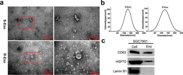
Fig. 1 Characteristics of exosomes derived from SGC-7901 and SGC-7901/5-FU GC cells. a The transmission electron micrograph showed round-shaped vesicles with bilayer membranes ranging from 30 nm to 180 nm in diameter released by SGC-7901 (S-Exo) and SGC-7901/5-FU (R-Exo) cells. Scale bar = 500 nm and 200 nm, respectively. b Dynamic light scattering analysis (DLS) indicated that the dominant size of S- and R-Exo was about 120 nm. c The positive markers of exosomes, CD63 and HSP70, were detected in S- and R-Exo by Western blot
Fig. 1 Characteristics of exosomes derived from SGC-7901 and SGC-7901/5-FU GC cells. a The transmission electron micrograph showed round-shaped vesicles with bilayer membranes ranging from 30 nm to 180 nm in diameter released by SGC-7901 (S-Exo) and SGC-7901/5-FU (R-Exo) cells. Scale bar = 500 nm and 200 nm, respectively. b Dynamic light scattering analysis (DLS) indicated that the dominant size of S- and R-Exo was about 120 nm. c The positive markers of exosomes, CD63 and HSP70, were detected in S- and R-Exo by Western blotCC BY · PubMed Central
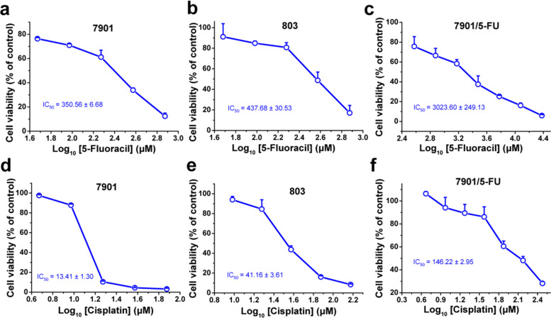
Fig. 2 The sensitivity of GC cells to the chemotherapy drugs 5-FU and CDDP. The cell viability of SGC-7901 (a and d), MGC-803 (b and e) sensitive cell strains and SGC-7901/5-FU (c and f) resistant cell strains was determined after treated with 5-FU for 48 h (a, b, c) and CDDP (d, e, f) for 24 h, respectively. Cell viability was determined by MTT assay. Cells treated with vehicle serve as a blank control. All experiments were conducted in quintuplicates and data were expressed as the mean ± SD (n = 5)
Fig. 2 The sensitivity of GC cells to the chemotherapy drugs 5-FU and CDDP. The cell viability of SGC-7901 (a and d), MGC-803 (b and e) sensitive cell strains and SGC-7901/5-FU (c and f) resistant cell strains was determined after treated with 5-FU for 48 h (a, b, c) and CDDP (d, e, f) for 24 h, respectively. Cell viability was determined by MTT assay. Cells treated with vehicle serve as a blank control. All experiments were conducted in quintuplicates and data were expressed as the mean ± SD (n = 5)CC BY · PubMed Central
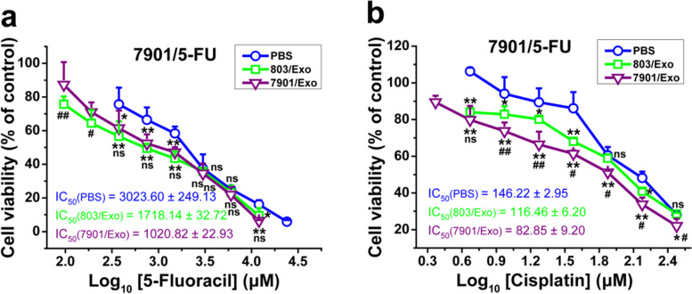
Fig. 3 The exosomes isolated from sensitive GC cells increased drug sensitivity of drug-resistant GC cells. The cell viability of SGC-7901/5-FU cells after treated with 5-FU (a) or CDDP (b) with or without the exosomes isolated from SGC-7901 or MGC-803 cells for 48 or 24 h, respectively. Cell viability was determined by MTT assay. Cells treated with vehicle serve as a blank control. Abbreviations: Exo, exosomes. All experiments were conducted in quintuplicates and data were expressed as the mean ± SD (n = 5). Statistical significances were determined using one-way ANOVA followed by Dunnett’s test. *P < 0.05,**P < 0.01, compared with the PBS control group;#P < 0.05,##P < 0.01, compared with the 803/Exo group; ns, no significance (P > 0.05), compared with the PBS control or 803/Exo groups
Fig. 3 The exosomes isolated from sensitive GC cells increased drug sensitivity of drug-resistant GC cells. The cell viability of SGC-7901/5-FU cells after treated with 5-FU (a) or CDDP (b) with or without the exosomes isolated from SGC-7901 or MGC-803 cells for 48 or 24 h, respectively. Cell viability was determined by MTT assay. Cells treated with vehicle serve as a blank control. Abbreviations: Exo, exosomes. All experiments were conducted in quintuplicates and data were expressed as the mean ± SD (n = 5). Statistical significances were determined using one-way ANOVA followed by Dunnett’s test. *P < 0.05,**P < 0.01, compared with the PBS control group;#P < 0.05,##P < 0.01, compared with the 803/Exo group; ns, no significance (P > 0.05), compared with the PBS control or 803/Exo groupsCC BY · PubMed Central
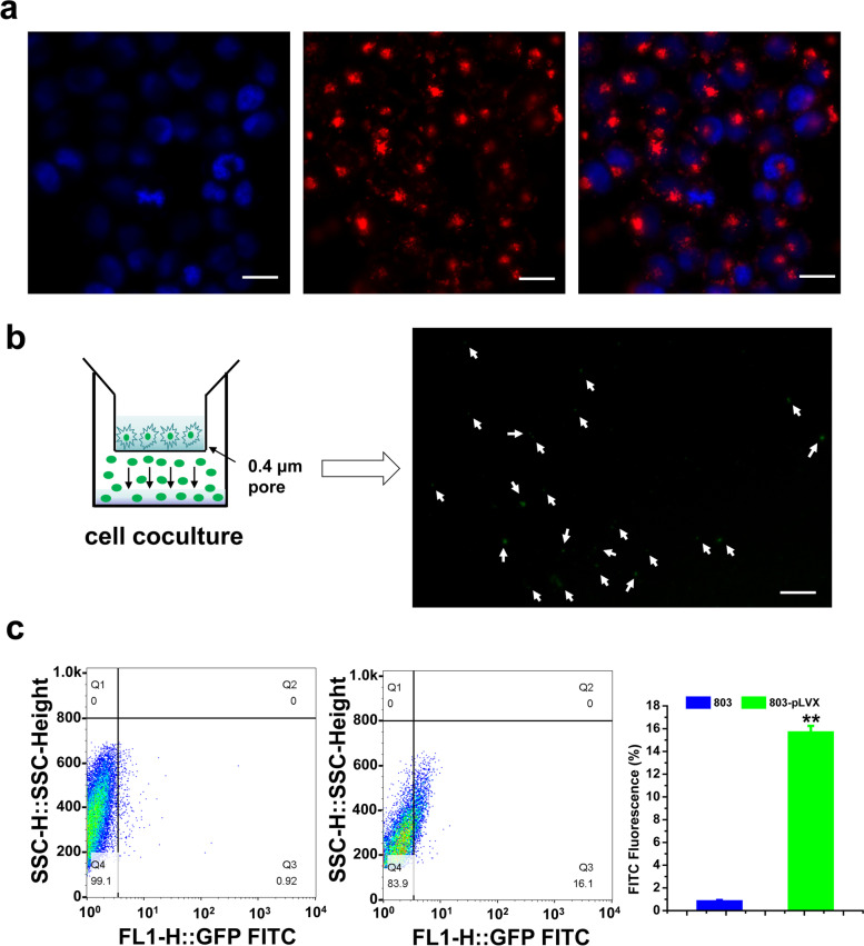
Fig. 4 Exosomes derived from sensitive GC cells were taken up by SGC-7901/5-FU cells. a Confocal microscopy showed exosome internalization by SGC-7901/5-FU recipient cells after co-incubation with PKH26-labeled SGC-7901 exosomes. Red: exosomes stained with PKH26, blue: DAPI. Scale Bars = 20 μm. b The MGC-803-pLVX stable cells were placed in the upper chamber and coincubated with SGC-7901/5-FU cells seeded in the lower chamber in a coculture system with a 0.4 μm pore membrane. Green fluorescence was observed in the SGC-7901/5-FU recipient cells under the fluorescence microscope. Scale Bars = 35 μm. c The percentage of green fluorescence signals in SGC-7901/5-FU resistant GC cells were determined by flow cytometry. Data were expressed as the mean ± SD (n = 3). Statistical significances were determined using one-way ANOVA followed by Dunnett’s test. **P < 0.01, compared with the group of MGC-803 cells
Fig. 4 Exosomes derived from sensitive GC cells were taken up by SGC-7901/5-FU cells. a Confocal microscopy showed exosome internalization by SGC-7901/5-FU recipient cells after co-incubation with PKH26-labeled SGC-7901 exosomes. Red: exosomes stained with PKH26, blue: DAPI. Scale Bars = 20 μm. b The MGC-803-pLVX stable cells were placed in the upper chamber and coincubated with SGC-7901/5-FU cells seeded in the lower chamber in a coculture system with a 0.4 μm pore membrane. Green fluorescence was observed in the SGC-7901/5-FU recipient cells under the fluorescence microscope. Scale Bars = 35 μm. c The percentage of green fluorescence signals in SGC-7901/5-FU resistant GC cells were determined by flow cytometry. Data were expressed as the mean ± SD (n = 3). Statistical significances were determined using one-way ANOVA followed by Dunnett’s test. **P < 0.01, compared with the group of MGC-803 cellsCC BY · PubMed Central
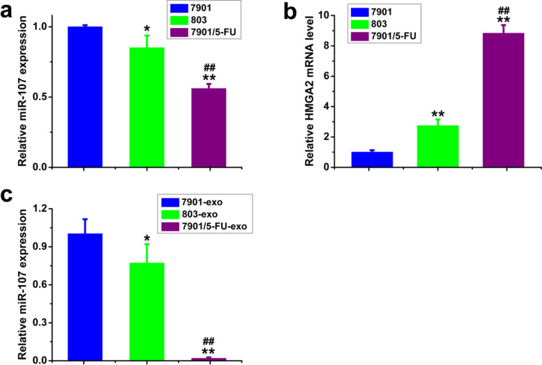
Fig. 5 The expression levels of miR-107 and HMGA2 mRNA in SGC-7901, MGC-803 and SGC-7901/5-FU cells and exosomes were detected by qPCR. the expression levels of miR-107 (a) and HMGA2 mRNA (b) in SGC-7901, MGC-803 sensitive and SGC-7901/5-FU resistant cells; the expression levels of miR-107 (c) in SGC-7901, MGC-803 sensitive and SGC-7901/5-FU resistant exosomes. mRNA and miRNA levels were determined by qPCR using GAPDH and U6 as the internal control, respectively. Data were expressed as the mean ± SD (n = 3). Statistical significances were determined using one-way ANOVA followed by Dunnett’s test. *P < 0.05, **P < 0.01, compared with SGC-7901 cells/exosomes; ##P < 0.01, compared with MGC-803 cells/exosomes
Fig. 5 The expression levels of miR-107 and HMGA2 mRNA in SGC-7901, MGC-803 and SGC-7901/5-FU cells and exosomes were detected by qPCR. the expression levels of miR-107 (a) and HMGA2 mRNA (b) in SGC-7901, MGC-803 sensitive and SGC-7901/5-FU resistant cells; the expression levels of miR-107 (c) in SGC-7901, MGC-803 sensitive and SGC-7901/5-FU resistant exosomes. mRNA and miRNA levels were determined by qPCR using GAPDH and U6 as the internal control, respectively. Data were expressed as the mean ± SD (n = 3). Statistical significances were determined using one-way ANOVA followed by Dunnett’s test. *P < 0.05, **P < 0.01, compared with SGC-7901 cells/exosomes; ##P < 0.01, compared with MGC-803 cells/exosomesCC BY · PubMed Central
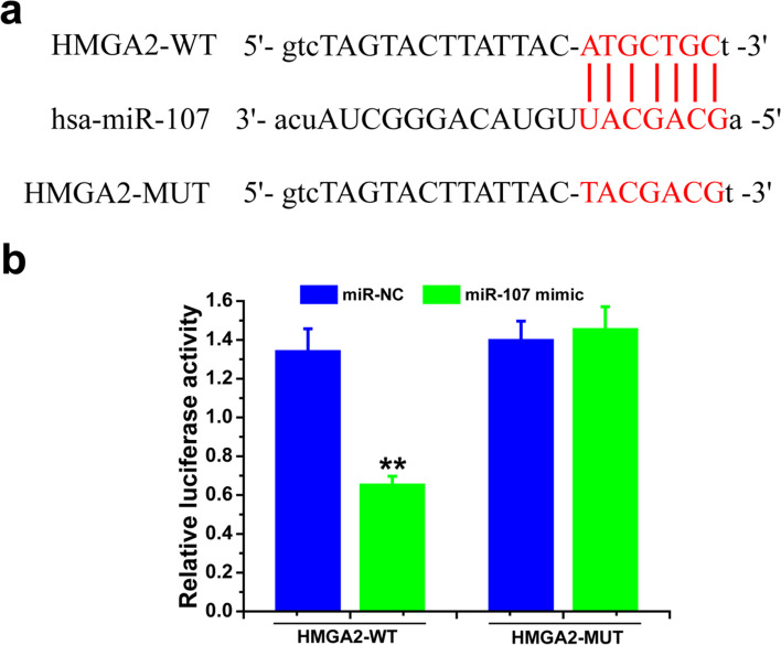
Fig. 6 Downstream target molecular of miR-107 was HMGA2. a Predicted binding sites of miR-107 in HMGA2 3′-UTR. b Plasmid vectors of human HMGA2 3′-UTR or its mutation were transfected into 293 T cells together with miR-107 mimic or NC. Data were expressed as fold change of the luciferase activity over the control from the NC group (mean ± SD, n = 3). Statistical significances were determined using one-way ANOVA followed by Dunnett’s test. **P < 0.01, compared with respective controls
Fig. 6 Downstream target molecular of miR-107 was HMGA2. a Predicted binding sites of miR-107 in HMGA2 3′-UTR. b Plasmid vectors of human HMGA2 3′-UTR or its mutation were transfected into 293 T cells together with miR-107 mimic or NC. Data were expressed as fold change of the luciferase activity over the control from the NC group (mean ± SD, n = 3). Statistical significances were determined using one-way ANOVA followed by Dunnett’s test. **P < 0.01, compared with respective controlsCC BY · PubMed Central
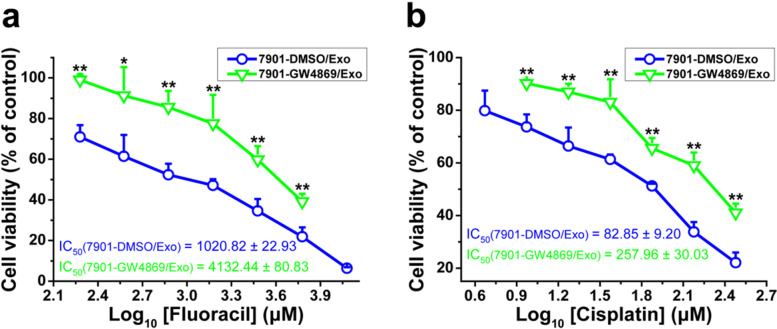
Fig. 7 Exosomes transfer increased drug sensitivity of SGC-7901/5-FU cells. The cell viability of SGC-7901/5-FU cells after treated with 5-FU (a) or CDDP (b) combined with exosomes extracted from SGC-7901 cells (with or without the exosome inhibitor GW4869 treatment) for 48 or 24 h, respectively. Cell viability was determined by MTT assay. Cells treated with vehicle serve as a blank control. Abbreviations: Exo, exosomes. All experiments were conducted in quintuplicates and data were expressed as the mean ± SD (n = 5). Statistical significances were determined using one-way ANOVA followed by Dunnett’s test. *P < 0.05,**P < 0.01, compared with the DMSO/Exo control group
Fig. 7 Exosomes transfer increased drug sensitivity of SGC-7901/5-FU cells. The cell viability of SGC-7901/5-FU cells after treated with 5-FU (a) or CDDP (b) combined with exosomes extracted from SGC-7901 cells (with or without the exosome inhibitor GW4869 treatment) for 48 or 24 h, respectively. Cell viability was determined by MTT assay. Cells treated with vehicle serve as a blank control. Abbreviations: Exo, exosomes. All experiments were conducted in quintuplicates and data were expressed as the mean ± SD (n = 5). Statistical significances were determined using one-way ANOVA followed by Dunnett’s test. *P < 0.05,**P < 0.01, compared with the DMSO/Exo control groupCC BY · PubMed Central
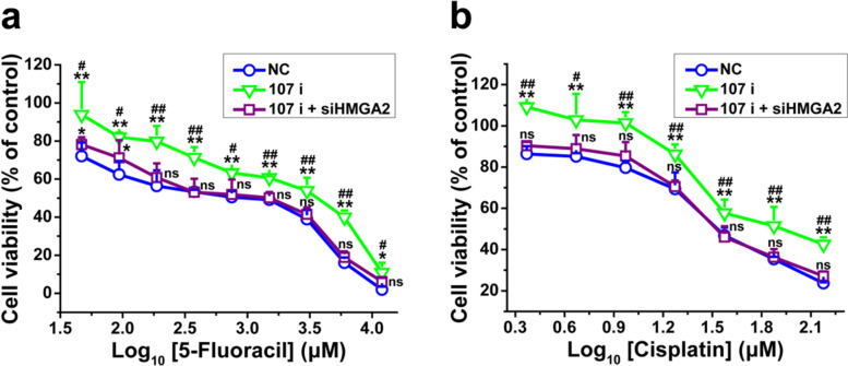
Fig. 8 SGC-7901-secreted exosomal miR-107 reversed drug resistance of SGC-7901/5-FU cells by inhibiting HMGA2. The SGC-7901 cells were transfected with miRNA-107 inhibitor, co-transfection with miRNA-107 inhibitor and siRNA traget for HMGA2 and miRNA-107 inhibitor/siHMGA2 negative control for 24 h, and the cell viability of SGC-7901/5-FU cells after treated with different concentrations of 5-FU (a) or CDDP (b) combined with exosomes extracted from SGC-7901 (NC, 107 i, 107 i + siHMGA2) cells for 48 or 24 h, respectively. Cell viability was determined by MTT assay. Cells treated with vehicle serve as a blank control. Abbreviations: Exo, exosomes. All experiments were conducted in quintuplicates and data were expressed as the mean ± SD (n = 5). Statistical significances were determined using one-way ANOVA followed by Dunnett’s test. *P < 0.05, **P < 0.01, compared with negative control transfection group; #P < 0.05, ##P < 0.01, compared with miRNA-107 inhibitor/siHMGA2 co-transfection group; ns, no significance (P > 0.05), compared with the negative control transfection and miRNA-107 inhibitor/siHMGA2 co-transfection groups
Fig. 8 SGC-7901-secreted exosomal miR-107 reversed drug resistance of SGC-7901/5-FU cells by inhibiting HMGA2. The SGC-7901 cells were transfected with miRNA-107 inhibitor, co-transfection with miRNA-107 inhibitor and siRNA traget for HMGA2 and miRNA-107 inhibitor/siHMGA2 negative control for 24 h, and the cell viability of SGC-7901/5-FU cells after treated with different concentrations of 5-FU (a) or CDDP (b) combined with exosomes extracted from SGC-7901 (NC, 107 i, 107 i + siHMGA2) cells for 48 or 24 h, respectively. Cell viability was determined by MTT assay. Cells treated with vehicle serve as a blank control. Abbreviations: Exo, exosomes. All experiments were conducted in quintuplicates and data were expressed as the mean ± SD (n = 5). Statistical significances were determined using one-way ANOVA followed by Dunnett’s test. *P < 0.05, **P < 0.01, compared with negative control transfection group; #P < 0.05, ##P < 0.01, compared with miRNA-107 inhibitor/siHMGA2 co-transfection group; ns, no significance (P > 0.05), compared with the negative control transfection and miRNA-107 inhibitor/siHMGA2 co-transfection groupsCC BY · PubMed Central
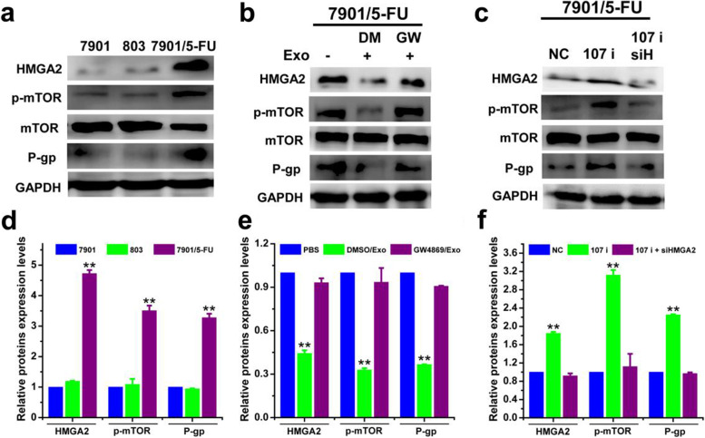
Fig. 9 Exosomal miR-107 reversed drug resistance of SGC-7901/5-FU cells by downregulating HMGA2/mTOR/P-gp pathway. The protein expression levels of HMGA2, p-mTOR/mTOR, P-gp (a-c) and corresponding quantitative analysis (d-f) of SCG7901, MGC-803 and SCG7901/5-FU cells treated with or without exosomal inhibitor GW4869 and transfected with miR-107 inhibitor, co-transfected with miRNA-107 inhibitor/siHMGA2 or negative control were determined. The protein expression levels were detected by western blotting analysis using GAPDH as internal control. Cells treated with vehicle, or transfected with control miRNA or siRNA serve as control. Abbreviations: Exo, exosome; DM, DMSO; GW, GW4869; NC, negative control. Data in a-c are the representative of two independent experiments. Statistical significances in d-f were determined using one-way ANOVA followed by Dunnett’s test. **P < 0.01, compared with the respective controls or sensitive cells
Fig. 9 Exosomal miR-107 reversed drug resistance of SGC-7901/5-FU cells by downregulating HMGA2/mTOR/P-gp pathway. The protein expression levels of HMGA2, p-mTOR/mTOR, P-gp (a-c) and corresponding quantitative analysis (d-f) of SCG7901, MGC-803 and SCG7901/5-FU cells treated with or without exosomal inhibitor GW4869 and transfected with miR-107 inhibitor, co-transfected with miRNA-107 inhibitor/siHMGA2 or negative control were determined. The protein expression levels were detected by western blotting analysis using GAPDH as internal control. Cells treated with vehicle, or transfected with control miRNA or siRNA serve as control. Abbreviations: Exo, exosome; DM, DMSO; GW, GW4869; NC, negative control. Data in a-c are the representative of two independent experiments. Statistical significances in d-f were determined using one-way ANOVA followed by Dunnett’s test. **P < 0.01, compared with the respective controls or sensitive cellsCC BY · PubMed Central
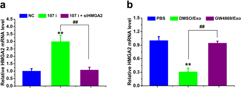
Fig. 10 Exosomal miR-107 increased drug sensitivity of SCG-7901/5-FU cells by regulating the mRNA level of HMGA2. mRNA level of HMGA2 of SCG-7901/5-FU cells treated with exosomes isolated from SGC-7901 (transfected with NC, 107 i, 107 i + siHMGA2) cells (a) and treated by exosomes isolated from SGC-7901 cells (with or without exosomal inhibitor GW4869) (b) were detected. mRNA levels were determined by qPCR using GAPDH as the internal control. Cells treated with vehicle, or transfected with control miRNA or siRNA serve as control. Abbreviations: Exo, exosome; DM, DMSO; GW, GW4869; NC, negative control. Data were expressed as the mean ± SD (n = 3). Statistical significances were determined using one-way ANOVA followed by Dunnett’s test. **P < 0.01, compared with the respective controls; ##P < 0.01, comparison between the two columns
Fig. 10 Exosomal miR-107 increased drug sensitivity of SCG-7901/5-FU cells by regulating the mRNA level of HMGA2. mRNA level of HMGA2 of SCG-7901/5-FU cells treated with exosomes isolated from SGC-7901 (transfected with NC, 107 i, 107 i + siHMGA2) cells (a) and treated by exosomes isolated from SGC-7901 cells (with or without exosomal inhibitor GW4869) (b) were detected. mRNA levels were determined by qPCR using GAPDH as the internal control. Cells treated with vehicle, or transfected with control miRNA or siRNA serve as control. Abbreviations: Exo, exosome; DM, DMSO; GW, GW4869; NC, negative control. Data were expressed as the mean ± SD (n = 3). Statistical significances were determined using one-way ANOVA followed by Dunnett’s test. **P < 0.01, compared with the respective controls; ##P < 0.01, comparison between the two columnsCC BY · PubMed Central
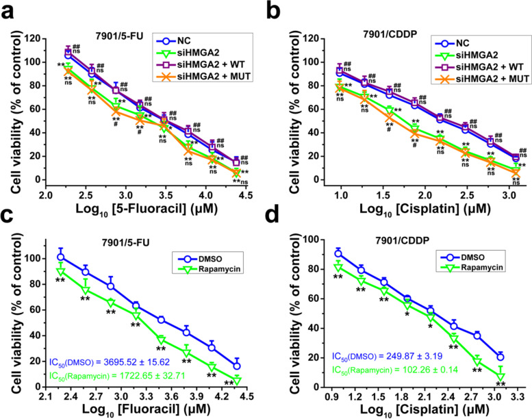
Fig. 11 Inhibition of HMGA2/mTOR pathway increased drug sensitivity of SGC-7901/5-FU and SGC-7901/CDDP cells. The cell viability of SGC-7901/5-FU (a and c) and SGC-7901/CDDP (b and d) cells was determined after cells were transfected with siHMGA2/NC together with pEX-HMGA2-WT/MUT43 plasmid vectors (a and b) or treated with or without rapamycin (10 μM) (c and d) and then treated with 5-FU (a and c) or CDDP (b and d) for 48 or 24 h, respectively. Cell viability was determined by MTT assay. Cells treated with vehicle serve as a blank control. All experiments were conducted in quintuplicates and data were expressed as the mean ± SD (n = 5). Statistical significances were determined using one-way ANOVA followed by Dunnett’s test. *P < 0.05, **P < 0.01, compared with negative control transfection/DMSO group; #P < 0.05, ##P < 0.01, compared with siHMGA2 transfection group; ns, no significance (P > 0.05), compared with the negative control transfection and siHMGA2 transfection groups
Fig. 11 Inhibition of HMGA2/mTOR pathway increased drug sensitivity of SGC-7901/5-FU and SGC-7901/CDDP cells. The cell viability of SGC-7901/5-FU (a and c) and SGC-7901/CDDP (b and d) cells was determined after cells were transfected with siHMGA2/NC together with pEX-HMGA2-WT/MUT43 plasmid vectors (a and b) or treated with or without rapamycin (10 μM) (c and d) and then treated with 5-FU (a and c) or CDDP (b and d) for 48 or 24 h, respectively. Cell viability was determined by MTT assay. Cells treated with vehicle serve as a blank control. All experiments were conducted in quintuplicates and data were expressed as the mean ± SD (n = 5). Statistical significances were determined using one-way ANOVA followed by Dunnett’s test. *P < 0.05, **P < 0.01, compared with negative control transfection/DMSO group; #P < 0.05, ##P < 0.01, compared with siHMGA2 transfection group; ns, no significance (P > 0.05), compared with the negative control transfection and siHMGA2 transfection groupsCC BY · PubMed Central
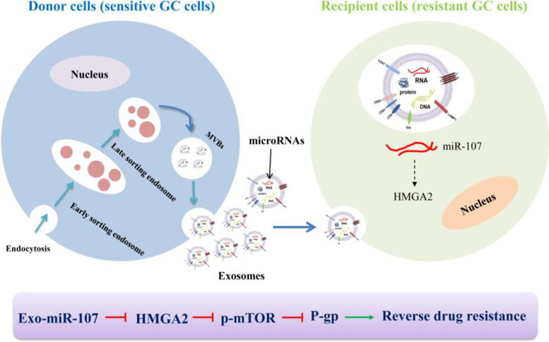
Fig. 12 The possible mechanism of how exosomal miR-107 reversed chemotherapeutic drug resistance of SGC-7901/5-FU cells. Exosomal miR-107 was transmitted from sensitive GC cells to recipient resistant GC cells, and drug sensitivity of SGC-7901/5-FU cells was improved through inhibiting HMGA2/mTOR/P-gp signal
Fig. 12 The possible mechanism of how exosomal miR-107 reversed chemotherapeutic drug resistance of SGC-7901/5-FU cells. Exosomal miR-107 was transmitted from sensitive GC cells to recipient resistant GC cells, and drug sensitivity of SGC-7901/5-FU cells was improved through inhibiting HMGA2/mTOR/P-gp signalCC BY · PubMed Central

Verification

Claims the paper makes

The claims and statistics of this paper haven’t been checked yet.

Run the check — free, no account needed. It reads the open-access text and checks the sources it cites.

A few free checks a day · papers already verified open instantly · results are public

Claims other papers make about this one

Each is checked against this paper’s own text: does it say what the citing paper says it does?

Self-check

Percentages, sums and reported differences re-computed from the paper’s own numbers.

The self-check needs the paper’s full text.

Citations

Cites

Research works printed but not linked: 39

  1. Arbyn M, Weiderpass E, Bruni L, de Sanjosé S, Saraiya M, Ferlay J, et al. Estimates of incidence and mortality of cervical cancer in 2018: a worldwide analysis. Lancet Glob Health. 2020;8(2):e191–203. https://doi.org/10.1016/S2214-109X(19)30482-6. · DOI
  2. Smyth EC, Nilsson M, Grabsch HI, van Grieken NC, Lordick F. Gastric cancer. Lancet. 2020;396(10251):635–48. https://doi.org/10.1016/S0140-6736(20)31288-5. · DOI
  3. Liu X, Lu Y, Xu YC, Hou SZ, Huang JL, Wang B, et al. Exosomal transfer of miR-501 confers doxorubicin resistance and tumorigenesis via targeting of BLID in gastric cancer. Cancer Lett. 2019;459:122–34. https://doi.org/10.1016/j.canlet.2019.05.035. · DOI
  4. He ZY, Li Z, Zhang X, Yin K, Wang WZ, Xu ZP, et al. MiR-422a regulates cellular metabolism and malignancy by targeting pyruvate dehydrogenase kinase 2 in gastric cancer. Cell Death Dis. 2018;9:505. https://doi.org/10.1038/s41419-018-0564-3. · DOI
  5. Pastan I, Gottesman MM. Multidrug resistance. Annu Rev Med. 1991;42:277–86 Https://doi.org/10.1146/annurev.me.42.020191.001425. · DOI
  6. Mowla M, Hashemi A. Functional roles of exosomal miRNAs in multi-drug resistance in cancer chemotherapeutics. Exp Mol Pathol. 2021;118:104592. https://doi.org/10.1016/j.yexmp.2020.104592. · DOI
  7. Santos P, Almeida F. Role of exosomal miRNAs and the tumor microenvironment in drug resistance. Cells. 2020;9(6):1450. https://doi.org/10.3390/cells9061450. · DOI
  8. Mashouri L, Yousefi H, Aref AR, Ahadi AM, Molaei F, Alahari SK. Exosomes: composition, biogenesis, and mechanisms in cancer metastasis and drug resistance. Mol Cancer. 2019;18(1):75. https://doi.org/10.1186/s12943-019-0991-5. · DOI · Paper page
  9. Liu T, Zhang X, Du LT, Wang YS, Liu XM, Tian H, et al. Exosome-transmitted miR-128-3p increase chemosensitivity of oxaliplatin-resistant colorectal cancer. Mol Cancer. 2019;18(1):43. https://doi.org/10.1186/s12943-019-0981-7. · DOI
  10. Ma YZ, Yuwen DL, Chen JW, Zheng BF, Gao J, Fan MM, et al. Exosomal transfer of cisplatin-induced miR-425-3p confers cisplatin resistance in NSCLC through activating autophagy. Int J Nanomedicine. 2019;14:8121–32. https://doi.org/10.2147/IJN.S221383. · DOI
  11. Liang Y, Zhu DX, Hou LD, Wang Y, Huang X, Zhou C, et al. MiR-107 confers chemoresistance to colorectal cancer by targeting calcium-binding protein 39. Br J Cancer. 2020;122(5):705–14. https://doi.org/10.1038/s41416-019-0703-3. · DOI
  12. Jiao YN, Kong L, Yao YJ, Li SH, Tao ZY, Yan YH, et al. Osthole decreases beta amyloid levels through up-regulation of miR-107 in Alzheimer's disease. Neuropharmacology. 2016;108:332–44. https://doi.org/10.1016/j.neuropharm.2016.04.046. · DOI
  13. Lin SS, Yuan LJ, Niu CC, Tu YK, Yang CY, Ueng SWN. Hyperbaric oxygen inhibits the HMGB1/RAGE signaling pathway by upregulating Mir-107 expression in human osteoarthritic chondrocytes. Osteoarthr Cartil. 2019;27(9):1372–81. https://doi.org/10.1016/j.joca.2019.05.011. · DOI
  14. Lu CJ, Xie ZB, Peng QZ. miRNA-107 enhances chemosensitivity to paclitaxel by targeting antiapoptotic factor Bcl-w in non small cell lung cancer. Am J Cancer Res. 2017;7:1863–73.
  15. Wang GS, Ma CP, Shi XJ, Guo WC, Niu JX. miR-107 enhances the sensitivity of breast cancer cells to paclitaxel. Open Med (Wars). 2019;14:456–66. https://doi.org/10.1515/med-2019-0049. · DOI
  16. Inoue T, Iinuma H, Ogawa E, Inaba T, Fukushima R. Clinicopathological and prognostic significance of microRNA-107 and its relationship to DICER1 mRNA expression in gastric cancer. Oncol Rep. 2012;27(6):1759–64. https://doi.org/10.3892/or.2012.1709. · DOI
  17. Vignali R, Marracci S. HMGA genes and proteins in development and evolution. Int J Mol Sci. 2020;21(2):654. https://doi.org/10.3390/ijms21020654. · DOI
  18. Wang XH, Huang W, Liu GS, Cai WF, Millard RW, Wang YG, et al. Cardiomyocytes mediate anti-angiogenesis in type 2 diabetic rats through the exosomal transfer of miR-320 into endothelial cells. J Mol Cell Cardiol. 2014;(74):139–50. https://doi.org/10.1016/j.yjmcc.2014.05.001. · DOI
  19. Li M, Lu Y, Xu YC, Wang JW, Zhang CH, Du Y, et al. Horizontal transfer of exosomal CXCR4 promotes murine hepatocarcinoma cell migration, invasion and lymphangiogenesis. Gene. 2018;676:101–9. https://doi.org/10.1016/j.gene.2018.07.018. · DOI
  20. Noro B, Licheri B, Sgarra R, Rustighi A, Tessari MA, Chau K-Y, et al. Molecular dissection of the architectural transcription factor HMGA2. 2003;42(15):4569–77. https://doi.org/10.1021/bi026605k. · DOI
  21. Jiang L, Wang P, Sun YJ, Wu YJ. Ivermectin reverses the drug resistance in cancer cells through EGFR/ERK/Akt/NF-κB pathway. J Exp Clin Cancer Res. 2019;38(1):265. https://doi.org/10.1186/s13046-019-1251-7. · DOI
  22. Tan SM, Xia LZ, Yi P, Han YQ, Tang L, Pan Q, et al. Exosomal miRNAs in tumor microenvironment. J Exp Clin Cancer Res. 2020;39(1):67. https://doi.org/10.1186/s13046-020-01570-6. · DOI
  23. Cao C, Wang B, Tang JN, Zhao JM, Guo JC, Guo QQ, et al. Circulating exosomes repair endothelial cell damage by delivering miR-193a-5p. J Cell Mol Med. 2021;25(4):2176–89. https://doi.org/10.1111/jcmm.16202. · DOI
  24. Fu X, Liu MJ, Qu SY, Ma JQ, Zhang YM, Shi TT, et al. Exosomal microRNA-32-5p induces multidrug resistance in hepatocellular carcinoma via the PI3K/Akt pathway. J Exp Clin Cancer Res. 2018;37(1):52. https://doi.org/10.1186/s13046-018-0677-7. · DOI
  25. Zhu XL, Shen HL, Yin XM, Yang ML, Wei H, Chen Q, et al. Macrophages derived exosomes deliver miR-223 to epithelial ovarian cancer cells to elicit a chemoresistant phenotype. J Exp Clin Cancer Res. 2019;38(1):81. https://doi.org/10.1186/s13046-019-1095-1. · DOI · Paper page
  26. Wang M, Qiu R, Yu SR, Xu XY, Li G, Gu RM, et al. Paclitaxel-resistant gastric cancer MGC-803 cells promote epithelial-to-mesenchymal transition and chemoresistance in paclitaxel-sensitive cells via exosomal delivery of miR-155-5p. Int J Oncol. 2018;54:326–38. https://doi.org/10.3892/ijo.2018.4601. · DOI
  27. Lin H, Zhang L, Zhang CH, Liu PP. Exosomal MiR-500a-3p promotes cisplatin resistance and stemness via negatively regulating FBXW7 in gastric cancer. J Cell Mol Med. 2020;24(16):8930–41. https://doi.org/10.1111/jcmm.15524. · DOI · Paper page
  28. Yang S, Gu YL, Wang GJ, Hu QZ, Chen SX, Wang Y, et al. HMGA2 regulates acute myeloid leukemia progression and sensitivity to daunorubicin via Wnt/β-catenin signaling. Int J Mol Med. 2019;44(2):427–36. https://doi.org/10.3892/ijmm.2019.4229. · DOI
  29. Yang Y, Zhang P, Zhao YF, Yang J, Jiang GN, Fan J. Decreased MicroRNA-26a expression causes cisplatin resistance in human non-small cell lung cancer. Cancer Biol Ther. 2016;17(5):515–25. https://doi.org/10.1080/15384047.2015.1095405. · DOI
  30. Yang XY, Zhao Q, Yin HL, Lei XY, Gan RL. MiR-33b-5p sensitizes gastric cancer cells to chemotherapy drugs via inhibiting HMGA2 expression. J Drug Target. 2017;25(7):653–60. https://doi.org/10.1080/1061186X.2017.1323220. · DOI
  31. Wang Y, Chen FQ, Zhao M, Yang Z, Zhang SQ, Ye LH, et al. MiR-107 suppresses proliferation of hepatoma cells through targeting HMGA2 mRNA 3'UTR. Biochem Biophys Res Commun. 2016;480(3):455–60. https://doi.org/10.1016/j.bbrc.2016.10.070. · DOI
  32. Liu XZ, Yidayitula Y, Zhao H, Luo Y, Ma XQ, Xu MH. LncRNA LINC00152 promoted glioblastoma progression through targeting the miR-107 expression. Environ Sci Pollut Res Int. 2018;25(18):17674–81. https://doi.org/10.1007/s11356-018-1784-x. · DOI
  33. Mansoori B, Mohammadi A, Ditzel HJ, Duijf PHG, Khaze V, Gjerstorff MF, et al. HMGA2 as a critical regulator in Cancer development. Genes (Basel). 2021;12(2):269. https://doi.org/10.3390/genes12020269. · DOI · Paper page
  34. Tan L, Wei XP, Zheng LX, Zeng JC, Liu HB, Yang SJ, et al. Amplified HMGA2 promotes cell growth by regulating Akt pathway in AML. J Cancer Res Clin Oncol. 2016;142(2):389–99 Https://doi.org/10.1007/s00432-015-2036-9. · DOI
  35. Wang YD, Mao JD, Wang JF, Xu MQ. MiR-590 suppresses proliferation and induces apoptosis in pancreatic cancer by targeting high mobility group A2. Technol Cancer Res Treat. 2020;19:1533033820928143. https://doi.org/10.1177/1533033820928143. · DOI
  36. Robey RW, Pluchino KM, Hall MD, Fojo AT, Bates SE, Gottesman MM. Revisiting the role of ABC transporters in multidrug-resistant cancer. Nat Rev Cancer. 2018;18(7):452–64. https://doi.org/10.1038/s41568-018-0005-8. · DOI · Paper page
  37. Song L, Zhou ZG, Gan YB, Li P, Xu Y, Zhang ZH, et al. Long noncoding RNA OIP5-AS1 causes cisplatin resistance in osteosarcoma through inducing the LPAATβ/PI3K/AKT/mTOR signaling pathway by sponging the miR-340-5p. J Cell Biochem. 2019;120(6):9656–66. https://doi.org/10.1002/jcb.28244. · DOI
  38. Ma Q, Chang ZH, Wang W, Wang BM. Rapamycin-mediated mTOR inhibition reverses drug resistance to adriamycin in colon cancer cells. Hepatogastroenterology. 2015;62(140):880–6.
  39. Wang SF, Chou YC, Mazumder N, Kao FJ, Nagy LD, Guengerich FP, et al. 7-Ketocholesterol induces P-glycoprotein through PI3K/mTOR signaling in hepatoma cells. Biochem Pharmacol. 2013;86(4):548–60. https://doi.org/10.1016/j.bcp.2013.06.006. · DOI

Cited by

Sorted by citations

See all 59

Metadata

Authors
Jiang, L., Zhang, Y., Guo, L., Liu, C., Wang, P., & Ren, W.
Venue
BMC Cancer
Published
2021-12
Volume
21
Issue
1
ISSN
1471-2407
Cited by
59
References
2
Language
en

About these checks

Registry, retraction, Cites and statistics results are computed, not judged, with one exception: a printed reference that no index lists is read once by a model before it counts as a citation error. Claims are extracted by a model and kept only when their quote appears verbatim in the text; each cited source is then checked, with a model’s help, for whether it says what the claim relies on. Those verdicts can be wrong — read the claim’s quote and the cited source before relying on one.

Statistics and arithmetic are recomputed, not judged.

Confidence bands are shown only for fields calibrated on our annotated test set. None is calibrated yet, so no band is shown.

What counts as a claim ›How we measure confidence ›How we check this ›

Are you an author, or do you see a problem with this page? Request a review