

The DOI matches its Crossref record; 1 of the 1 references it cites were found in a registry or an index.
TUBB2B regulates epithelial-mesenchymal transition via interaction with Vimentin to promote glioma migration and invasion
Open accessPublished version of a preprint
Abstract
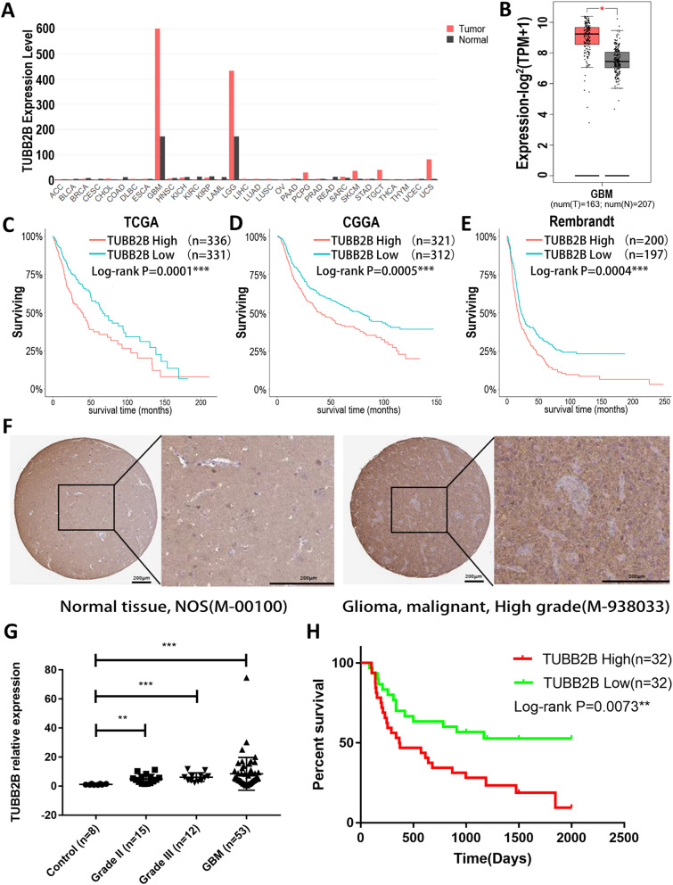
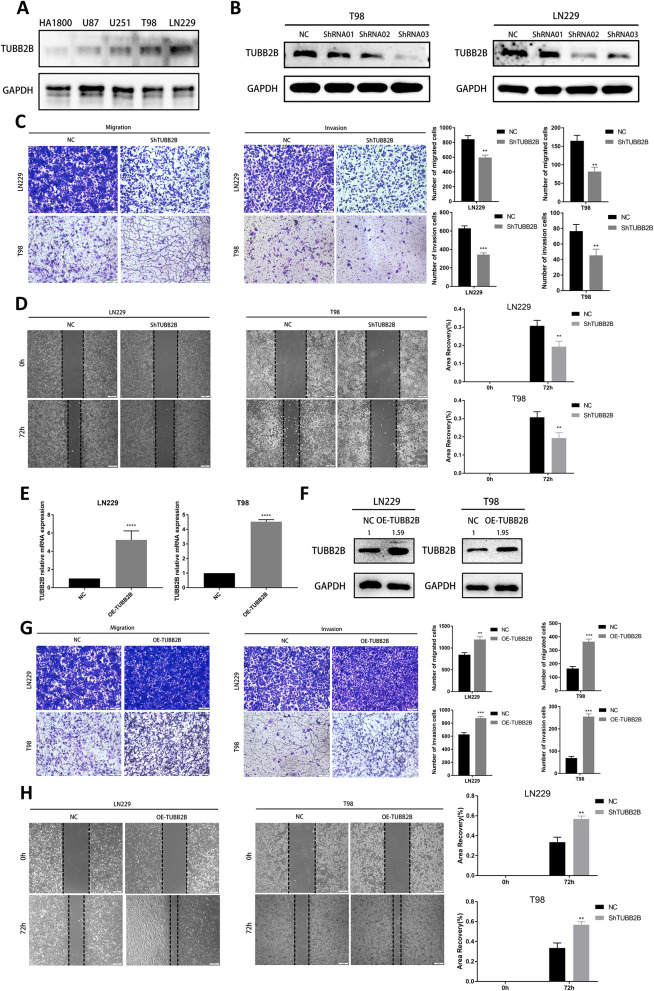
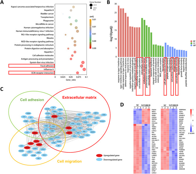
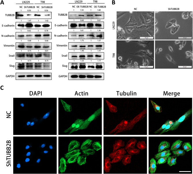
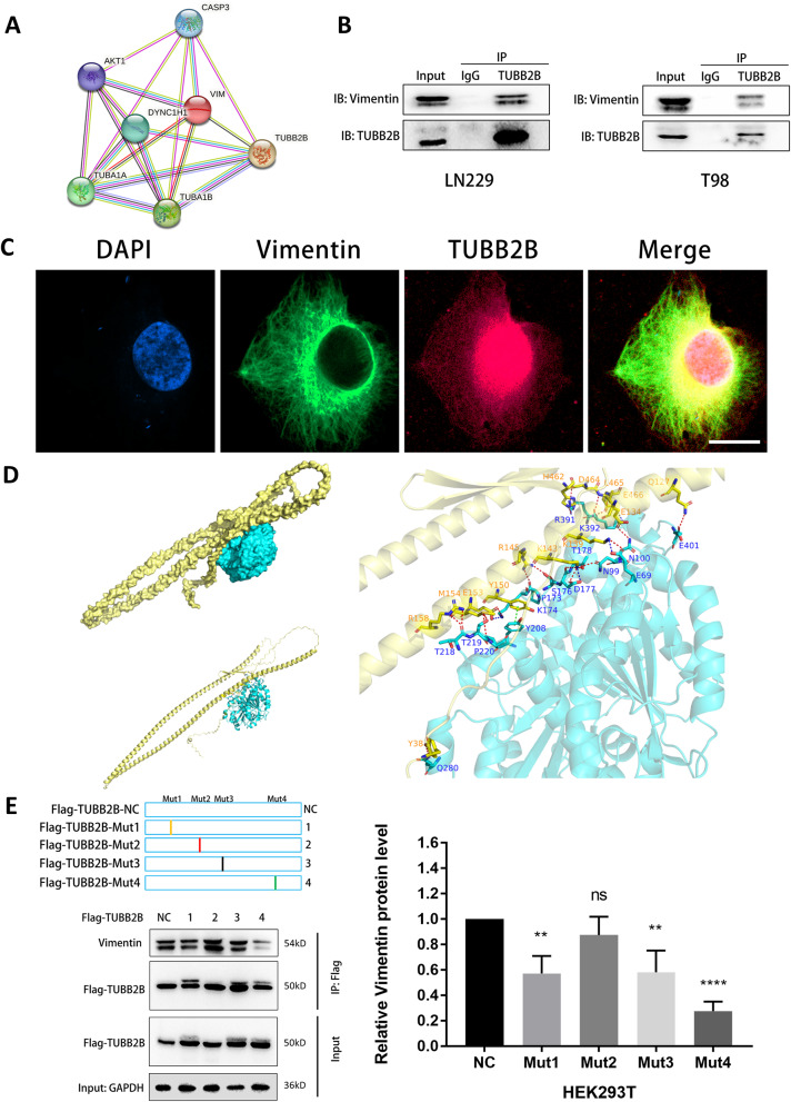
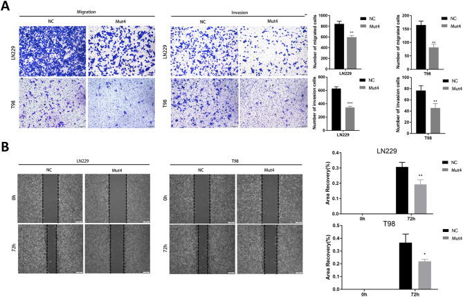
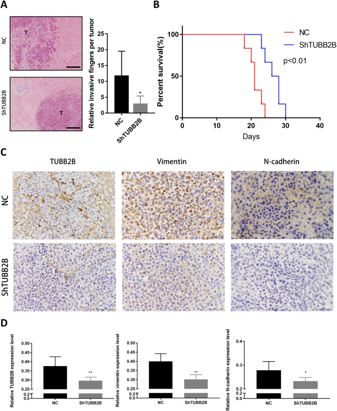
BACKGROUND: Epithelial-mesenchymal transition (EMT) plays a crucial role in the migration and invasion capabilities of glioblastoma (GBM) cells. Several studies have established tubulin as a significant regulator of the EMT process. Tubulin beta 2B class IIb (TUBB2B), a critical component of microtubules, has been linked to the prognosis of various tumors. However, the specific biological function and mechanism of TUBB2B in GBM remain unclear. METHODS: In vitro experiments demonstrated that TUBB2B knockdown inhibited the migration and invasion of GBM cells, while its overexpression enhanced these capabilities. Western blot, immunofluorescence (IF) and co-immunoprecipitation (Co-IP) assays revealed that TUBB2B interacts with Vimentin. Molecular docking and residue mutation scanning indicated that TUBB2B interacts with Vimentin at the R391/K392/A393/F394 sites. In vivo experiments using nude mice confirmed that TUBB2B knockdown inhibited GBM cell invasion and migration. RESULTS: TUBB2B was upregulated in GBM tissue samples compared with normal tissues. The sites of TUBB2B(R391/K392/A393/F394) physically interacts with Vimentin to induce EMT, which promotes migration and invasion. CONCLUSION: TUBB2B may regulate EMT and promote the migration and invasion of GBM cells through its interaction with Vimentin, highlighting TUBB2B as a potential therapeutic target for GBM.
Overview
The claims and statistics of this paper haven’t been checked yet.
Run the check — free, no account needed. It reads the open-access text and checks the sources it cites.
Integrity signals
Registry
Cites
Statistics
Figures
Verification
Claims the paper makes
The claims and statistics of this paper haven’t been checked yet.
Run the check — free, no account needed. It reads the open-access text and checks the sources it cites.
Self-check
Percentages, sums and reported differences re-computed from the paper’s own numbers.
The self-check needs the paper’s full text.
Citations
Cites
Highly accurate protein structure prediction with AlphaFold
Jumper, J., Evans, R., Pritzel, A., Green, T., Figurnov, M., Ronneberger, O., Tunyasuvunakool, K., Bates, R., Žídek, A., Potapenko, A., et al. (2021) · Nature · doi:10.1038/s41586-021-03819-2 · Cited by 43,764
Research works printed but not linked: 38
- Rivera M, Bander ED, Cisse B. Perspectives on Microglia-based Immune therapies against Glioblastoma. World Neurosurg. 2021;154:228–31. · DOI
- Weller M, van den Bent M, Preusser M, Le Rhun E, Tonn JC, Minniti G, et al. EANO guidelines on the diagnosis and treatment of diffuse gliomas of adulthood. Nat Reviews Clin Oncol. 2021;18(3):170–86. · DOI · Paper page
- Nørøxe DS, Poulsen HS, Lassen U. Hallmarks of glioblastoma: a systematic review. ESMO Open 2016;1(6). · DOI
- Yang J, Antin P, Berx G, Blanpain C, Brabletz T, Bronner M, et al. Guidelines and definitions for research on epithelial–mesenchymal transition. Nat Rev Mol Cell Biol. 2020;21(6):341–52. · DOI · Paper page
- Chi M, Liu J, Mei C, Shi Y, Liu N, Jiang X et al. TEAD4 functions as a prognostic biomarker and triggers EMT via PI3K/AKT pathway in bladder cancer. J Experimental Clin Cancer Res. 2022;41(1). · DOI
- Aiello NM, Kang Y. Context-dependent EMT programs in cancer metastasis. J Exp Med. 2019;216(5):1016–26. · DOI
- Zeisberg M, Neilson EG. Biomarkers for epithelial-mesenchymal transitions. J Clin Investig. 2009;119(6):1429–37. · DOI · Paper page
- Shibue T, Brooks MW, Weinberg RA. An integrin-linked machinery of cytoskeletal regulation that enables experimental tumor initiation and metastatic colonization. Cancer Cell. 2013;24(4):481–98. · DOI
- McConkey DJ, Choi W, Marquis L, Martin F, Williams MB, Shah J, et al. Role of epithelial-to-mesenchymal transition (EMT) in drug sensitivity and metastasis in bladder cancer. Cancer Metastasis Rev. 2009;28(3):335–44. · DOI
- Admoni-Elisha L, Elbaz T, Chopra A, Shapira G, Bedford Mark T, Fry Christopher J, et al. TWIST1 methylation by SETD6 selectively antagonizes LINC-PINT expression in glioma. Nucleic Acids Res. 2022;50(12):6903–18. · DOI
- Colas E, Pedrola N, Devis L, Ertekin T, Campoy I, Martínez E, et al. The EMT signaling pathways in endometrial carcinoma. Clin Transl Oncol. 2012;14(10):715–20. · DOI
- van Zijl F, Zulehner G, Petz M, Schneller D, Kornauth C, Hau M, et al. Epithelial–mesenchymal transition in hepatocellular carcinoma. Future Oncol. 2009;5(8):1169–79. · DOI
- Bates RC, Mercurio A. The epithelial-mesenchymal tansition (EMT) and colorectal cancer progression. Cancer Biol Ther. 2005;4(4):371–6. · DOI
- Babu D, Mudiraj A, Yadav N, Y.B.V.K C, Panigrahi M, Prakash Babu P. Rabeprazole has efficacy per se and reduces resistance to temozolomide in glioma via EMT inhibition. Cell Oncol. 2021;44(4):889–905. · DOI
- Ruksha K, Mezheyeuski A, Nerovnya A, Bich T, Tur G, Gorgun J et al. Over-expression of βII-Tubulin and especially its localization in cell nuclei correlates with poorer outcomes in Colorectal Cancer. Cells. 2019;8(1). · DOI
- Wang X, Shi J, Huang M, Chen J, Dan J, Tang Y et al. TUBB2B facilitates progression of hepatocellular carcinoma by regulating cholesterol metabolism through targeting HNF4A/CYP27A1. Cell Death Dis. 2023;14(3). · DOI
- Liotti A, La Civita E, Cennamo M, Crocetto F, Ferro M, Guadagno E, et al. Periprostatic adipose tissue promotes prostate cancer resistance to docetaxel by paracrine IGF-1 upregulation of TUBB2B beta‐tubulin isoform. Prostate. 2021;81(7):407–17. · DOI
- Wang Z, Wang H, Wang Z, He S, Jiang Z, Yan C, et al. Associated analysis of PER1/TUBB2B with endometrial cancer development caused by circadian rhythm disorders. Med Oncol. 2020;37(10):90. · DOI
- Maillard C, Roux CJ, Charbit-Henrion F, Steffann J, Laquerriere A, Quazza F, et al. Tubulin mutations in human neurodevelopmental disorders. Semin Cell Dev Biol. 2023;137:87–95. · DOI
- Kozakov D, Hall DR, Xia B, Porter KA, Padhorny D, Yueh C, et al. The ClusPro web server for protein–protein docking. Nat Protoc. 2017;12(2):255–78. · DOI
- Schymkowitz J, Borg J, Stricher F, Nys R, Rousseau F, Serrano L. The FoldX web server: an online force field. Nucleic Acids Res. 2005;33(suppl2):W382–8. · DOI · Paper page
- Sadrkhanloo M, Entezari M, Orouei S, Ghollasi M, fathi N, Rezaei S, et al. STAT3-EMT axis in tumors: modulation of cancer metastasis, stemness and therapy response. Pharmacol Res. 2022;182:106311. · DOI
- Kar R, Jha NK, Jha SK, Sharma A, Dholpuria S, Asthana N et al. A NOTCH deeper into the Epithelial-To-Mesenchymal transition (EMT) program in breast Cancer. Genes. 2019;10(12). · DOI
- Xu J, Lamouille S, Derynck R. TGF-β-induced epithelial to mesenchymal transition. Cell Res. 2009;19(2):156–72. · DOI · Paper page
- Atiya HI, Gorecki G, Garcia GL, Frisbie LG, Baruwal R, Coffman L. Stromal-modulated epithelial-to-mesenchymal transition in Cancer cells. Biomolecules [Internet]. 2023; 13(11). · DOI
- Lin Y-T, Wu K-J. Epigenetic regulation of epithelial-mesenchymal transition: focusing on hypoxia and TGF-β signaling. J Biomed Sci. 2020;27(1):39. · DOI
- Stanton RA, Gernert KM, Nettles JH, Aneja R. Drugs that target dynamic microtubules: a new molecular perspective. Med Res Rev. 2011;31(3):443–81. · DOI
- Wen J, Wang Q, Zhang W, Wang W. TUBA1A licenses APC/C-mediated mitotic progression to drive glioblastoma growth by inhibiting PLK3. FEBS Lett. 2023;597(24):3072–86. · DOI
- Sobierajska K, Wieczorek K, Ciszewski WM, Sacewicz-Hofman I, Wawro ME, Wiktorska M, et al. β-III tubulin modulates the behavior of snail overexpressed during the epithelial-to-mesenchymal transition in colon cancer cells. Biochimica et Biophysica Acta (BBA) -. Mol Cell Res. 2016;1863(9):2221–33.
- Gui S, Chen P, Liu Y, Chen Q, Cheng T, Lv S, et al. TUBA1C expression promotes proliferation by regulating the cell cycle and indicates poor prognosis in glioma. Biochem Biophys Res Commun. 2021;577:130–8. · DOI
- Liu J, Li Y. Upregulation of MAPK10, TUBB2B and RASL11B may contribute to the development of neuroblastoma. Mol Med Rep. 2019. · DOI
- Strouhalova K, Přechová M, Gandalovičová A, Brábek J, Gregor M, Rosel D. Vimentin intermediate filaments as potential target for Cancer Treatment. Cancers [Internet]. 2020; 12(1). · DOI
- Sannino G, Marchetto A, Kirchner T, Grünewald TGP. Epithelial-to-mesenchymal and mesenchymal-to-epithelial transition in mesenchymal tumors: a Paradox in Sarcomas? Cancer Res. 2017;77(17):4556–61. · DOI
- Huang X, Xiang L, Wang B, Hu J, Liu C, Ren A et al. CMTM6 promotes migration, invasion, and EMT by interacting with and stabilizing vimentin in hepatocellular carcinoma cells. J Translational Med. 2021;19(1). · DOI
- Preitner N, Quan J, Nowakowski Dan W, Hancock Melissa L, Shi J, Tcherkezian J, et al. APC is an RNA-Binding protein, and its Interactome provides a link to neural development and Microtubule Assembly. Cell. 2014;158(2):368–82. · DOI
- Lu Q, Luduena RF. Removal of β III isotype enhances Taxol Induced Microtubule Assembly. Cell Struct Funct. 1993;18(3):173–82. · DOI
- Hendrix MJ, Seftor EA, Seftor RE, Trevor KT. Experimental co-expression of vimentin and keratin intermediate filaments in human breast cancer cells results in phenotypic interconversion and increased invasive behavior. Am J Pathol. 1997;150(2):483–95.
- Liu C-Y, Lin H-H, Tang M-J, Wang Y-K. Vimentin contributes to epithelial-mesenchymal transition cancer cell mechanics by mediating cytoskeletal organization and focal adhesion maturation. Oncotarget; Vol 6, No 18. 2015. · DOI
Metadata
- Authors
- Li, J., Zhou, Z., Zhang, J., Wang, M., Luan, X., Zhao, M., Jiang, G., Wang, G., Li, S., Xiang, W., Chen, L., & Zhou, J.
- Affiliations
- Southwest Medical University (CN)
- Corresponding author
- Xiang, Wei
- Chen, Ligang
- Zhou, Jie
- Venue
- Cancer Cell International
- Published
- 2024-12-20
- Volume
- 24
- Issue
- 1
- Pages
- 423-423
- ISSN
- 1475-2867 (ISSN-L)
- PMC
- PMC11662515
- OpenAlex
- W4405636637
- Cited by
- 8
- References
- 1
- Citations by year
- 2026: 6 · 2025: 2
- FWCI
- 2.25
- Type
- journal article
- Language
- en
- Open access
- gold
- doi.org
- Licence
- Locations
- Cancer Cell International (publishedVersion, cc-by-nc-nd)
- Research Square (acceptedVersion, cc-by)
- PubMed Central (publishedVersion, cc-by-nc-nd)
- PubMed (publishedVersion)
- DOAJ (DOAJ: Directory of Open Access Journals) (submittedVersion)
- Funders
- The research was supported by National Natural Science Foundation project (82072780 and 82372825)
- the Science and Technology Department of Sichuan Province (2022YFS0630)
- the Sichuan Medical Youth Innovation Research Project (Q22044)
- the Luzhou Science and Technology Program (2023JYJ028)
- the University Science and Technology Strategic Cooperation Project (2024SNXNYD03)
- Topics
- Health Sciences › Medicine › Oncology › Glioma Diagnosis and Treatment
- Health Sciences › Medicine › Oncology › Cancer Cells and Metastasis
- Life Sciences › Biochemistry, Genetics and Molecular Biology › Cell Biology › Microtubule and mitosis dynamics
- Keywords
- TUBB2B, epithelial-mesenchymal transition, vimentin, glioblastoma, cell migration, tumor invasion, protein-protein interaction, microtubules, therapeutic target
About these checks
Registry, retraction, Cites and statistics results are computed, not judged, with one exception: a printed reference that no index lists is read once by a model before it counts as a citation error. Claims are extracted by a model and kept only when their quote appears verbatim in the text; each cited source is then checked, with a model’s help, for whether it says what the claim relies on. Those verdicts can be wrong — read the claim’s quote and the cited source before relying on one.
Statistics and arithmetic are recomputed, not judged.
Confidence bands are shown only for fields calibrated on our annotated test set. None is calibrated yet, so no band is shown.
What counts as a claim ›How we measure confidence ›How we check this ›
Are you an author, or do you see a problem with this page? Request a review
How to cite
Cite CoChat for the checks on this page, and the paper for its own findings.
- This page
- CoChat. (2026). Citation check: TUBB2B regulates epithelial-mesenchymal transition via interaction with Vimentin to promote glioma migration and invasion. https://app.cochat.ai/papers/10.1186/s12935-024-03618-5
- The paper
- Li, J., Zhou, Z., Zhang, J., Wang, M., Luan, X., Zhao, M., Jiang, G., Wang, G., Li, S., Xiang, W., Chen, L., & Zhou, J. (2024). TUBB2B regulates epithelial-mesenchymal transition via interaction with Vimentin to promote glioma migration and invasion. Cancer Cell International. https://doi.org/10.1186/s12935-024-03618-5
