

The DOI matches its Crossref record; 191 of the 192 references it cites were found in a registry or an index.
Three-dimensional cardiac computational modelling: methods, features and applications.
Open accessAbstract
The combination of computational models and biophysical simulations can help to interpret an array of experimental data and contribute to the understanding, diagnosis and treatment of complex diseases such as cardiac arrhythmias. For this reason, three-dimensional (3D) cardiac computational modelling is currently a rising field of research. The advance of medical imaging technology over the last decades has allowed the evolution from generic to patient-specific 3D cardiac models that faithfully represent the anatomy and different cardiac features of a given alive subject. Here we analyse sixty representative 3D cardiac computational models developed and published during the last fifty years, describing their information sources, features, development methods and online availability. This paper also reviews the necessary components to build a 3D computational model of the heart aimed at biophysical simulation, paying especial attention to cardiac electrophysiology (EP), and the existing approaches to incorporate those components. We assess the challenges associated to the different steps of the building process, from the processing of raw clinical or biological data to the final application, including image segmentation, inclusion of substructures and meshing among others. We briefly outline the personalisation approaches that are currently available in 3D cardiac computational modelling. Finally, we present examples of several specific applications, mainly related to cardiac EP simulation and model-based image analysis, showing the potential usefulness of 3D cardiac computational modelling into clinical environments as a tool to aid in the prevention, diagnosis and treatment of cardiac diseases.
Overview
The claims and statistics of this paper haven’t been checked yet.
Run the check — free, no account needed. It reads the open-access text and checks the sources it cites.
Integrity signals
Registry
Cites
Statistics
Figures
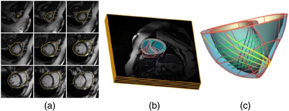
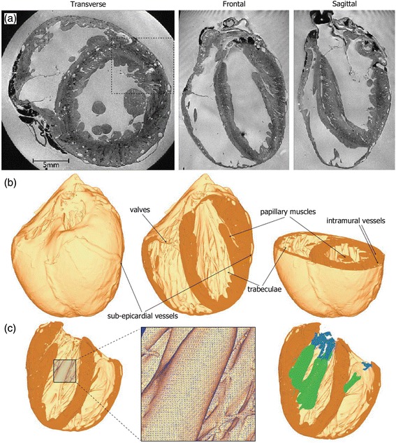
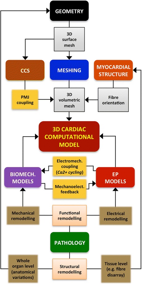
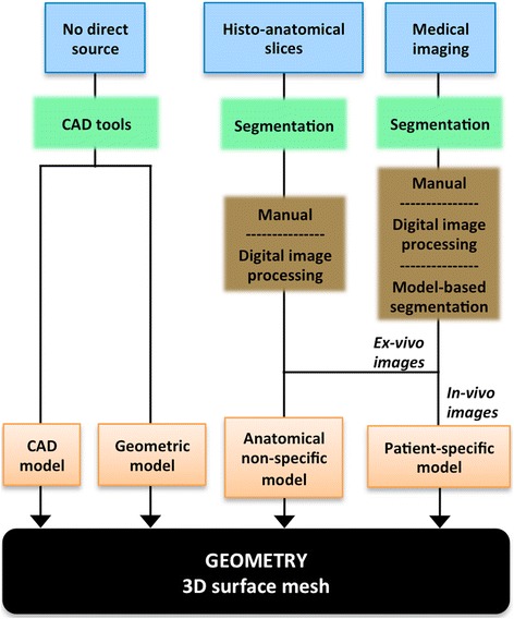
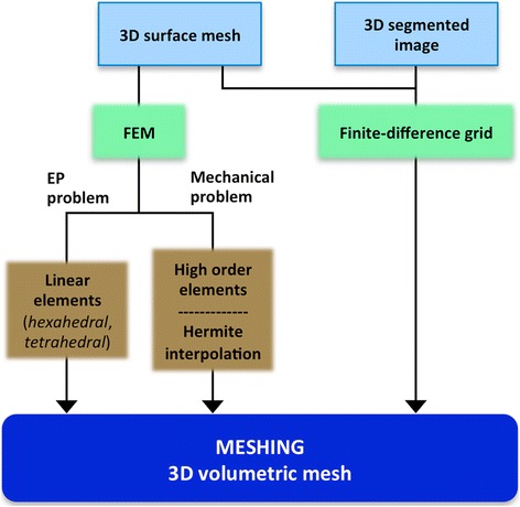
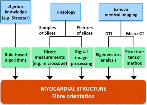
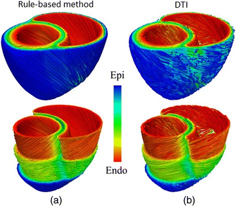
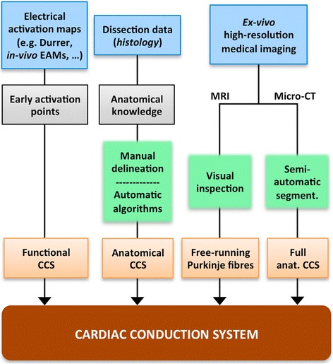
Verification
Claims the paper makes
The claims and statistics of this paper haven’t been checked yet.
Run the check — free, no account needed. It reads the open-access text and checks the sources it cites.
Claims other papers make about this one
Each is checked against this paper’s own text: does it say what the citing paper says it does?
The source backs part of the claim, not all of it — or backs it for a different population, condition or comparison.
In the citing paper
“However, when imaging data is noisy, an alternative rule-based methodology can be utilized to simulate electrical wave propagation and mechanical contraction in the heart (Bayer et al., 2012; Lopez-Perez et al., 2015; Nguyen et al., 2020).”
From Thoroughly Calibrated Modular Agent-Based Model of the Human Cardiovascular and Renal Systems for Blood Pressure Regulation in Health and Disease (2021) · cites this paper as Lopez-Perez et al., 2015
Experimental — accuracy not yet measured
Self-check
Percentages, sums and reported differences re-computed from the paper’s own numbers.
The self-check needs the paper’s full text.
Citations
Cites
Hodgkin, A. L., & Huxley, A. F. (1952) · The Journal of Physiology · doi:10.1113/jphysiol.1952.sp004764 · Cited by 23,191
Impulses and Physiological States in Theoretical Models of Nerve Membrane
Fitzhugh, R. (1961) · Biophysical Journal · doi:10.1016/s0006-3495(61)86902-6 · Cited by 6,199
Kim, R. J., Fieno, D. S., Parrish, T. B., Harris, K. R., Chen, E.-L., Simonetti, O. P., Bundy, J. M., Finn, J. P., Klocke, F. J., & Judd, R. M. (1999) · Circulation · doi:10.1161/01.cir.100.19.1992 · Cited by 2,583
Total Excitation of the Isolated Human Heart
DURRER, D., van Dam, R. T., FREUD, G. E., Janse, M. J., MEIJLER, F. L., & ARZBAECHER, R. C. (1970) · Circulation · doi:10.1161/01.cir.41.6.899 · Cited by 1,779
Fiber Orientation in the Canine Left Ventricle during Diastole and Systole
Streeter, D. D., Spotnitz, H. M., PATEL, D. P., Ross, J., & Sonnenblick, E. H. (1969) · Circulation Research · doi:10.1161/01.res.24.3.339 · Cited by 1,457
A computer model of atrial fibrillation
Moe, G. K., Rheinboldt, W. C., & Abildskov, J. A. (1964) · American Heart Journal · doi:10.1016/0002-8703(64)90371-0 · Cited by 1,349
O'Hara, T., Virág, L., Varró, A., & Rudy, Y. (2011) · PLoS Computational Biology · doi:10.1371/journal.pcbi.1002061 · Cited by 1,196
Assessment of Myocardial Fibrosis With Cardiovascular Magnetic Resonance
Mewton, N., Liu, C. Y., Croisille, P., Bluemke, D. A., & Lima, J. A. C. (2011) · Journal of the American College of Cardiology · doi:10.1016/j.jacc.2010.11.013 · Cited by 987
Fenton, F. H., & Karma, A. (1998) · Chaos An Interdisciplinary Journal of Nonlinear Science · doi:10.1063/1.166311 · Cited by 868
LeGrice, I. J., Smaill, B. H., Chai, L. Z., Edgar, S. G., Gavin, J. B., & Hunter, P. J. (1995) · American Journal of Physiology-Heart and Circulatory Physiology · doi:10.1152/ajpheart.1995.269.2.h571 · Cited by 841
Cited by
Sorted by citations
Items 101–151 of 151
Dewi, D. E. O., & Yusof, N. S. M. (2019) · Series in bioengineering · doi:10.1007/978-981-10-8405-8_1 · Cited by 3
Pre-cath Laboratory Planning for Left Atrial Appendage Occlusion – Optional or Essential?
Devgun, J. K., Potter, T. J. R. D., Fabbricatore, D., & Wang, D. D. (2023) · Cardiac Electrophysiology Clinics · doi:10.1016/j.ccep.2023.01.009 · Cited by 3
ISACHI: Integrated Segmentation and Alignment Correction for Heart Images
Villard, B., Zacur, E., & Grau, V. (2019) · Lecture notes in computer science · doi:10.1007/978-3-030-12029-0_19 · Cited by 3
Berg, L. A., Oliveira, R. S., Camps, J., de Lima, L. M. R., Campos, J. D. O., Wang, Z. J., Doste, R., Bueno‐Orovio, A., dos Santos, R. W., & Rodríguez, B. (2026) · Scientific Reports · doi:10.1038/s41598-025-33709-w · Cited by 2
A dynamic statistical cardiac atlas with adjustable shape and motion features
Zhao, P., Alghalban, H. Y. Y., Zhu, Y., Chong, Y., & Wang, H. (2022) · Digital Medicine · doi:10.4103/digm.digm_1_22 · Cited by 2
Smoothed finite element methods in simulation of active contraction of myocardial tissue samples
Martonová, D., Holz, D., Dương, M. T., & Leyendecker, S. (2023) · Journal of Biomechanics · doi:10.1016/j.jbiomech.2023.111691 · Cited by 2
Ganem, G. C. A., Coelho, R. C., & Godoy, C. (2019) · World Congress on Medical Physics and Biomedical Engineering, September 7 - 12, 2009, Munich, Germany · doi:10.1007/978-3-030-30648-9_90 · Cited by 2
The Evolution of 3D Modeling in Cardiac Disease
Schievano, S., & Taylor, A. M. (2019) · Elsevier eBooks · doi:10.1016/b978-0-323-65391-6.00001-6 · Cited by 2
Serra, D., Romero, P., Lozano, M., García‐Fernández, I., Liberos, A., Rodrigo, M., Berruezo, A., Bueno‐Orovio, A., & Sebastián, R. (2021) · Lecture notes in computer science · doi:10.1007/978-3-030-78710-3_51 · Cited by 2
Serra, D., Romero, P., Lozano, M., García‐Fernández, I., Liberos, A., Rodrigo, M., Berruezo, A., Bueno‐Orovio, A., & Sebastián, R. (2021) · Computing in cardiology · doi:10.23919/cinc53138.2021.9662767 · Cited by 1
Darayi, M., Kara, D., Le, H., Fan, Y., Poddar, A., Chen, D., Nguyen, C. T., & Goswami, D. (2026) · Comput. Methods Programs Biomed. · doi:10.1016/j.cmpb.2026.109465 · Cited by 1
Proteomics as a Tool to Decipher Novel Mechanistic Candidates in Cardiac Pathophysiology
Sarkar, S. (2015) · JOURNAL OF BIOINFORMATICS AND PROTEOMICS REVIEW · doi:10.15436/2381-0793.15.013 · Cited by 1
Finite Element Modelling Of Pacemaker Electrode For Time Varying Excitation
Kalra, S., & Nabi, M. U. (2018) · European Conference on Modelling and Simulation (ECMS) · doi:10.7148/2018-0288 · Cited by 1
Metrics for motion and MR quality assessment
Tisdall, M. D., & Küstner, T. (2022) · Advances in magnetic resonance technology and applications · doi:10.1016/b978-0-12-824460-9.00033-9 · Cited by 1
Cellular Automata for Fast Simulations of Arrhythmogenic Atrial Substrate
Romitti, G. S., Liberos, A., Romero, P., Serra, D., García‐Fernández, I., Lozano, M., Sebastián, R., & Rodrigo, M. (2023) · Lecture notes in computer science · doi:10.1007/978-3-031-35302-4_11 · Cited by 1
Methods and tools for three-dimensional cardiac modeling
Yarovoy, A. V. (2024) · Cardiometry · doi:10.18137/cardiometry.2024.31.163167 · Cited by 1
Vasilev, E., Lachinov, D., Grishin, A., & Турлапов, В. (2018) · Russian Journal of Numerical Analysis and Mathematical Modelling · doi:10.1515/rnam-2018-0026 · Cited by 1
A novel approach to visualization of the right ventricular outflow tract
Kaczyńska, A., Kosiński, A., Kamiński, R., Zajączkowski, M. A., Nowicka, E., & Gleinert-Rożek, M. (2019) · European Journal of Translational and Clinical Medicine · doi:10.31373/ejtcm/100603 · Cited by 1
Luo, C., Xing-Ji, C., & Chen, M.-H. (2019) · Mathematical Biosciences & Engineering · doi:10.3934/mbe.2020095 · Cited by 1
Gómez, J. F., Trénor, B., & Sebastián, R. (2020) · Lecture notes in computer science · doi:10.1007/978-3-030-39074-7_36 · Cited by 1
Xing-Ji, C., Luo, C., & Chen, M.-H. (2020) · Medical & Biological Engineering & Computing · doi:10.1007/s11517-020-02220-x · Cited by 1
Pires, C. W. S., Vasconcellos, E. C., & Clua, E. (2020) · Lecture notes in computer science · doi:10.1007/978-3-030-58799-4_28 · Cited by 1
Imaging and modeling the cardiovascular system
Maksuti, E. (2016) · KTH Publication Database DiVA (KTH Royal Institute of Technology) · doi:10.13140/rg.2.2.31756.51846 · Cited by 1
He, J., Pertsov, A. M., Cherry, E. M., Fenton, F. H., Roney, C. H., Niederer, S., Zang, Z., & Mangharam, R. (2023) · PubMed · Cited by 1
EWE: Toward electro‐mechanical cardiac simulations with MOOSE
Ruprecht, D., Winkel, M., & Krause, R. (2015) · PAMM · doi:10.1002/pamm.201510331
Biffi, C., Cerrolaza, J. J., Tarroni, G., Bai, W., Oktay, O., Folgoc, L. L., Kamnitsas, K., de Marvao, A., Doumou, G., Duan, J., et al. (2019) · arXiv (Cornell University) · Cited by 1
Computational Human Models in Cardiovascular Imaging: From Design to Generations
Salih, N. M., & Dewi, D. E. O. (2019) · Series in bioengineering · doi:10.1007/978-981-10-8405-8_3
Piazzese, C., Carminati, C., Krause, R., Auricchio, A., Weinert, L., Gripari, P., Tamborini, G., Pontone, G., Andreini, D., Lang, R. M., et al. (2020) · Biomedical Signal Processing and Control · doi:10.1016/j.bspc.2020.101866
Inverse estimation of the cardiac purkinje system from electroanatomical maps
Miralles, F. B. (2020) · Dialnet (Universidad de la Rioja)
Godoy, C., Selusniacki, M. C., dos Santos, V. S., de Godoy, C. C. F., dos Santos, G. M., & Coelho, R. C. (2021) · Lecture notes in computer science · doi:10.1007/978-3-030-89029-2_25
Modeling of Congenital Heart Malformations with a Focus on Topology
Haraguchi, R., Ueki, W., Morita, Y., & Matsuyama, T. (2021) · Springer eBooks · doi:10.1007/978-981-16-4325-5_15
Papaioannou, T. G. (2019) · Series in bioengineering · doi:10.1007/978-981-10-5092-3_12
Serra, D., Franco-Ocaña, P., Romero, P., García‐Fernández, I., Lozano, M., Soto, D., Penela, D., Berruezo, A., Cámara, Ó., & Sebastián, R. (2022) · Lecture notes in computer science · doi:10.1007/978-3-031-23443-9_6
Development of Three-dimensional Human Motion Modeling Framework and Its Computational Techniques
Li, C. (2022) · Advances in computer science research · doi:10.2991/978-94-6463-108-1_8
Effects of Long- and Short-term Memory on Action Potential Duration for Atrial Cellular Automata
Romitti, G. S., de Antonio, P. R., Liberos, A., Serra, D., Fernandez, I. G., Ibáñez, M. L., Rafael, S., & Rodrigo, M. (2022) · Computing in cardiology · doi:10.22489/cinc.2022.258
Duchateau, N., Cámara, Ó., Sebastián, R., & King, A. P. (2023) · Springer eBooks · doi:10.1007/978-3-031-05071-8_10
Romitti, G. S., Liberos, A., Romero, P., Serra, D., García, I., Lozano, M., Sebastián, R., & Rodrigo, M. (2023) · Annual International Conference of the IEEE Engineering in Medicine and Biology Society (EMBC) · doi:10.1109/embc40787.2023.10340415
Ganta, S., Ryan, J. R., Lewis, M. J., & Nigro, J. J. (2025) · World Journal for Pediatric and Congenital Heart Surgery · doi:10.1177/21501351241305129
Soft Tissue Deformation Modeling Based on a Complex Variable Meshless Approach
Du, H., Li, J., Song, X., Zhang, Z., & Wang, X. (2025) · Recent Patents on Engineering · doi:10.2174/0118722121362528250313024008
Genesis of the T Wave and Repolarization Properties
Tsutsumi, T. (2025) · Japanese Journal of Electrocardiology · doi:10.5105/jse.45.171
Uncertainty analysis, sensitivity analysis, and machine learning in cardiovascular biomechanics
Rapadamnaba, R. (2020) · theses.fr (ABES) · doi:10.70675/93986445z1128z48b2z90b2z1459135a5ac8
Cepero, N. S., Narayanan, A., Kong, F., & Shadden, S. C. (2026) · JRSM Cardiovascular Disease · doi:10.1177/20480040261455944
Conformal Left Ventricular Mapping for Visualization of Cardiac Conduction
Hadjittoouli, N., Coleman, J., Garcia-Cabrera, C., Curran, K. M., Pitris, C., & Bueno‐Orovio, A. (2026) · Proceedings - IEEE Symposium on Computer-Based Medical Systems · doi:10.1109/cbms69103.2026.00193
Kariman, M., Augustin, C. M., Schrotter, T., Gsell, M. A. F., Vigmond, E. J., & Plank, G. (2026) · Frontiers in Cardiovascular Medicine · doi:10.3389/fcvm.2026.1872859
From Reduced Order Models to ATLAS: A Real‐Time Platform for Hemodynamics
Mirzaiyan, Z., Oberto, D., Siena, P., & Rozza, G. (2026) · PAMM · doi:10.1002/pamm.70258
Metadata
- Authors
- Lopez-Perez, A., Sebastian, R., & Ferrero, J. M.
- Affiliations
- Universitat Politècnica de València (ES)
- Corresponding author
- Lopez-Perez, Alejandro
- Venue
- BioMedical Engineering OnLine
- Published
- 2015-04-17
- Volume
- 14
- Issue
- 1
- Pages
- 35-35
- ISSN
- 1475-925X (ISSN-L)
- PMC
- PMC4424572
- OpenAlex
- W2047658479
- Cited by
- 151
- References
- 192
- Citations by year
- 2026: 7 · 2025: 5 · 2024: 9 · 2023: 15 · 2022: 20 · 2021: 19 · 2020: 21 · 2019: 20 · 2018: 14 · 2017: 9 · 2016: 10 · 2015: 2
- FWCI
- 2.01
- Type
- journal article
- Language
- en
- Open access
- gold
- doi.org
- Locations
- BioMedical Engineering OnLine (publishedVersion, cc-by)
- Europe PMC (PubMed Central) (publishedVersion, cc-by)
- PubMed Central (publishedVersion, cc-by)
- PubMed (publishedVersion)
- RiuNet (Universitat Politècnica de València) (submittedVersion, cc-by-nc-nd)
- Funders
- European Commission
- Ministerio de Economía y Competitividad (BES-2013, TIN2011-28067, TIN2012-37546-C03-01)
- Generalitat Valenciana
- European Regional Development Fund
- Topics
- Health Sciences › Medicine › Cardiology and Cardiovascular Medicine › Cardiac electrophysiology and arrhythmias
- Health Sciences › Medicine › Cardiology and Cardiovascular Medicine › Cardiac Arrhythmias and Treatments
- Physical Sciences › Engineering › Biomedical Engineering › Anatomy and Medical Technology
- Keywords
- patient-specific cardiac models, cardiac electrophysiology simulation, cardiac arrhythmias, image segmentation, mesh generation, model-based image analysis, biophysical simulation
- MeSH terms
- Animals, Biomechanical Phenomena, Cardiology, Computer Simulation, Decision Making, Computer-Assisted, Dogs, Electrophysiology, Heart Conduction System, Heart Diseases, Humans, Image Processing, Computer-Assisted, Models, Cardiovascular, Myocardium, Rabbits, Myocytes, Cardiac, Biophysical Phenomena, Precision Medicine, Cardiac Resynchronization Therapy
About these checks
Registry, retraction, Cites and statistics results are computed, not judged, with one exception: a printed reference that no index lists is read once by a model before it counts as a citation error. Claims are extracted by a model and kept only when their quote appears verbatim in the text; each cited source is then checked, with a model’s help, for whether it says what the claim relies on. Those verdicts can be wrong — read the claim’s quote and the cited source before relying on one.
Statistics and arithmetic are recomputed, not judged.
Confidence bands are shown only for fields calibrated on our annotated test set. None is calibrated yet, so no band is shown.
What counts as a claim ›How we measure confidence ›How we check this ›
Are you an author, or do you see a problem with this page? Request a review
