

The DOI matches its Crossref record; 34 of the 34 references it cites were found in a registry or an index.
Lung cancer-derived exosomal miR-132-3p contributed to interstitial lung disease development
Abstract
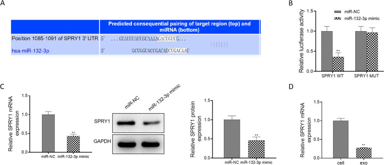
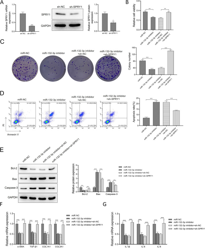
Purpose: Interstitial lung diseases (ILDs) have high morbidity and mortality and poor prognosis. The significance of microRNAs (miRNAs) was highlighted in ILDs development. Currently, we attempted to confirm the functions of lung cancer-derived exosomal miR-132-3p and reveal the underlying mechanism. Method: Characteristics of exosomes were verified by transmission electron microscope (TEM), nanoparticle tracking analysis, and Western blot assay. Exosome uptake for the normal human lung fibroblasts (NHLF) was assessed using a PKH67 staining assay. MTT and colony formation assays were applied to examine the proliferation abilities of NHLF. The interaction between miR-132-3p and sprouty1 (SPRY1) was confirmed by a luciferase reporter assay. Results: Lung cancer-derived exosomes promoted normal human lung fibroblast activation. Exosome inhibitor GW4869 reversed the effects of Exo on NHLF. Subsequently, miR-132-3p in lung cancer-derived exosomes activated the normal human lung fibroblast and promoted interstitial lung disease development ex vivo. Next, SPRY1 was verified to be the binding protein of miR-132-3p, and sh-SPRY1 abrogated the effects of the miR-132-3p inhibitor on NHLF. Conclusion: Exosomal miR-132-3p from A549 cells accelerated the development of interstitial lung disease through binding to SPRY1, which might serve as an important target for ILDs.
Overview
The claims and statistics of this paper haven’t been checked yet.
Run the check — free, no account needed. It reads the open-access text and checks the sources it cites.
Integrity signals
Registry
Cites
Statistics
Figures
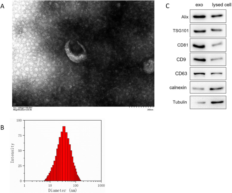
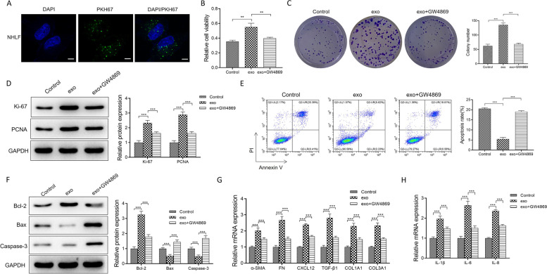
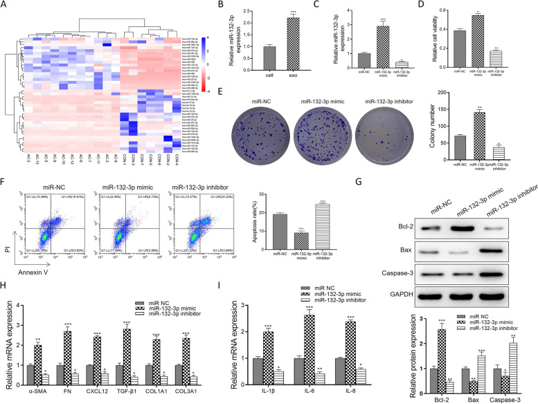
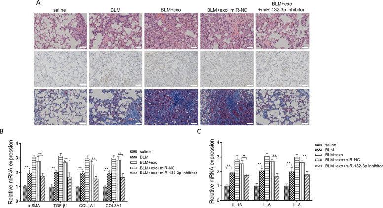
Verification
Claims the paper makes
The claims and statistics of this paper haven’t been checked yet.
Run the check — free, no account needed. It reads the open-access text and checks the sources it cites.
Claims other papers make about this one
Each is checked against this paper’s own text: does it say what the citing paper says it does?
The cited source says what the claim attributes to it.
In the citing paper
“Additionally, multiple of these core miR-132 targets (e.g. FOXO1, DPYSL3, SPRY1, BMI1, and CITED2 via P300) have previously been identified as miR-132 targets in other cell types, supporting the notion that they represent true miR-132 targets [2, 72–79].”
From microRNA-132 attenuates inflammation in induced pluripotent stem cell-derived microglia from Alzheimer’s disease patients (2026) · cites this paper as [77]
Self-check
Percentages, sums and reported differences re-computed from the paper’s own numbers.
The self-check needs the paper’s full text.
Citations
Cites
Communication by Extracellular Vesicles: Where We Are and Where We Need to Go
Tkach, M., & Théry, C. (2016) · Cell · doi:10.1016/j.cell.2016.01.043 · Cited by 3,444
MicroRNA-21 contributes to myocardial disease by stimulating MAP kinase signalling in fibroblasts
Thum, T., Gross, C., Fiedler, J., Fischer, T., Kissler, S., Bussen, M., Galuppo, P., Just, S., Rottbauer, W., Frantz, S., et al. (2008) · Nature · doi:10.1038/nature07511 · Cited by 2,349
Jin, X., Chen, Y., Chen, H., Fei, S., Chen, D., Cai, X., Liu, L., Lin, B., Su, H., Zhao, L., et al. (2017) · Clinical Cancer Research · doi:10.1158/1078-0432.ccr-17-0577 · Cited by 558
Inhalation of lung spheroid cell secretome and exosomes promotes lung repair in pulmonary fibrosis
Dinh, P.-U. C., Paudel, D., Brochu, H., Popowski, K. D., Gracieux, M. C., Cores, J., Huang, K., Hensley, M. T., Harrell, E., Vandergriff, A. C., et al. (2020) · Nature Communications · doi:10.1038/s41467-020-14344-7 · Cited by 435
Progression of fibrosing interstitial lung disease
Wong, A. W., Ryerson, C. J., & Guler, S. A. (2020) · Respiratory Research · doi:10.1186/s12931-020-1296-3 · Cited by 194
Guiot, J., Cambier, M., Boeckx, A., Henket, M. A., Nivelles, O., Gester, F., Louis, E. J., Malaise, M., Dequiedt, F., Louis, R. E., et al. (2020) · Thorax · doi:10.1136/thoraxjnl-2019-214077 · Cited by 154
Sepsis plasma-derived exosomal miR-1-3p induces endothelial cell dysfunction by targeting SERP1
Gao, M., Yu, T., Liu, D., Shi, Y., Yang, P., Zhang, J., Wang, J., Liu, Y., & Zhang, X. (2021) · Clinical Science · doi:10.1042/cs20200573 · Cited by 126
The Anti-fibrotic Effects and Mechanisms of MicroRNA-486-5p in Pulmonary Fibrosis
Ji, X., Wu, B., Fan, J., Han, R., Luo, C., Wang, T., Yang, J., Han, L., Zhu, B., Wei, D., et al. (2015) · Scientific Reports · doi:10.1038/srep14131 · Cited by 124
Self-citationp53 and miR-34a Feedback Promotes Lung Epithelial Injury and Pulmonary Fibrosis
Shetty, S. K., Tiwari, N., Marudamuthu, A. S., Puthusseri, B., Bhandary, Y. P., Fu, J., Levin, J. L., Idell, S., & Shetty, S. (2017) · American Journal Of Pathology · doi:10.1016/j.ajpath.2016.12.020 · Cited by 112
The CDR1as/miR-7/TGFBR2 Axis Modulates EMT in Silica-Induced Pulmonary Fibrosis
Yao, W., Li, Y., Han, L., Ji, X., Pan, H., Liu, Y., Yuan, J., Yan, W., & Ni, C. (2018) · Toxicological Sciences · doi:10.1093/toxsci/kfy221 · Cited by 112
Bijkerk, R., de Bruin, R. G., Solingen, C. V., van Gils, J. M., Duijs, J. M. G. J., van der Veer, E. P., Rabelink, T. J., Humphreys, B. D., & van Zonneveld, A. J. (2016) · Kidney International · doi:10.1016/j.kint.2016.01.029 · Cited by 110
Ning, Z., Luo, X., Wang, G., Yang, L., Pan, M., Yang, R., Ling, X., Huang, S., Ma, X., Jin, S., et al. (2016) · Antioxidants and Redox Signaling · doi:10.1089/ars.2016.6669 · Cited by 109
Zhang, S., Jia, X., Zhang, Q., Zhang, L., Yang, J., Hu, C., Shi, J., Jiang, X., Lu, J., & Shen, H. (2019) · Journal of Cellular and Molecular Medicine · doi:10.1111/jcmm.14858 · Cited by 99
Ren, J., Liu, Y., Yao, Y., Feng, L., Zhao, X., Li, Z., & Yang, L. (2020) · International Immunopharmacology · doi:10.1016/j.intimp.2020.107288 · Cited by 91
Application of small RNA sequencing to identify microRNAs in acute kidney injury and fibrosis
Pellegrini, K. L., Gerlach, C. V., Craciun, F. L., Ramachandran, K., Bijol, V., Kissick, H. T., & Vaidya, V. S. (2015) · Toxicology and Applied Pharmacology · doi:10.1016/j.taap.2015.12.002 · Cited by 66
Ding, J., Tang, Q., Luo, B., Zhang, L., Li, L., Han, L., Hao, M., Li, M., Yu, L., & Li, M. (2019) · European Journal of Pharmacology · doi:10.1016/j.ejphar.2019.172549 · Cited by 65
Ko, W., Hong, C.-Y., Hou, S., Lin, C., Ong, E.-T., Lee, C.-F., Tsai, C., & Lai, L. (2011) · Circulation Journal · doi:10.1253/circj.cj-10-0892 · Cited by 55
Cai, Y., Huang, G., Ma, L., Dong, L., Chen, S., Shen, X., Zhang, S., Xue, R., Sun, D., & Zhang, S. (2017) · Biochimica et Biophysica Acta (BBA) - Molecular Cell Research · doi:10.1016/j.bbamcr.2017.10.011 · Cited by 54
MicroRNA-132 attenuated cardiac fibrosis in myocardial infarction-induced heart failure rats
Wang, G., Wang, R., Ruan, Z., Liu, L., Li, Y., & Zhu, L. (2020) · Bioscience Reports · doi:10.1042/bsr20201696 · Cited by 42
Downregulation of exosomal let-7d and miR-16 in idiopathic pulmonary fibrosis
Lacedonia, D., Scioscia, G., Soccio, P., Conese, M., Catucci, L., Palladino, G. P., Simone, F., Quarato, C. M. I., Gioia, S. D., Rana, R. L., et al. (2021) · BMC Pulmonary Medicine · doi:10.1186/s12890-021-01550-2 · Cited by 42
Fathi, E., Mesbah‐Namin, S. A., Vietor, I., & Farahzadi, R. (2022) · PubMed · doi:10.22038/ijbms.2022.66737.14633 · Cited by 38
Farahzadi, R., Valipour, B., Anakök, Ö. F., Fathi, E., & Montazersaheb, S. (2023) · Transplant Immunology · doi:10.1016/j.trim.2023.101797 · Cited by 34
Eskildsen, T. V., Schneider, M., Sandberg, M. B., Skov, V., Brønnum, H., Thomassen, M., Kruse, T. A., Andersen, D. C., & Sheikh, S. P. (2014) · Journal of the Renin-Angiotensin-Aldosterone System · doi:10.1177/1470320314539367 · Cited by 33
miR-132 in atrial fibrillation directly targets connective tissue growth factor
Qiao, G., Xia, D., Cheng, Z., & Zhang, G. (2017) · Molecular Medicine Reports · doi:10.3892/mmr.2017.7045 · Cited by 32
Update in Interstitial Lung Disease 2019
Montesi, S. B., Fisher, J. H., Martínez, F. J., Selman, M., Pardo, A., & Johannson, K. A. (2020) · American Journal of Respiratory and Critical Care Medicine · doi:10.1164/rccm.202002-0360up · Cited by 32
Jiang, Z., Tao, J.-H., Zuo, T., Xm, L., Wang, G.-S., Fang, X., Xu, X.-L., & Xp, L. (2016) · Scandinavian Journal of Rheumatology · doi:10.3109/03009742.2016.1167950 · Cited by 27
Li, Y., Huang, J., Hu, C., Zhou, J., Xu, D., Hou, Y., Wu, C., Zhao, J., Li, M., Zeng, X., et al. (2021) · Arthritis Research & Therapy · doi:10.1186/s13075-020-02411-9 · Cited by 24
Song, Y., Cao, J., Jin, Z., Hu, W., Wu, R., Lu-hai, T., Yang, B., Wang, J., Xiao, Y., & Huang, C. (2018) · Journal of Cellular Biochemistry · doi:10.1002/jcb.28190 · Cited by 23
Ortíz-Quintero, B., Buendía-Roldán, I., Ramírez-Salazar, E. G., Balderas-Martínez, Y. I., Ramírez-Rodriguez, S. L., Martínez‐Espinosa, K., & Selman, M. (2020) · Cells · doi:10.3390/cells9061556 · Cited by 23
Shi, S., & Li, H. (2020) · American Journal of Physiology-Cell Physiology · doi:10.1152/ajpcell.00479.2019 · Cited by 19
miR-1 is a novel biomarker for polymyositis/dermatomyositis-associated interstitial lung disease
Sugiyama, Y., Yoshimi, R., Takeno, M., Kunishita, Y., Kishimoto, D., Kamiyama, R., Kirino, Y., Ohno, S., & Nakajima, H. (2019) · Modern Rheumatology · doi:10.1080/14397595.2019.1661584 · Cited by 12
Role of Sprouty1 (Spry1) in the pathogenesis of atrial fibrosis
Qiao, G., Xia, D., Cheng, Z., et al. (2017) · Pathology - Research and Practice · doi:10.1016/j.prp.2017.04.021 · Cited by 11
Diagnostic and Therapeutic Applications of Extracellular Vesicles in Interstitial Lung Diseases
Ibrahim, A., Ibrahim, A., & Parimon, T. (2021) · Diagnostics · doi:10.3390/diagnostics11010087 · Cited by 10
Sun, X., Kang, Y. J., Xue, S., Zou, J., Xu, J., Tang, D., & Qin, H. (2020) · The Korean Journal of Internal Medicine · doi:10.3904/kjim.2019.098 · Cited by 9
Cited by
Penning, A., Snoeck, S., Ormaechea, O. R., Ayyildiz, D., Polzer, O., Buitrago-Arango, M., Capobianco, R., de Winter, F., Balusu, S., Verhaagen, J., et al. (2026) · Acta Neuropathologica Communications · doi:10.1186/s40478-026-02228-8 · Cited by 6
Metadata
- Authors
- Fang, S., Wang, T., Weng, L., Han, X., Zheng, R., & Zhang, H.
- Venue
- World Journal of Surgical Oncology
- Published
- 2023-07-15
- Volume
- 21
- Issue
- 1
- ISSN
- 1477-7819
- PMC
- PMC10349414
- Cited by
- 13
- References
- 34
- Language
- en
- Licence
- creativecommons.org/licenses/by/4.0 (tdm)
- creativecommons.org/licenses/by/4.0 (vor)
About these checks
Registry, retraction, Cites and statistics results are computed, not judged, with one exception: a printed reference that no index lists is read once by a model before it counts as a citation error. Claims are extracted by a model and kept only when their quote appears verbatim in the text; each cited source is then checked, with a model’s help, for whether it says what the claim relies on. Those verdicts can be wrong — read the claim’s quote and the cited source before relying on one.
Statistics and arithmetic are recomputed, not judged.
Confidence bands are shown only for fields calibrated on our annotated test set. None is calibrated yet, so no band is shown.
What counts as a claim ›How we measure confidence ›How we check this ›
Are you an author, or do you see a problem with this page? Request a review
Data sources
Bibliographic data: Semantic Scholar, OpenAlex, Crossref.
Contains information from the Semantic Scholar Academic Graph Datasets, made available under the ODC Attribution License (ODC-BY 1.0).
Semantic Scholar’s data is described in: Kinney, R., et al. (2023). The Semantic Scholar Open Data Platform. arXiv:2301.10140.
