

The DOI matches its Crossref record; 31 of the 31 references it cites were found in a registry or an index.
Mesenchymal stem cells and their secreted molecules predominantly ameliorate fulminant hepatic failure and chronic liver fibrosis in mice respectively
Open accessAbstract
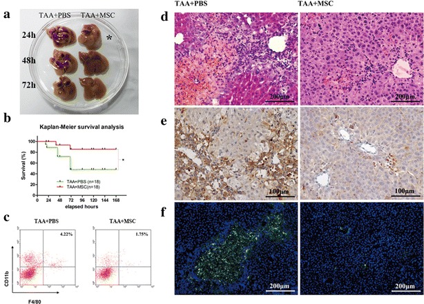
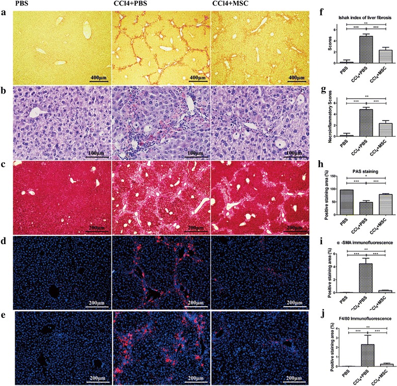
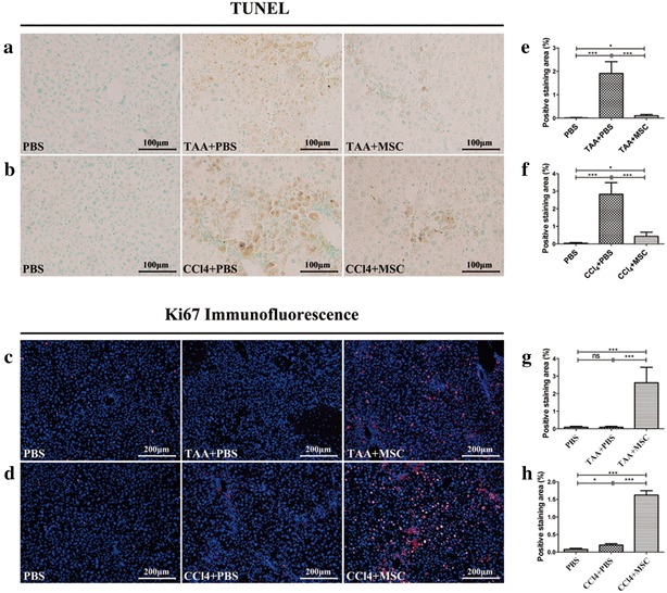
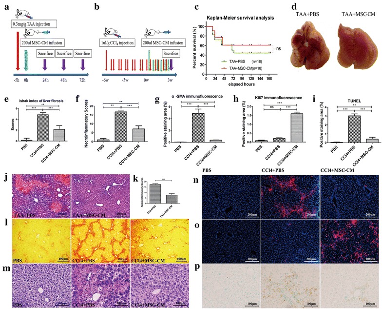
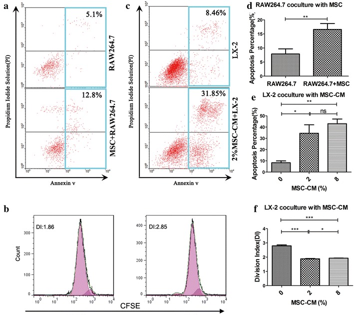
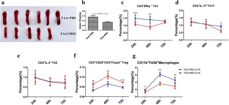
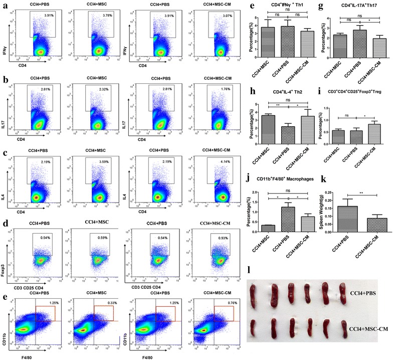
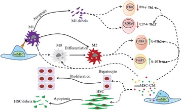
BACKGROUND: Orthotopic liver transplantation is the only effective treatment for liver failure but limited with shortage of available donor organs. Recent studies show promising results of mesenchymal stem cells (MSCs)-based therapies. METHODS: We systematically investigate the therapeutic effects of MSCs or MSC-conditioned medium (MSC-CM) in ameliorating fulminant hepatic failure (FHF) and chronic liver fibrosis in mice. In addition, extensive flow cytometry analysis of spleens from vehicle and MSC- and MSC-CM-treated mice was applied to reveal the alteration of inflammatory state. RESULTS: In FHF model, MSCs treatment reduced remarkably the death incidents; the analysis of gross histopathology showed that control livers were soft and shrunken with extensive extravasated blood, which was gradually reduced at later time points, while MSC-treated livers showed gross pathological changes, even 24 h after MSC infusion, and hematoxylin and eosin staining revealed dramatical hepatocellular death with cytoplasmic vacuolization suppressed by MSCs treatment; flow cytometry analysis of total lymphocytes showed that macrophages (F4/80) infiltrated into control livers more than MSC-treated livers; by contrast, MSC-CM partially ameliorates FHF. In chronic liver injury model, MSC and MSC-CM both suppressed fibrogenesis and necroinflammatory, and the later was better; activation of hepatic stellate cells (α-SMA) was inhibited; glycogen synthesis and storage (indicated by periodic acid-Schiff -staining) was improved; liver regeneration (Ki67) was promoted while liver apoptosis (TUNEL) was reduced. In the in vitro, MSCs promote macrophage line RAW264.7 apoptosis and MSC-CM promotes apoptosis and inhibits proliferation of HSC line LX-2. We also found that MSCs and MSC-CM could improve spleen; MSC-CM increased levels of Th2 and Treg cells, and reduced levels of Th17 cells, whereas levels of Th1 cells were unchanged; comparatively, MSC treatment did not affect Th17 and Treg cells and only slightly alters inflammatory state; MSC and MSC-CM treatment both substantially down-regulated macrophages in the spleens. CONCLUSION: Both MSCs and MSC-CM exert therapeutic effects by acting on various key cells during the pathogenesis of FHF and chronic fibrosis, stimulating hepatocyte proliferation and suppressing apoptosis, down-regulating infiltrating macrophages, converting CD4(+) T lymphocyte system into an anti-inflammatory state, and facilitating hepatic stellate cell death.
Overview
The claims and statistics of this paper haven’t been checked yet.
Run the check — free, no account needed. It reads the open-access text and checks the sources it cites.
Integrity signals
Registry
Cites
Statistics
Figures
Verification
Claims the paper makes
The claims and statistics of this paper haven’t been checked yet.
Run the check — free, no account needed. It reads the open-access text and checks the sources it cites.
Claims other papers make about this one
Each is checked against this paper’s own text: does it say what the citing paper says it does?
The source backs part of the claim, not all of it — or backs it for a different population, condition or comparison.
In the citing paper
“Preclinical studies [22–25] and clinical trials [26–30] demonstrated that MSC transplantation significantly improves liver function and is safe and effective in the treatment of liver failure.”
From From signaling pathways to clinical trials: mesenchymal stem cells as multimodal regenerative architects in liver cirrhosis therapy (2025) · cites this paper as [23]
Experimental — accuracy not yet measured
Self-check
Percentages, sums and reported differences re-computed from the paper’s own numbers.
The self-check needs the paper’s full text.
Citations
Cites
The chemokine system in diverse forms of macrophage activation and polarization
MANTOVANI, A., SICA, A., SOZZANI, S., ALLAVENA, P., VECCHI, A., & LOCATI, M. (2004) · Trends in Immunology · doi:10.1016/j.it.2004.09.015 · Cited by 6,534
Histological grading and staging of chronic hepatitis.
Ishak, K., Baptista, A., Bianchi, L., Callea, F., Groote, J. D., Gudat, F., Denk, H., Desmet, V., Korb, G., Macsween, R. N., et al. (1995) · Journal of Hepatology · doi:10.1016/0168-8278(95)80226-6 · Cited by 5,290
Mesenchymal stem cells in health and disease
Uccelli, A., Moretta, L., & Pistoia, V. (2008) · Nature reviews. Immunology · doi:10.1038/nri2395 · Cited by 3,838
Bone Marrow as a Potential Source of Hepatic Oval Cells
Petersen, B. E., Bowen, W. C., Patrene, K. D., Mars, W. M., Sullivan, A. K., Murase, N., Boggs, S. S., Greenberger, J. S., & Goff, J. P. (1999) · Science · doi:10.1126/science.284.5417.1168 · Cited by 2,377
Mechanisms involved in the therapeutic properties of mesenchymal stem cells
da Silva Meirelles, L., Fontes, A. M., Covas, D. T., & Caplan, A. I. (2009) · Cytokine & Growth Factor Reviews · doi:10.1016/j.cytogfr.2009.10.002 · Cited by 1,436
Interactions Between Human Mesenchymal Stem Cells and Natural Killer Cells
Sotiropoulou, P. A., Perez, S. A., Gritzapis, A. D., Baxevanis, C. N., & Papamichail, M. (2006) · Stem Cells · doi:10.1634/stemcells.2004-0359 · Cited by 956
Gupta, N., Su, X., Popov, B. V., Lee, J. W., Serikov, V. B., & Matthay, M. A. (2007) · The Journal of Immunology · doi:10.4049/jimmunol.179.3.1855 · Cited by 933
Mesenchymal Stem/Stromal Cells (MSCs): Role as Guardians of Inflammation
Prockop, D. J., & Youn Oh, J. (2012) · Molecular Therapy · doi:10.1038/mt.2011.211 · Cited by 816 · cited 1×
“For example, MSCs mediate a switch from pro-inflammatory M1-type macrophages to anti-inflammatory M2-type macrophages [17, 18].”
Myofibroblasts revert to an inactive phenotype during regression of liver fibrosis
Kisseleva, T., Cong, M., Paik, Y., Scholten, D., Jiang, C., Benner, C., Iwaisako, K., Moore-Morris, T., Scott, B., Tsukamoto, H., et al. (2012) · Proceedings of the National Academy of Sciences · doi:10.1073/pnas.1201840109 · Cited by 805
Mesenchymal-Stem-Cell-Induced Immunoregulation Involves FAS-Ligand-/FAS-Mediated T Cell Apoptosis
Akiyama, K., Chen, C., Wang, D., Xu, X., Qu, C., Yamaza, T., Cai, T., Chen, W., Sun, L., & Shi, S. (2012) · Cell stem cell · doi:10.1016/j.stem.2012.03.007 · Cited by 707
Self-citation
Cited by
Sorted by citations
Items 101–184 of 184
Huang, Y.-L., Gregorio, C. D., Silva, V., Elorza, Á. A., Léniz, P., Aliaga-Tobar, V., Maracaja‐Coutinho, V., Budini, M., Ezquer, F. E., & Ezquer, M. (2023) · Cells · doi:10.3390/cells12040636 · Cited by 15
Ravan, A. P., Goudarzi, F., Rafieemehr, H., Bahmani, M., Rad, F., Jafari, M., & Mahmoodi, M. (2019) · Toxin Reviews · doi:10.1080/15569543.2019.1590849 · Cited by 13
Wang, Q., Li, Y., Yuan, H., Peng, L., Dai, Z., Sun, Y., Li, W., Li, W., Li, J., & Zhu, C. (2024) · Tissue and Cell · doi:10.1016/j.tice.2024.102326 · Cited by 12
Gasanov, V. A. O., Kashirskikh, D. A., Khotina, V. A., Kuzmina, D. M., Nikitochkina, S. Y., Mukhina, I. V., Vorotelyak, E. A., & Vasiliev, A. V. (2025) · Cells · doi:10.3390/cells14050341 · Cited by 12
Mo, Y., Kang, S., Bang, J., Kim, Y., Jeong, J., Jeong, E. M., Kim, H. Y., Cho, S., & Kang, H. (2022) · Molecules and Cells · doi:10.14348/molcells.2022.0038 · Cited by 12
Stem Cell-based Therapy Strategy for Hepatic Fibrosis by Targeting Intrahepatic Cells
Deng, Y., Xia, B., Chen, Z., Wang, F., Lv, Y., & Chen, G. (2021) · Stem Cell Reviews and Reports · doi:10.1007/s12015-021-10286-9 · Cited by 12
microRNAs ‐based diagnostic and therapeutic applications in liver fibrosis
Zhao, X., Xue, X., Cui, Z., Amevor, F. K., Wan, Y., Fu, K., Wang, C., Peng, C. Y., & Li, Y. (2022) · Wiley Interdisciplinary Reviews - RNA · doi:10.1002/wrna.1773 · Cited by 12
Shang, L., Wang, M., Liu, Y., Zhu, X., & Wang, S. (2023) · Stem Cell Reviews and Reports · doi:10.1007/s12015-023-10566-6 · Cited by 11
Han, J. H., Kim, O., Lee, S. C., Kim, K., Park, J. H., Lee, J. I., Lee, K. H., Hong, H., Seo, H., Choi, H. J., et al. (2019) · International Journal of Molecular Sciences · doi:10.3390/ijms20246302 · Cited by 11
Sitbon, A., Delmotte, P.-R., Goumard, C., Turco, C., Gautheron, J., Conti, F., Aoudjehane, L., Scatton, O., & Monsel, A. (2023) · Minerva Anestesiologica · doi:10.23736/s0375-9393.23.17265-8 · Cited by 11
Arfianti, A., Ulfah, U., Hutabarat, L. S., Agnes Ivana, G., Budiarti, A. D., Sahara, N. S., & Saputra, N. P. K. (2023) · Biomedicine · doi:10.37796/2211-8039.1416 · Cited by 10
Kao, Y. H., Chang, C.-Y., Lin, Y., Chen, P., Lee, P., Chang, H., Chang, W.-Y., Chang, Y., Wun, S.-F., & Sun, C. (2023) · Current Stem Cell Research & Therapy · doi:10.2174/1574888x19666230918102826 · Cited by 11
Götze, T., Krueger, M., Meutsch, J. K., Dörfel, M. J., Born, S., Sowa, J., & Canbay, A. E. (2019) · Clinical and Translational Gastroenterology · doi:10.14309/ctg.0000000000000095 · Cited by 10
Dilogo, I. H., Rahmatika, D., Pawitan, J. A., Liem, I. K., Kurniawati, T., Kispa, T. D., & Mujadid, F. (2020) · European Journal of Orthopaedic Surgery & Traumatology · doi:10.1007/s00590-020-02765-5 · Cited by 10
Bozorgi, V., Babaahmadi, M., Salehi, M., Vafaeimanesh, J., & Hajizadeh-Saffar, E. (2025) · Stem Cell Research & Therapy · doi:10.1186/s13287-025-04535-8 · Cited by 11
Pretreatment of UC-MSCs with IFN-α2 improves treatment of liver fibrosis by recruiting neutrophils
Xie, Y., Yao, J., Yan, M., Lin, Y., Wei, J., Wang, H., Mao, Y., Liu, P., & Li, X. (2023) · Journal of Translational Medicine · doi:10.1186/s12967-023-04732-0 · Cited by 9
Self-citationCzekaj, P., Król, M., Limanówka, Ł., Skubis-Sikora, A., Kolanko, E., Bogunia, E., Hermyt, M., Michalik, M., Sikora, B., Prusek, A., et al. (2022) · Frontiers in Bioscience-Landmark · doi:10.31083/j.fbl2708237 · Cited by 12
Evaluating effects of L-carnitine on human bone-marrow-derived mesenchymal stem cells
Fujisawa, K., Takami, T., Fukui, Y., Quintanilha, L. F., Matsumoto, T., Yamamoto, N., & Sakaida, I. (2017) · Cell and Tissue Research · doi:10.1007/s00441-017-2569-0 · Cited by 9
Hurdles to overcome for mesenchymal stem cell translation from bench to bedside
Mayeen, N. F., Salma, U., Kasim, N. H. A., Mahmoud, O., & Haque, N. (2025) · World Journal of Stem Cells · doi:10.4252/wjsc.v17.i12.114349 · Cited by 8
Xiang, W., Wang, X., Yu, X., Xie, Y., Zhang, L., Lu, N., & Jiang, W. (2023) · Stem Cells International · doi:10.1155/2023/6890299 · Cited by 8
Yao, Y., Zhang, L., Cheng, F., Jiang, Q., Ye, Y., Ren, Y., He, Y., Su, D., Cheng, L., Shi, G., et al. (2023) · Stem Cell Research & Therapy · doi:10.1186/s13287-023-03416-2 · Cited by 8
Kweon, M., Kim, J. Y., Jun, J. H., & Kim, G. J. (2020) · International Journal of Molecular Sciences · doi:10.3390/ijms22010239 · Cited by 9
Noorwali, A. A., Faidah, M., Ahmed, N., & Bima, A. I. H. (2019) · Bioinformation · doi:10.6026/97320630015001 · Cited by 8
Redko, O., Dovgalyuk, A., Довбуш, А. В., Небесна, З. М., Yakubyshyna, L. V., & Krynytska, І. (2021) · Cell and Organ Transplantology · doi:10.22494/cot.v9i2.130 · Cited by 8
Molaei, S., Amiri, F., Salimi, R., Ferdowsi, S., & Bahadori, M. (2022) · Molecular Biology Reports · doi:10.1007/s11033-022-07785-4 · Cited by 8
Exploring the roles of non-coding RNAs in liver regeneration
Li, P., Ma, X., Huang, D., & Gu, X. (2024) · Non-coding RNA Research · doi:10.1016/j.ncrna.2024.04.003 · Cited by 8
Ghufran, H., Azam, M., Mehmood, A., Umair, M., Baig, M. T., Tasneem, S., Butt, H., & Riazuddin, S. A. (2024) · Journal of Clinical and Experimental Hepatology · doi:10.1016/j.jceh.2024.101364 · Cited by 7
Ali, S. A., & Datusalia, A. K. (2024) · Toxicology and Applied Pharmacology · doi:10.1016/j.taap.2024.117129 · Cited by 6
Vištějnová, L., Liška, V., Kumar, A., Křečková, J., Vyčítal, O., Brůha, J., Beneš, J., Kolinko, Y., Blassová, T., Tonar, Z., et al. (2021) · International Journal of Molecular Sciences · doi:10.3390/ijms22094304 · Cited by 6
Abdelbaset‐Ismail, A., Tharwat, A., Ahmed, A. E., Khamis, T., El-Rahim, I. H. A., Alhag, S. K., & Dowidar, M. (2022) · Biomedicine & Pharmacotherapy · doi:10.1016/j.biopha.2022.113805 · Cited by 6
Fathi-Kazerooni, M., & Tavoosidana, G. (2019) · Europe PMC (PubMed Central) · Cited by 6
Morimoto, K., Nakashima, A., Ishiuchi, N., Miyasako, K., Tanaka, Y., Sasaki, K., Matsuda, G., Maeda, S., Miyaki, S., & Masaki, T. (2025) · Stem Cells · doi:10.1093/stmcls/sxaf022 · Cited by 5
Stem Cell Delivery Routes: From Preclinical Models to Clinical Applications
Fagoonee, S. (2021) · BENTHAM SCIENCE PUBLISHERS eBooks · doi:10.2174/97898150401041210101 · Cited by 5
Bahmani, M., Ziamajidi, N., Hashemnia, M., & Abbasalipourkabir, R. (2020) · Toxin Reviews · doi:10.1080/15569543.2020.1813779 · Cited by 5
Luo, X., Li, H., Sun, X., Zuo, Q., Li, B., Zhu, Y., Wei, W., & Gu, X. Q. (2021) · BioMed Research International · doi:10.1155/2021/5529276 · Cited by 5
RetractedAttia, N., Khalifa, Y. H., Rostom, D. M., & Mashal, M. (2021) · Medical Research Archives · doi:10.18103/mra.v9i2.2318 · Cited by 5
Gilglioni, E. H., Bansal, M., St‐Pierre‐Wijckmans, W., Talamantes, S., Kasarinaite, A., Hay, D. C., & Gurzov, E. N. (2025) · Obesity Reviews · doi:10.1111/obr.13899 · Cited by 4
Mesenchymal stromal cell-dependent immunoregulation in chemically-induced acute liver failure
Zhou, J., Lü, X., Yan, C., Sheng, X., & Cao, H. (2021) · World Journal of Stem Cells · doi:10.4252/wjsc.v13.i3.208 · Cited by 4
Khosravi-Farsani, S., Zaminy, A., Kazemi, S., & Chaleshtori, M. H. (2022) · PubMed Central · doi:10.22038/ijbms.2022.62642.13860 · Cited by 4
Application of Modified Mesenchymal Stem Cells Transplantation in the Treatment of Liver Injury
Liu, L., & Yang, F. (2021) · Physiological Research · doi:10.33549/physiolres.934623 · Cited by 4
Stem Cell Therapy and Its Products Such as Exosomes: Modern Regenerative Medicine Approach
Dehghani, L., Kheirkhah, A. H., Jalili, A., Partan, A. S., Nikukar, H., & Sadeghian‐Nodoushan, F. (2023) · Biochemistry · doi:10.5772/intechopen.111574 · Cited by 3
Xu, S.-J., Ye, L., Wang, W., Chen, Y., Dong, J. W., Mao, X., & Li, S.-W. (2021) · Stem Cells International · doi:10.1155/2021/5513309 · Cited by 3
Yan, J., Zhang, L., Ye, J., Yan, S., Han, X., & Han, Z. (2020) · World Academy of Sciences Journal · doi:10.3892/wasj.2020.81 · Cited by 2
Wei, N., Chen, X., Liu, D., Bu, X., Wang, G., Sun, X., & Zhang, J. (2024) · Journal of Materials Chemistry B · doi:10.1039/d4tb00914b · Cited by 2
Lin, S., Luo, Y., Mao, X., He, W., Xu, C., & Zeng, M. (2024) · The American Journal of the Medical Sciences · doi:10.1016/j.amjms.2024.05.015 · Cited by 1
Dexamethasone-boosted mesenchymal stem cell secretome: insight into hepatic protection
Adly, E. M., Diab, T. A., & Hessien, M. (2025) · BMC Biotechnology · doi:10.1186/s12896-025-00980-8 · Cited by 1
Payushina, O. V., Tsomartova, D. A., Васильевна, Ч. Е., Ivanova, M. Y., & Kuznetsov, S. L. (2021) · Biology Bulletin Reviews · doi:10.1134/s2079086421010059 · Cited by 1
Yu, C., Chen, Y., Xu, X., Dou, X., Liu, S., Wang, H., Wei, X., Xu, Z., Wang, B., Cui, X., et al. (2021) · Research Square · doi:10.21203/rs.3.rs-699167/v1 · Cited by 1
Self-citationChen, Y. M., Yu, C., Xu, X., Dou, X., Liu, S., Wang, H., Wei, X., Xu, Z., Wang, B., Cui, X., et al. (2024) · doi:10.22541/au.170666991.17444752/v1
Self-citationAfsharzadeh, D. (2020) · University of Groningen research database (University of Groningen / Centre for Information Technology) · doi:10.33612/diss.121499227
Shakerian, E., Yaghooti, H., Kheirollah, A., & Mohammadtaghvaei, N. (2020) · Jentashapir Journal of Cellular and Molecular Biology · doi:10.5812/jjcmb.110136
Stem cell therapy in children with acute liver failure: The dream could come true
Issa, R. R., Sira, A. M., & Sira, M. M. (2021) · Annals of Clinical Gastroenterology and Hepatology · doi:10.29328/journal.acgh.1001024
Amiri, F., Molaei, S., Salimi, R., Ferdowsi, S., & Bahadori, M. (2022) · Research Square · doi:10.21203/rs.3.rs-1284048/v1
Meng, W., Liu, Y., Zhu, Z., Liu, S., Shen, Y., & Liu, S. (2022) · Journal of Biomaterials and Tissue Engineering · doi:10.1166/jbt.2022.3051
Donmez, H., & Akan, H. S. (2022) · Hacettepe Journal of Biology and Chemistry · doi:10.15671/hjbc.1001427
Mahmoudi, A. A., Meidany, P., Almahmeed, W. A., Jamialahmadi, T., & Sahebkar, A. (2024) · Current Stem Cell Reports · doi:10.1007/s40778-024-00241-y · Cited by 1
Bone marrow mesenchymal stem cell transplantation for treatment of liver cirrhosis: Recent advances
Ruan, M.-F., Yin, Y.-H., Shao, X., & Qi, X. (2025) · World Chinese Journal of Digestology · doi:10.11569/wcjd.v33.i2.106
Dexamethasone marginally surpasses MSC-secretome in resolving acute liver failure in mice
Diab, T. A., Adly, E. M., & Hessien, M. (2024) · The Animal Biology · doi:10.15407/animbiol26.04.012
Cell Therapy for Liver Cirrhosis
Liêm, N. T., & Dao, L. T. M. (2025) · Springer eBooks · doi:10.1007/978-981-96-1261-1_13
Gugjoo, M. B. (2025) · doi:10.1002/9781394253289.ch10
Septiana, W. L., Azzahra, R., Ahmad, A., Nurvianto, D., Utomo, M. F. B., Junaidi, H., & Astuti, S. W. (2025) · Journal of Cellular Biotechnology · doi:10.1177/23523689251359483
Hendrawan, S., Lheman, J., Marcelina, O., Tantoso, L., Baer, H. U., & Gunawan, S. (2025) · World Academy of Sciences Journal · doi:10.3892/wasj.2025.404
Hendrawan, S., Lheman, J., Dewi, F. N., Nuraeni, N., Permanawati, & Baer, H. U. (2025) · Regenerative Medicine · doi:10.1080/17460751.2025.2574178
Zhang, L., Wei, Y., Chi, Y., Liu, D., Yang, S., Han, Z., & Li, Z. (2021) · Figshare · doi:10.6084/m9.figshare.13532107
Xie, Y., Yao, J., Yan, M., Lin, Y., Wei, J., Wang, H., Mao, Y., Liu, P., & Li, X. (2023) · Figshare · doi:10.6084/m9.figshare.24587682
Self-citationZheng, W., Bian, S., Qiu, S., Bishop, C. E., Wan, M., Xu, N., Sun, X., Sequeira, R. C., Atala, A., Gu, Z., et al. (2023) · Figshare · doi:10.6084/m9.figshare.24257464
Ichinohe, N., Masayuki, I., Tanimizu, N., Toru, M., Yoshioka, Y., Ochiya, T., Suzuki, H., & Mitaka, T. (2021) · Figshare · doi:10.6084/m9.figshare.14702251
Yao, Y., Zhang, L., Cheng, F., Jiang, Q., Ye, Y., Ren, Y., He, Y., Su, D., Cheng, L., Shi, G., et al. (2024) · Figshare · doi:10.6084/m9.figshare.26607010
Zhang, L., Wei, Y., Chi, Y., Liu, D., Yang, S., Han, Z., & Li, Z. (2021) · Figshare · doi:10.6084/m9.figshare.13532104
Zhang, L., Wei, Y., Chi, Y., Liu, D., Yang, S., Han, Z., & Li, Z. (2021) · Figshare · doi:10.6084/m9.figshare.13532116
Liu, Q.-W., Ying, Y.-M., Zhou, J.-X., Zhang, W.-J., Liu, Z., Jia, B.-B., Gu, H.-C., Zhao, C.-Y., Guan, X.-H., Deng, K., et al. (2022) · Figshare · doi:10.6084/m9.figshare.20003259
Zheng, W., Bian, S., Qiu, S., Bishop, C. E., Wan, M., Xu, N., Sun, X., Sequeira, R. C., Atala, A., Gu, Z., et al. (2024) · Figshare · doi:10.6084/m9.figshare.24257624
Zhang, L., Wei, Y., Chi, Y., Liu, D., Yang, S., Han, Z., & Li, Z. (2021) · Figshare · doi:10.6084/m9.figshare.13532113
Yao, Y., Zhang, L., Cheng, F., Jiang, Q., Ye, Y., Ren, Y., He, Y., Su, D., Cheng, L., Shi, G., et al. (2024) · Figshare · doi:10.6084/m9.figshare.26607013
Zhang, L., Wei, Y., Chi, Y., Liu, D., Yang, S., Han, Z., & Li, Z. (2021) · Figshare · doi:10.6084/m9.figshare.13532119
Bi, Y., Guo, X., Zhang, M., Zhu, K., Shi, C., Fan, B., Wu, Y., Yang, Z., & Ji, G. (2021) · Figshare · doi:10.6084/m9.figshare.17162925
Zhang, L., Wei, Y., Chi, Y., Liu, D., Yang, S., Han, Z., & Li, Z. (2021) · Figshare · doi:10.6084/m9.figshare.13532110
Yao, Y., Zhang, L., Cheng, F., Jiang, Q., Ye, Y., Ren, Y., He, Y., Su, D., Cheng, L., Shi, G., et al. (2024) · Figshare · doi:10.6084/m9.figshare.26607016
Wang, Z., Li, M., Yan, X., Liu, Y., Yang, P., Liu, W., & Guan, W. (2025) · International Journal of Molecular Sciences · doi:10.3390/ijms262411798
Mise au point et développement de modèles hépatiques complexes par bio-impression 3D
Cuvellier, M. (2020) · theses.fr (ABES) · doi:10.70675/b6c577cdzcaffz460az8214z9681de8f3941
Mesenchymal stem cells and extracellular vesicles in liver regeneration
Karnawat, K., Puthiyattil, N., Parthasarathy, R., Sekar, J. A., Elangbam, P., Roy, N., Gupta, K., Balachander, G. M., & Pai, R. R. (2026) · Elsevier eBooks · doi:10.1016/b978-0-443-45056-3.00017-0
Xiong, W., 杨忠霞, Qian, Z., 彭雪彬, Ying, L., & Xiaorong, M. (2026) · Liver Research · doi:10.1016/j.livres.2026.08.003
Exosomes Derived From Mesenchymal Stem Cells: The Next Frontiers in Regenerative Therapies
Rasouli, A., Yousefi, M. H., Kavianpour, M., Farsad, S., Afkhami, H., & Sheykhhasan, M. (2026) · Current Treatment Options in Cardiovascular Medicine · doi:10.1007/s11936-026-01160-3
Shahin, R. K., Elazazy, O., Wahdan, S. A., & AbdelHamid, S. G. (2026) · Molecular Medicine · doi:10.1186/s10020-026-01655-6
Metadata
- Authors
- Huang, B., Cheng, X., Wang, H., Huang, W., la Ga hu, Z., Wang, D., Zhang, K., Zhang, H., Xue, Z., Da, Y., Zhang, N., Hu, Y., Yao, Z., Qiao, L., Gao, F., & Zhang, R.
- Affiliations
- Tianjin Medical University (CN)
- Tianjin Hospital (CN)
- The University of Sydney (AU)
- Chinese Academy of Sciences (CN)
- Corresponding author
- Gao, Fei
- Zhang, Rongxin
- ORCID
- Venue
- Journal of Translational Medicine
- Published
- 2016-02-09
- Volume
- 14
- Issue
- 1
- Pages
- 45-45
- ISSN
- 1479-5876 (ISSN-L)
- PMC
- PMC4746907
- OpenAlex
- W2254654454
- Cited by
- 183
- References
- 31
- Citations by year
- 2026: 3 · 2025: 14 · 2024: 22 · 2023: 21 · 2022: 20 · 2021: 35 · 2020: 21 · 2019: 22 · 2018: 9 · 2017: 10 · 2016: 6
- FWCI
- 11.37
- Type
- journal article
- Language
- en
- Open access
- gold
- doi.org
- Licence
- cc-by
- Locations
- Journal of Translational Medicine (publishedVersion, cc-by)
- PubMed Central (publishedVersion, cc-by)
- PubMed (publishedVersion)
- Funders
- Ministry of Science and Technology of China (2012CB932503)
- Ministry of Science and Technology of the People's Republic of China (CN) (2011CB933100)
- National Natural Science Foundation of China (CN) (91029705, 81172864, 81272317)
- Scientific Innovation Project of the Chinese Academy of Science (XDA 01040106)
- Ministry of Education (Tianjin) (TD12-5025)
- Natural Science Foundation of Tianjin City (CN) (14JCTPJC00487)
- Topics
- Life Sciences › Biochemistry, Genetics and Molecular Biology › Cell Biology › Mesenchymal stem cell research
- Health Sciences › Medicine › Hepatology › Liver physiology and pathology
- Health Sciences › Medicine › Hepatology › Liver Disease and Transplantation
- Keywords
- mesenchymal stem cells, MSC-conditioned medium, acute liver failure, chronic liver fibrosis, hepatic stellate cells, macrophage infiltration, Th17/Treg balance, hepatocyte apoptosis, liver regeneration, hepatocyte proliferation, liver fibrosis, MSC-based therapy
- MeSH terms
- RAW 264.7 Cells, Animals, Carbon Tetrachloride, Chronic Disease, Female, Inflammation, Liver, Liver Cirrhosis, Macrophages, Mice, Inbred C57BL, Mice, Inbred ICR, Regeneration, Down-Regulation, Survival Analysis, Culture Media, Conditioned, Liver Failure, Acute, Apoptosis, Mesenchymal Stem Cell Transplantation, Cell Proliferation, Mice, Hepatic Stellate Cells, Mesenchymal Stem Cells
About these checks
Registry, retraction, Cites and statistics results are computed, not judged, with one exception: a printed reference that no index lists is read once by a model before it counts as a citation error. Claims are extracted by a model and kept only when their quote appears verbatim in the text; each cited source is then checked, with a model’s help, for whether it says what the claim relies on. Those verdicts can be wrong — read the claim’s quote and the cited source before relying on one.
Statistics and arithmetic are recomputed, not judged.
Confidence bands are shown only for fields calibrated on our annotated test set. None is calibrated yet, so no band is shown.
What counts as a claim ›How we measure confidence ›How we check this ›
Are you an author, or do you see a problem with this page? Request a review
