

The DOI matches its Crossref record; 48 of the 49 references it cites were found in a registry or an index.
Deep learning-based predictive biomarker of pathological complete response to neoadjuvant chemotherapy from histological images in breast cancer
Open accessAbstract
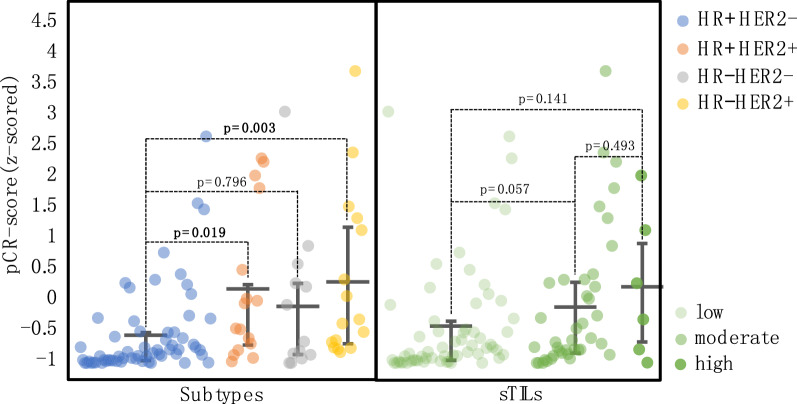
Background: Pathological complete response (pCR) is considered a surrogate endpoint for favorable survival in breast cancer patients treated with neoadjuvant chemotherapy (NAC). Predictive biomarkers of treatment response are crucial for guiding treatment decisions. With the hypothesis that histological information on tumor biopsy images could predict NAC response in breast cancer, we proposed a novel deep learning (DL)-based biomarker that predicts pCR from images of hematoxylin and eosin (H&E)-stained tissue and evaluated its predictive performance. Methods: In total, 540 breast cancer patients receiving standard NAC were enrolled. Based on H&E-stained images, DL methods were employed to automatically identify tumor epithelium and predict pCR by scoring the identified tumor epithelium to produce a histopathological biomarker, the pCR-score. The predictive performance of the pCR-score was assessed and compared with that of conventional biomarkers including stromal tumor-infiltrating lymphocytes (sTILs) and subtype. Results: The pCR-score derived from H&E staining achieved an area under the curve (AUC) of 0.847 in predicting pCR directly, and achieved accuracy, F1 score, and AUC of 0.853, 0.503, and 0.822 processed by the logistic regression method, respectively, higher than either sTILs or subtype; a prediction model of pCR constructed by integrating sTILs, subtype and pCR-score yielded a mean AUC of 0.890, outperforming the baseline sTIL-subtype model by 0.051 (0.839, P = 0.001). Conclusion: The DL-based pCR-score from histological images is predictive of pCR better than sTILs and subtype, and holds the great potentials for a more accurate stratification of patients for NAC.
Overview
The claims and statistics of this paper haven’t been checked yet.
Run the check — free, no account needed. It reads the open-access text and checks the sources it cites.
Integrity signals
Registry
Cites
Statistics
Figures
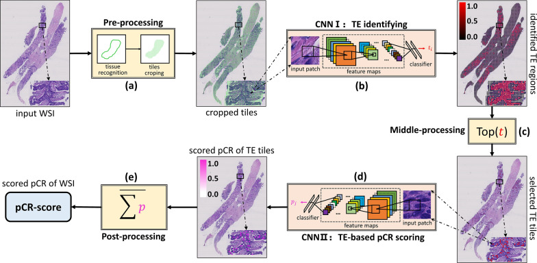
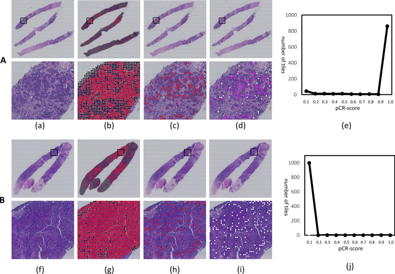
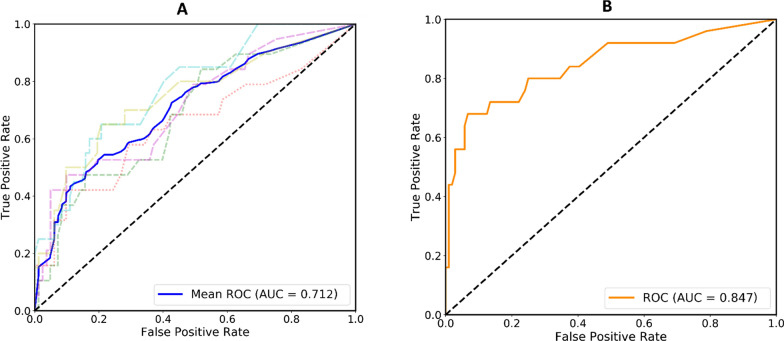
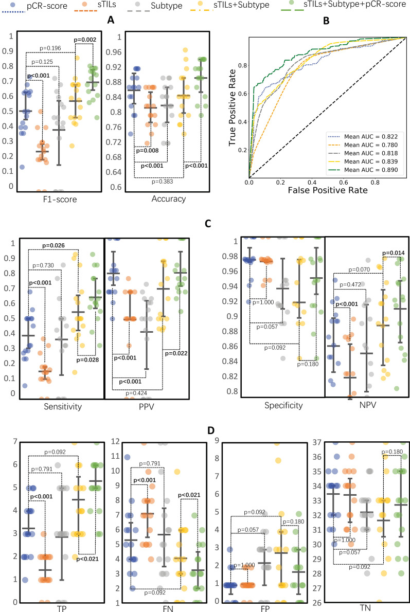
Verification
Claims the paper makes
The claims and statistics of this paper haven’t been checked yet.
Run the check — free, no account needed. It reads the open-access text and checks the sources it cites.
Claims other papers make about this one
Each is checked against this paper’s own text: does it say what the citing paper says it does?
The source backs part of the claim, not all of it — or backs it for a different population, condition or comparison.
In the citing paper
“WSI-based deep learning can improve efficiency in predicting prognosis and treatment response fields (14–16).”
From A multimodal and fully automated system for prediction of pathological complete response to neoadjuvant chemotherapy in breast cancer (2025) · cites this paper as [15]
Experimental — accuracy not yet measuredThe cited source says what the claim attributes to it.
In the citing paper
“Emerging evidence has shown that histopathology whole slide image (WSI) provides information that reflects molecular characteristics or genetic patterns.”
From A multimodal and fully automated system for prediction of pathological complete response to neoadjuvant chemotherapy in breast cancer (2025) · cites this paper as [15]
The cited source says what the claim attributes to it.
In the citing paper
“WSI-based deep learning biomarkers show potential in pCR prediction for breast cancer (15, 16, 26).”
From A multimodal and fully automated system for prediction of pathological complete response to neoadjuvant chemotherapy in breast cancer (2025) · cites this paper as [15]
Self-check
Percentages, sums and reported differences re-computed from the paper’s own numbers.
The self-check needs the paper’s full text.
Citations
Cites
2017 IEEE Conference on Computer Vision and Pattern Recognition (CVPR)
(2017) · doi:10.1109/cvpr35066.2017 · Cited by 7,284
Cortazar, P., Zhang, L., Untch, M., Mehta, K. M., Costantino, J. P., Wolmark, N., Bonnefoi, H. R., Cameron, D., Gianni, L., Valagussa, P., et al. (2014) · The Lancet · doi:10.1016/s0140-6736(13)62422-8 · Cited by 4,777
Goldhirsch, A., Winer, E. P., Coates, A. S., Gelber, R. D., Piccart, M. J., Thurlimann, B. J. K., Senn, H.-J., Albain, K. S., André, F., Bergh, J. C. S., et al. (2013) · Annals of Oncology · doi:10.1093/annonc/mdt303 · Cited by 3,848
Supervised Contrastive Learning
Khosla, P., Teterwak, P., Wang, C., Sarna, A., Tian, Y., Isola, P., Maschinot, A., Liu, C., & Krishnan, D. (2020) · arXiv · doi:10.48550/arxiv.2004.11362 · Cited by 3,569
Ehteshami Bejnordi, B., Veta, M., Johannes van Diest, P., van Ginneken, B., Karssemeijer, N., Litjens, G., van der Laak, J. A. W. M., and the CAMELYON16 Consortium, Hermsen, M., Manson, Q. F., et al. (2017) · JAMA · doi:10.1001/jama.2017.14585 · Cited by 3,293
Salgado, R., Denkert, C., Demaria, S., Sirtaine, N., Klauschen, F., Pruneri, G., Wienert, S., den Eynden, G. V., Baehner, F. L., Penault-Llorca, F. M., et al. (2014) · Annals of Oncology · doi:10.1093/annonc/mdu450 · Cited by 3,142
Coudray, N., Ocampo, P. S., Sakellaropoulos, T., Narula, N., Snuderl, M., Fenyö, D., Moreira, A. L., Razavian, N., & Tsirigos, A. (2018) · Nature Medicine · doi:10.1038/s41591-018-0177-5 · Cited by 2,930
Wolff, A. C., Hammond, M. E. H., Allison, K. H., Harvey, B. E., Mangu, P. B., Bartlett, J. M. S., Bilous, M., Ellis, I. O., Fitzgibbons, P. L., Hanna, W. M., et al. (2018) · Journal of Clinical Oncology · doi:10.1200/jco.2018.77.8738 · Cited by 2,449
Denkert, C., von Minckwitz, G., Darb-Esfahani, S., Lederer, B., Heppner, B. I., Weber, K. E., Budczies, J., Huober, J., Klauschen, F., Furlanetto, J., et al. (2018) · The Lancet Oncology · doi:10.1016/s1470-2045(17)30904-x · Cited by 2,157
Denkert, C., Loibl, S., Noske, A., Roller, M., Müller, B. M., Komor, M., Budczies, J., Darb‐Esfahani, S., Kronenwett, R., Hanusch, C. A., et al. (2009) · Journal of Clinical Oncology · doi:10.1200/jco.2009.23.7370 · Cited by 1,796
Cited by
Sorted by citations
Artificial intelligence in drug development
Zhang, K., Yang, X., Wang, Y., Yu, Y., Huang, N., Li, G. L., Li, X., Wu, J. C., & Yang, S. (2025) · Nature Medicine · doi:10.1038/s41591-024-03434-4 · Cited by 585
Artificial intelligence for digital and computational pathology
Song, A. H., Jaume, G., Williamson, D. F. K., Lu, M. Y., Vaidya, A., Miller, T. R., & Mahmood, F. (2023) · Nature Reviews Bioengineering · doi:10.1038/s44222-023-00096-8 · Cited by 356
van den Ende, N. S., Nguyen, A. H., Jager, A., Kok, M., Debets, R., & van Deurzen, C. H. M. (2023) · International Journal of Molecular Sciences · doi:10.3390/ijms24032969 · Cited by 220
Deep Learning in Breast Cancer Imaging: A Decade of Progress and Future Directions
Luo, L., Wang, X., Lin, Y., Ma, X., Tan, A., Chan, R., Vardhanabhuti, V., Chu, C. W., Cheng, K.-T. T., & Chen, H. (2024) · IEEE Reviews in Biomedical Engineering · doi:10.1109/rbme.2024.3357877 · Cited by 192
Dolezal, J. M., Srisuwananukorn, A., Karpeyev, D., Ramesh, S., Kochanny, S., Cody, B., Mansfield, A. S., Rakshit, S., Bansal, R., Bois, M. C., et al. (2022) · Nature Communications · doi:10.1038/s41467-022-34025-x · Cited by 147
Metadata
- Authors
- Li, F., Yang, Y., Wei, Y., He, P., Chen, J., Zheng, Z., & Bu, H.
- Affiliations
- Sichuan University (CN)
- Corresponding author
- Zheng, Zhongxi
- Bu, Hong
- Venue
- Journal of Translational Medicine
- Published
- 2021-08-16
- Volume
- 19
- Issue
- 1
- Pages
- 348-348
- ISSN
- 1479-5876 (ISSN-L)
- PMC
- PMC8365907
- OpenAlex
- W3211240094
- Cited by
- 113
- References
- 49
- Citations by year
- 2026: 20 · 2025: 27 · 2024: 34 · 2023: 20 · 2022: 11 · 2021: 1
- FWCI
- 5.83
- Type
- journal article
- Language
- en
- Open access
- gold
- doi.org
- Locations
- Journal of Translational Medicine (publishedVersion, cc-by)
- PubMed Central (publishedVersion, cc-by)
- PubMed (publishedVersion)
- DOAJ (DOAJ: Directory of Open Access Journals) (submittedVersion, cc-by-sa)
- Funders
- 1·3·5 project for disciplines of excellence (ZYGD18012)
- the Technological Innovation Project of Chengdu New Industrial Technology Research Institute (2017-CY02–00026-GX)
- the Sichuan Science and Technology Program (2020YFS0088)
- the 1·3·5 project for disciplines of excellence Clinical Research Incubation Project (2019HXFH036)
- Topics
- Physical Sciences › Computer Science › Artificial Intelligence › AI in cancer detection
- Life Sciences › Biochemistry, Genetics and Molecular Biology › Cancer Research › Breast Cancer Treatment Studies
- Health Sciences › Medicine › Radiology, Nuclear Medicine and Imaging › Radiomics and Machine Learning in Medical Imaging
- Keywords
- breast cancer, neoadjuvant chemotherapy, pathological complete response, predictive biomarkers, deep learning, stromal tumor-infiltrating lymphocytes, tumor subtypes, treatment response prediction, patient stratification
- MeSH terms
- Deep Learning, Antineoplastic Combined Chemotherapy Protocols, Breast Neoplasms, Female, Humans, Biomarkers, Lymphocytes, Tumor-Infiltrating, Treatment Outcome, Neoadjuvant Therapy
About these checks
Registry, retraction, Cites and statistics results are computed, not judged, with one exception: a printed reference that no index lists is read once by a model before it counts as a citation error. Claims are extracted by a model and kept only when their quote appears verbatim in the text; each cited source is then checked, with a model’s help, for whether it says what the claim relies on. Those verdicts can be wrong — read the claim’s quote and the cited source before relying on one.
Statistics and arithmetic are recomputed, not judged.
Confidence bands are shown only for fields calibrated on our annotated test set. None is calibrated yet, so no band is shown.
What counts as a claim ›How we measure confidence ›How we check this ›
Are you an author, or do you see a problem with this page? Request a review
