

The DOI matches its Crossref record; 69 of the 70 references it cites were found in a registry or an index.
Exposure to gestational diabetes mellitus induces neuroinflammation, derangement of hippocampal neurons, and cognitive changes in rat offspring
Open accessAbstract
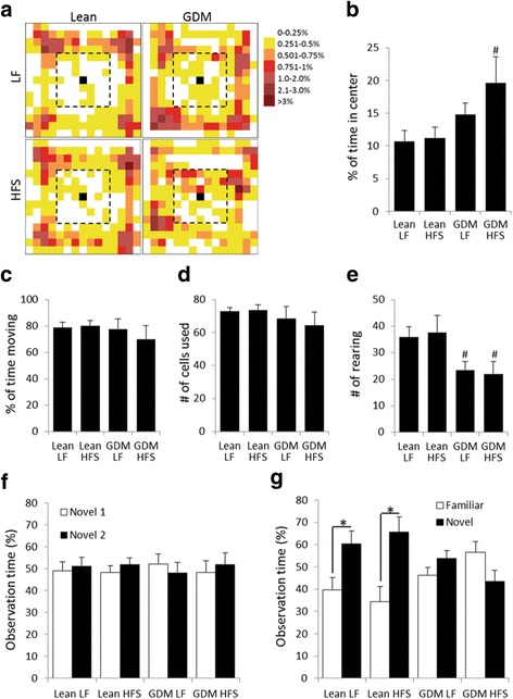
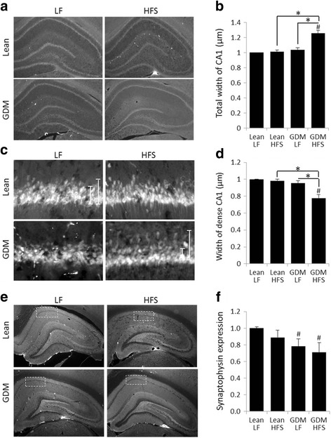
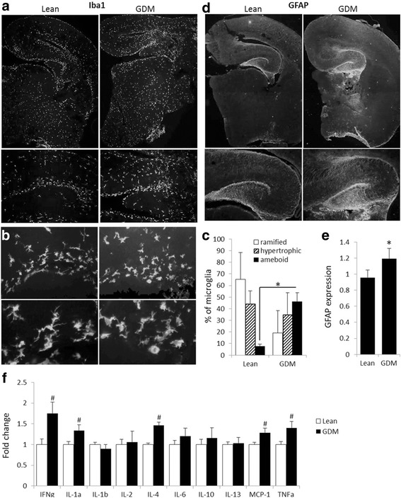
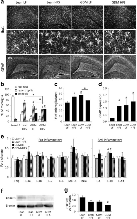
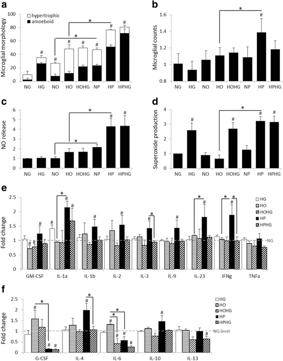
BACKGROUND: Birth cohort studies link gestational diabetes mellitus (GDM) with impaired cognitive performance in the offspring. However, the mechanisms involved are unknown. We tested the hypothesis that obesity-associated GDM induces chronic neuroinflammation and disturbs the development of neuronal circuitry resulting in impaired cognitive abilities in the offspring. METHODS: In rats, GDM was induced by feeding dams a diet high in sucrose and fatty acids. Brains of neonatal (E20) and young adult (15-week-old) offspring of GDM and lean dams were analyzed by immunohistochemistry, cytokine assay, and western blotting. Young adult offspring of GDM and lean dams went also through cognitive assessment. Cultured microglial responses to elevated glucose and/or fatty acids levels were analyzed. RESULTS: In rats, impaired recognition memory was observed in the offspring of GDM dams. GDM exposure combined with a postnatal high-fat and sucrose diet resulted in atypical inattentive behavior in the offspring. These cognitive changes correlated with reduced density and derangement of Cornu Ammonis 1 pyramidal neuronal layer, decreased hippocampal synaptic integrity, increased neuroinflammatory status, and reduced expression of CX3CR1, the microglial fractalkine receptor regulating microglial pro-inflammatory responses and synaptic pruning. Primary microglial cultures that were exposed to high concentrations of glucose and/or palmitate were transformed into an activated, amoeboid morphology with increased nitric oxide and superoxide production, and altered their cytokine release profile. CONCLUSIONS: These findings demonstrate that GDM stimulates microglial activation and chronic inflammatory responses in the brain of the offspring that persist into young adulthood. Reactive gliosis correlates positively with hippocampal synaptic decline and cognitive impairments. The elevated pro-inflammatory cytokine expression at the critical period of hippocampal synaptic maturation suggests that neuroinflammation might drive the synaptic and cognitive decline in the offspring of GDM dams. The importance of microglia in this process is supported by the reduced Cx3CR1 expression as an indication of the loss of microglial control of inflammatory responses and phagocytosis and synaptic pruning in GDM offspring.
Overview
The claims and statistics of this paper haven’t been checked yet.
Run the check — free, no account needed. It reads the open-access text and checks the sources it cites.
Integrity signals
Registry
Cites
Statistics
Figures
Verification
Claims the paper makes
The claims and statistics of this paper haven’t been checked yet.
Run the check — free, no account needed. It reads the open-access text and checks the sources it cites.
Claims other papers make about this one
Each is checked against this paper’s own text: does it say what the citing paper says it does?
The cited source says what the claim attributes to it.
In the citing paper
“Further studies using this GDM model have demonstrated that the offspring exhibit a hyperactive immune response [108] that manifests in neuroinflammation, which was correlated with cognitive impairment and reduced synaptic integrity [109].”
From Recent Experimental Studies of Maternal Obesity, Diabetes during Pregnancy and the Developmental Origins of Cardiovascular Disease (2022) · cites this paper as [109]
Self-check
Percentages, sums and reported differences re-computed from the paper’s own numbers.
The self-check needs the paper’s full text.
Citations
Cites
1 retracted
Fractalkine Attenuates Microglial Cell Activation Induced by Prenatal Stress
Ślusarczyk, J., Trojan, E., Głombik, K., Chamera, K., Roman, A., Budziszewska, B., & Basta‐Kaim, A. (2016) · Neural Plasticity · doi:10.1155/2016/7258201 · Cited by 17
RetractedPolazzi, E., Mengoni, I., Caprini, M., Peña‐Altamira, E., Kurtys, E., & Monti, B. (2012) · Neurosignals · doi:10.1159/000337115 · Cited by 9,023
Synaptic Pruning by Microglia Is Necessary for Normal Brain Development
Paolicelli, R. C., Bolasco, G., Pagani, F., Maggi, L., Scianni, M., Panzanelli, P., Giustetto, M., Ferreira, T. A., Guiducci, E., Dumas, L., et al. (2011) · Science · doi:10.1126/science.1202529 · Cited by 4,244
Obesity is associated with hypothalamic injury in rodents and humans.
Thaler, J. P., Yi, C.-X., Schur, E. A., Guyenet, S. J., Hwang, B. H., Dietrich, M. O., Zhao, X., Sarruf, D. A., Izgur, V., Maravilla, K. R., et al. (2012) · Journal of Clinical Investigation · doi:10.1172/jci59660 · Cited by 1,857
Control of microglial neurotoxicity by the fractalkine receptor
Cardona, A. E., Pioro, E. P., Sasse, M. E., Kostenko, V., Cardona, S. M., Dijkstra, I. M., Huang, D., Kidd, G., Dombrowski, S., Dutta, R., et al. (2006) · Nature Neuroscience · doi:10.1038/nn1715 · Cited by 1,531
Obstetric Complications and Schizophrenia: Historical and Meta-Analytic Review
Cannon, M. D., Jones, P. B., & Murray, R. M. (2002) · American Journal of Psychiatry · doi:10.1176/appi.ajp.159.7.1080 · Cited by 1,190
Milanski, M., Degasperi, G., Coope, A., Morari, J., Denis, R., Cintra, D. E., Tsukumo, D. M. L., Anhe, G., Amaral, M. E., Takahashi, H. K., et al. (2009) · The Journal of Neuroscience · doi:10.1523/jneurosci.2760-08.2009 · Cited by 1,125
Spatial memory, recognition memory, and the hippocampus
Broadbent, N. J., Squire, L. R., & Clark, R. E. (2004) · Proceedings of the National Academy of Sciences · doi:10.1073/pnas.0406344101 · Cited by 985
Microglia: Dynamic Mediators of Synapse Development and Plasticity
Wu, Y., Dissing-Olesen, L., MacVicar, B. A., & Stevens, B. (2015) · Trends in Immunology · doi:10.1016/j.it.2015.08.008 · Cited by 661
Valdearcos, M., Robblee, M. M., Benjamin, D. I., Nomura, D. K., Xu, A. W., & Koliwad, S. K. (2014) · Cell Reports · doi:10.1016/j.celrep.2014.11.018 · Cited by 629
Cited by
Sorted by citations
Items 101–147 of 147
Vilela, W. R., Platt, N., Bechara, L. R. G., de Paula, G. C., Ferreira, J. C. B., de Oliveira, J., Duarte, J. M. N., & Bem, A. F. D. (2025) · Molecular Neurobiology · doi:10.1007/s12035-025-05455-7 · Cited by 4
Antibodies against the flotillin-1/2 complex in patients with multiple sclerosis
Lleixà, C., Caballero‐Ávila, M., Pascual‐Goñi, E., Martín-Aguilar, L., Vidal, N., Tejada‐Illa, C., Valdés-Hevia, E., Zárate, E., Vesperinas, A., Collet‐Vidiella, R., et al. (2023) · Brain Communications · doi:10.1093/braincomms/fcad109 · Cited by 3
Belimezi, M., Kalliaropoulos, A., Mentis, A. A., & Chrousos, G. P. (2021) · Journal of Clinical Pathology · doi:10.1136/jclinpath-2021-207766 · Cited by 3
Huang, H., Apaijai, N., Thonusin, C., Suntornsaratoon, P., Chattipakorn, N., Charoenphandhu, N., & Chattipakorn, S. C. (2025) · Neuroscience · doi:10.1016/j.neuroscience.2025.01.056 · Cited by 2
Schipper, L., Schaap, N., Liu, Y., Li, H., Han, W., & Harvey, L. (2025) · Prostaglandins Leukotrienes and Essential Fatty Acids · doi:10.1016/j.plefa.2025.102700 · Cited by 2
Yan, Z., Pu, J., Li, D., Liu, M., Xu, Z., & Tang, J. (2026) · Journal of Personalized Medicine · doi:10.3390/jpm16010019 · Cited by 2
Torres, Y., Celis, C., Acurio, J., & Escudero, C. (2023) · Advances in experimental medicine and biology · doi:10.1007/978-3-031-32554-0_11 · Cited by 2
Bagheri, J., Fallahnezhad, S., Alipour, N., Babaloo, H., Tahmasebi, F., Kheradmand, H., Sazegar, G., & Haghir, H. (2023) · Journal of Chemical Neuroanatomy · doi:10.1016/j.jchemneu.2023.102326 · Cited by 2
Nazari, Z., Bahrehbar, K., Ghafari, S., & Golalipour, M. J. (2023) · PubMed Central · doi:10.22038/ijbms.2023.71300.15488 · Cited by 2
Aldhshan, M. S., Jhanji, G., & Mizuno, T. M. (2022) · Neuroreport · doi:10.1097/wnr.0000000000001820 · Cited by 2
Alipour, N., Fallahnezhad, S., Bagheri, J., Babaloo, H., Tahmasebi, F., Sazegar, G., & Haghir, H. (2023) · PubMed Central · doi:10.22038/ijbms.2023.69668.15171 · Cited by 2
LUO, S. H., Hsu, E., Lawrence, K. E., Adise, S., Pickering, T. A., Herting, M. M., Buchanan, T. A., Page, K. A., & Thompson, P. M. (2022) · medRxiv · doi:10.1101/2022.02.03.22270398 · Cited by 2
Groh, A. M. R., Afanasiev, E., Thevakumaran, R., Callahan‐Martin, L., Creeggan, F., Yaqubi, M., Zandee, S., Prat, A., Rudko, D. A., & Stratton, J. A. (2025) · bioRxiv (Cold Spring Harbor Laboratory) · doi:10.1101/2025.01.14.633055 · Cited by 3
Martins, M. G., Woodside, B. C., & Kiss, A. C. I. (2024) · Physiology & Behavior · doi:10.1016/j.physbeh.2024.114483 · Cited by 1
Martínez‐Carrillo, B. E., Aguilar-Rodríguez, E., Valdés‐Ramos, R., Reséndiz‐Albor, A. A., Rosales-Gómez, C., Luna, R. A. J., & López, A. L. G. (2025) · Exploration of Immunology · doi:10.37349/ei.2025.1003195 · Cited by 1
Zhang, Y., Wang, D., Su, J., Wu, F.-T., He, Y., Wang, C., Yang, F., & Yao, X. (2025) · Nutrition Reviews · doi:10.1093/nutrit/nuaf076 · Cited by 2
Qiu, H., Zhou, C.-Y., Hu, S., Li, L., Wang, X., Zhang, J., Tian, Y., Wang, B., & Yu, X. (2025) · World Journal of Pediatrics · doi:10.1007/s12519-025-00969-5 · Cited by 1
Mohyee, R. A., Elliott, B. L., Pike, M. R., Smith, E., Kring, A. M., Olson, I. R., Breen, E. C., Cohn, B. A., Cirillo, P. M., Krigbaum, N. Y., et al. (2025) · Proceedings of the National Academy of Sciences · doi:10.1073/pnas.2420188122 · Cited by 1
Khayat, S., Fanaei, H., & Safarzaei, A. (2025) · Metabolism Open · doi:10.1016/j.metop.2025.100422 · Cited by 1
Su, J., Cai, C., Zhang, Y., Wang, D., Yan, S., & Yao, X. (2025) · The Journal of Nutritional Biochemistry · doi:10.1016/j.jnutbio.2025.110232 · Cited by 1
Microglial morphology reflects cognitive status in the aging rat brain
Myers, S. J., Roseborough, A. D., Bayona, C. X., Carrese, C., Allman, B. L., & Whitehead, S. N. (2026) · Brain Behavior and Immunity · doi:10.1016/j.bbi.2026.106533 · Cited by 1
Al-Bakr, K. W., Alzahrani, A. M., & Ahmed, E. A. (2022) · Processes · doi:10.3390/pr10122551 · Cited by 1
Realinho, A. M., Boia, R., Paiva, B., Correia, R. G., Gaspar, R., Ambrósio, A. F., & Baptista, F. I. (2023) · Life Sciences · doi:10.1016/j.lfs.2023.121852 · Cited by 1
Gao, H., Zhou, Y., Jin, P.-S., Wu, D.-G., Wang, Y., Zhao, X., & Zhao, B. (2022) · Metabolic Brain Disease · doi:10.1007/s11011-022-01151-w · Cited by 1
The biology of schizophrenia as a neurodevelopmental disorder
van Berlekom, A. B. (2022) · Utrecht University Repository (Utrecht University) · doi:10.33540/1269 · Cited by 1
Keo, A., Dzyubachyk, O., van der Grond, J., Hafkemeijer, A., van de Berg, W. D. J., van Hilten, J. J., Reinders, M. J. T., & Mahfouz, A. E. (2019) · bioRxiv (Cold Spring Harbor Laboratory) · doi:10.1101/2019.12.17.875880
Borgenheimer, E., Zhang, Y., & Cvetanović, M. (2021) · bioRxiv (Cold Spring Harbor Laboratory) · doi:10.1101/2021.10.28.466301 · Cited by 1
Gao, H., Zhou, Y., Jin, P.-S., Wu, D.-G., Wang, Y., Zhao, X., & Zhao, B. (2022) · SSRN Electronic Journal · doi:10.2139/ssrn.4008171
Cystine/glutamate antiporter system Xc - deficiency impairs insulin secretionPreprint
Baat, A. D., Meier, D. T., Rachid, L., Fontana, A., Böni‐Schnetzler, M., & Donath, M. Y. (2023) · bioRxiv (Cold Spring Harbor Laboratory) · doi:10.1101/2023.01.19.524735
Aguilar-Rodríguez, E., Martínez‐Carrillo, B. E., Valdés‐Ramos, R., Albor, A. A. R., & Rosales-Gómez, C. (2023) · Revista Ciencia e Innovación Agroalimentaria de la Universidad de Guanajuato · doi:10.15174/cia.v4i2.9
TNF-alpha promotes cilia elongation via Mixed Lineage Kinases signalingPreprint
Kumari, A., Caliz, A. D., Kant, S., & Vertii, A. (2023) · bioRxiv (Cold Spring Harbor Laboratory) · doi:10.1101/2023.11.30.569502
Vanneau, T., Quiquempoix, M., Erkel, M.-C., Drogou, C., Trignol, A., Sauvet, F., Léger, D., Gomez-Mérino, D., & Chennaoui, M. (2024) · eNeuro · doi:10.1523/eneuro.0433-23.2023
Changes in microglia chromatin accessibility in aged female micePreprint
Ansere, V. A., Tooley, K. B., Srividya, K., Hoolehan, W., Pham, K. D., Ocañas, S. R., & Freeman, W. M. (2024) · bioRxiv (Cold Spring Harbor Laboratory) · doi:10.1101/2024.01.18.575944
Considering comorbidities: neurodevelopmental implications for children of diabetic mothers
Ottolini, K. M., & Andescavage, N. N. (2024) · Pediatric Research · doi:10.1038/s41390-024-03764-6
Early spontaneous movements in full-term infants exposed to gestational diabetes mellitus
Kepenek-Varol, B., Baştuğ, O., Özdemi̇r, A., & Menevşe, Ö. (2025) · Infant Behavior and Development · doi:10.1016/j.infbeh.2025.102156
Zhou, J., Tong, J., Ru, X., Teng, Y., Geng, M., Yan, S., Tao, F., & Huang, K. (2024) · Figshare · doi:10.6084/m9.figshare.24590474
Lang, G.-P., Ndongson‐Dongmo, B., Lajqi, T., Brodhun, M., Han, Y., Wetzker, R., Frasch, M. G., & Bauer, R. (2020) · Figshare · doi:10.6084/m9.figshare.13065620
Zhou, J., Tong, J., Ru, X., Teng, Y., Geng, M., Yan, S., Tao, F., & Huang, K. (2023) · Figshare · doi:10.6084/m9.figshare.24590349
Effect of Gestational Diabetes in Female Rats on Brain Astrocyte Functions in Their Offspring
Babkina, I., Savinkova, I. G., Artyukhov, A. S., & Горбачева, Л. Р. (2025) · Neurochemical Journal · doi:10.1134/s1819712425700837
Baradaran, R., Alipour, N., Anbarkeh, F. R., & Ghandy, N. (2026) · Neuroscience Letters · doi:10.1016/j.neulet.2026.138519
Dolores-SanJuan, J. L., Jaimes, O., de Gortari, P., & Alvarez‐Salas, E. (2026) · Behavioural Brain Research · doi:10.1016/j.bbr.2026.116134
Zhou, X., Li, L., & Lu, W. (2026) · Journal of Biosciences and Medicines · doi:10.4236/jbm.2026.145020
Research advances on the impact of gestational diabetes mellitus on offspring neurodevelopment
Liu, J., Gao, Y., Yi, Y., Wang, T., Chen, Y., & Ma, J. (2026) · Diabetology & Metabolic Syndrome · doi:10.1186/s13098-026-02175-3
Kawaharada, R., Kimura, R., Miyata, E., Noguchi, R., Toriumi, A., & Nakamura, A. (2026) · Nutrients · doi:10.3390/nu18111748
He, X., Duan, S., He, J., Sun, B., Li, T., Lan, M., Gu, X., Zhang, G., Lin, L., Li, D., et al. (2026) · Nutrients · doi:10.3390/nu18121840
Sun, X., Fang, X., Pan, Y., Zheng, Y., Lü, Y., Guo, X., Hu, X., & Xu, Z. (2026) · Chemico-Biological Interactions · doi:10.1016/j.cbi.2026.112295
Modifiers of Genetics in ASD Etiology
Chau, K., & LaSalle, J. M. (2026) · Current topics in behavioral neurosciences · doi:10.1007/7854_2026_618
Metadata
- Authors
- Vuong, B., Odero, G., Rozbacher, S., Stevenson, M., Kereliuk, S. M., Pereira, T. J., Dolinsky, V. W., & Kauppinen, T. M.
- Affiliations
- Health Sciences Centre (CA)
- Children's Hospital Research Institute of Manitoba (CA)
- Corresponding author
- Kauppinen, Tiina M.
- Venue
- Journal of Neuroinflammation
- Published
- 2017-04-07
- Volume
- 14
- Issue
- 1
- Pages
- 80-80
- ISSN
- 1742-2094 (ISSN-L)
- PMC
- PMC5384149
- OpenAlex
- W2604467698
- Cited by
- 147
- References
- 70
- Citations by year
- 2026: 11 · 2025: 19 · 2024: 13 · 2023: 25 · 2022: 22 · 2021: 17 · 2020: 17 · 2019: 11 · 2018: 9 · 2017: 2
- FWCI
- 12.55
- Type
- journal article
- Language
- en
- Open access
- gold
- doi.org
- Licence
- cc-by
- Locations
- Journal of Neuroinflammation (publishedVersion, cc-by)
- PubMed Central (publishedVersion, cc-by)
- PubMed (publishedVersion)
- DOAJ (DOAJ: Directory of Open Access Journals) (submittedVersion, cc-by-sa)
- Mspace (University of Manitoba) (submittedVersion)
- Funders
- Manitoba Health Research Council
- Children's Hospital Foundation Winnipeg, Manitoba
- Health Sciences Centre Foundation Winnipeg
- Canadian Institutes of Health Research (MOP136885)
- Research Manitoba
- Topics
- Health Sciences › Medicine › Obstetrics and Gynecology › Gestational Diabetes Research and Management
- Life Sciences › Neuroscience › Neurology › Neuroinflammation and Neurodegeneration Mechanisms
- Health Sciences › Medicine › Pediatrics, Perinatology and Child Health › Birth, Development, and Health
- Keywords
- gestational diabetes mellitus, neuroinflammation, hippocampus, cognitive impairment, microglial activation, CX3CR1, synaptic pruning, reactive gliosis, recognition memory, inattentive behavior, high-fat high-sugar diet, palmitate, nitric oxide, superoxide, inflammatory cytokines, cornu ammonis 1, synaptic integrity
- MeSH terms
- Animals, Cells, Cultured, Cognition, Female, Hippocampus, Inflammation, Male, Neurons, Pregnancy, Prenatal Exposure Delayed Effects, Random Allocation, Diabetes, Gestational, Rats, Sprague-Dawley, Inflammation Mediators, Dietary Sucrose, Rats, Diet, High-Fat
About these checks
Registry, retraction, Cites and statistics results are computed, not judged, with one exception: a printed reference that no index lists is read once by a model before it counts as a citation error. Claims are extracted by a model and kept only when their quote appears verbatim in the text; each cited source is then checked, with a model’s help, for whether it says what the claim relies on. Those verdicts can be wrong — read the claim’s quote and the cited source before relying on one.
Statistics and arithmetic are recomputed, not judged.
Confidence bands are shown only for fields calibrated on our annotated test set. None is calibrated yet, so no band is shown.
What counts as a claim ›How we measure confidence ›How we check this ›
Are you an author, or do you see a problem with this page? Request a review
