

The DOI matches its Crossref record; 103 of the 103 references it cites were found in a registry or an index.
Role of inflammasomes in multiple sclerosis and their potential as therapeutic targets
Open accessAbstract
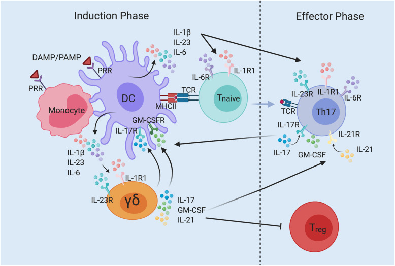
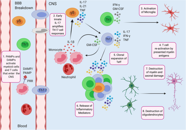
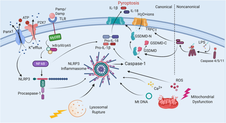
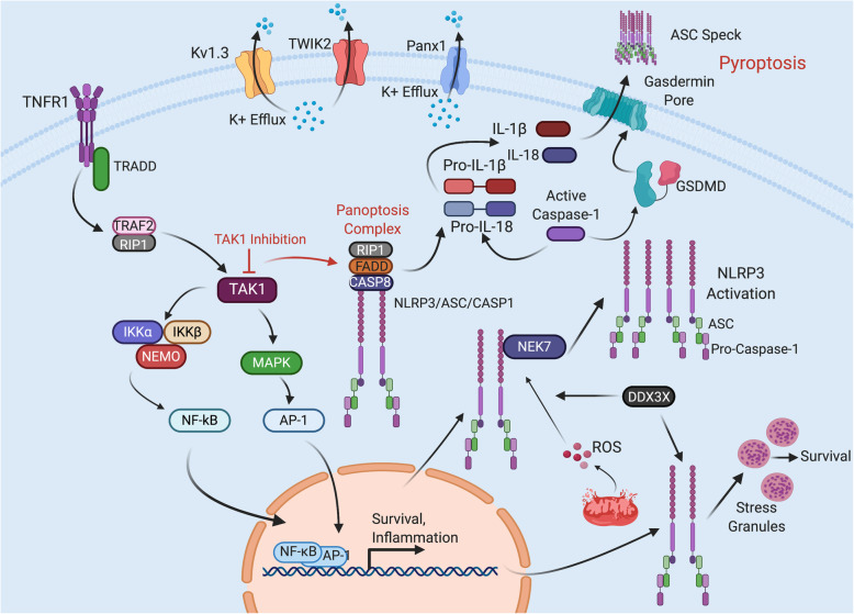
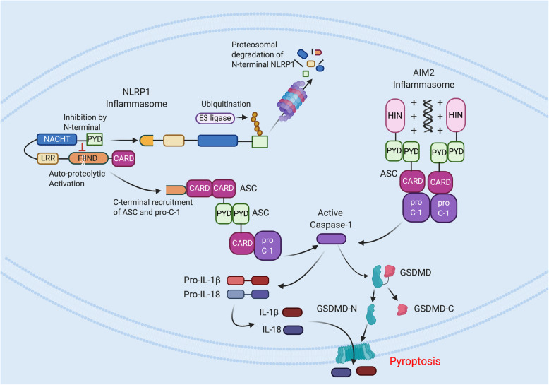
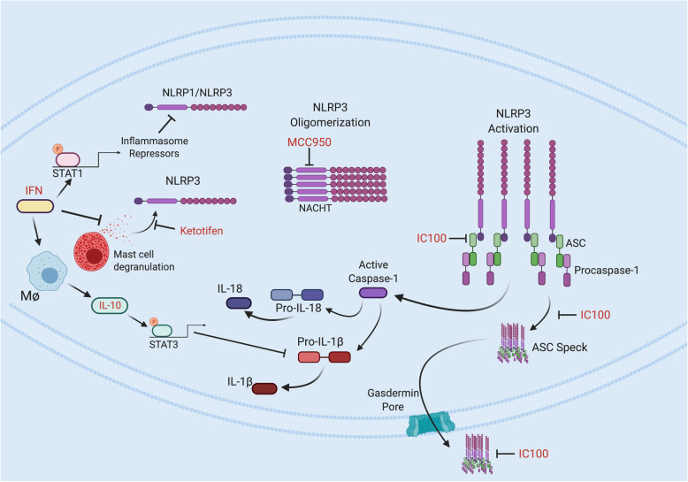
Multiple sclerosis (MS) is a demyelinating disease of the central nervous system (CNS), and it remains the most common immune-mediated disorder affecting the CNS. While the cause of MS is unclear, the underlying pathomechanisms are thought to be either destruction by autoimmune T cells or dysfunction of myelin-producing cells. Recent advances have indicated that inflammasomes contribute the etiology of MS. Inflammasomes are multiprotein complexes of the innate immune response involved in the processing of caspase-1, the activation of pro-inflammatory cytokines interleukin (IL)-1β and IL-18 as well as the cell death-mediated mechanism of pyroptosis and the activation of the adaptive immune response. Here we review the literature to date on the role of different inflammasome signaling pathways in the pathogenesis of MS and how these pathways may be targeted to reduce deleterious inflammatory processes and improve outcomes in this patient population.
Overview
The claims and statistics of this paper haven’t been checked yet.
Run the check — free, no account needed. It reads the open-access text and checks the sources it cites.
Integrity signals
Registry
Cites
Statistics
Figures
Verification
Claims the paper makes
The claims and statistics of this paper haven’t been checked yet.
Run the check — free, no account needed. It reads the open-access text and checks the sources it cites.
Claims other papers make about this one
Each is checked against this paper’s own text: does it say what the citing paper says it does?
The cited source says what the claim attributes to it.
In the citing paper
“Aberrant activation of the microglial NLRP3 inflammasome has been implicated in the onset and progression of multiple sclerosis (MS), an autoimmune demyelinating disease of the CNS [192, 193].”
From Microglial function, INPP5D/SHIP1 signaling, and NLRP3 inflammasome activation: implications for Alzheimer’s disease (2023) · cites this paper as [192]
Self-check
Percentages, sums and reported differences re-computed from the paper’s own numbers.
The self-check needs the paper’s full text.
Citations
Cites
Pattern Recognition Receptors and Inflammation
Takeuchi, O., & Akira, S. (2010) · Cell · doi:10.1016/j.cell.2010.01.022 · Cited by 8,895
Martinon, F., Burns, K., & Tschopp, J. (2002) · Molecular Cell · doi:10.1016/s1097-2765(02)00599-3 · Cited by 6,066
A role for mitochondria in nlrp3 inflammasome activation
Zhou, R., Yazdi, A. S., Menu, P., & Tschopp, J. (2011) · Nature · doi:10.1038/nature09663 · Cited by 5,602
The NLRP3 inflammasome: molecular activation and regulation to therapeutics
Swanson, K. V., Deng, M., & Ting, J. P.-Y. (2019) · Nature Reviews Immunology · doi:10.1038/s41577-019-0165-0 · Cited by 4,884
Caspase-11 cleaves gasdermin D for non-canonical inflammasome signalling
Kayagaki, N., Stowe, I. B., Lee, B. L., O’Rourke, K., Anderson, K., Warming, S., Cuellar, T., Haley, B., Roose-Girma, M., Phung, Q. T., et al. (2015) · Nature · doi:10.1038/nature15541 · Cited by 3,686
Activation and regulation of the inflammasomes
Latz, E., Xiao, T. S., & Stutz, A. (2013) · Nature Reviews Immunology · doi:10.1038/nri3452 · Cited by 2,884
Muñoz-Planillo, R., Kuffa, P., Martínez-Colón, G. J., Smith, B. L., Rajendiran, T. M., & Núñez, G. (2013) · Immunity · doi:10.1016/j.immuni.2013.05.016 · Cited by 2,167
Differential activation of the inflammasome by caspase-1 adaptors ASC and Ipaf
Mariathasan, S., Newton, K., Monack, D. M., Vucic, D., French, D. M., Lee, W. P., Roose-Girma, M., Erickson, S. L., & Dixit, V. M. (2004) · Nature · doi:10.1038/nature02664 · Cited by 1,864
NLRP3 inflammasome activation: the convergence of multiple signalling pathways on ROS production?
Tschopp, J., & Schroder, K. (2010) · Nature reviews. Immunology · doi:10.1038/nri2725 · Cited by 1,859
Unified Polymerization Mechanism for the Assembly of ASC-Dependent Inflammasomes
Lu, A., Magupalli, V. G., Ruan, J., Yin, Q., Atianand, M. K., Vos, M. R., Schröder, G. F., Fitzgerald, K. A., Wu, H., & Egelman, E. H. (2014) · Cell · doi:10.1016/j.cell.2014.02.008 · Cited by 1,353
Research works printed but not linked: 1
- Kesavardhana S, RKS M, Kanneganti T-D. Caspases in cell death, inflammation, and pyroptosis, vol. 38; 2020. p. 567–95.
Cited by
Sorted by citations
Inflammation and immune dysfunction in Parkinson disease
Tansey, M. G., Wallings, R. L., Houser, M. C., Herrick, M. K., Keating, C. E., & Joers, V. (2022) · Nature Reviews Immunology · doi:10.1038/s41577-022-00684-6 · Cited by 1,382
Anti-Inflammatory Effects of GLP-1 Receptor Activation in the Brain in Neurodegenerative Diseases
Diz-Chaves, Y., Mastoor, Z., Spuch, C., González-Matías, L. C., & Mallo, F. (2022) · International Journal of Molecular Sciences · doi:10.3390/ijms23179583 · Cited by 148
Terzioglu, G., & Young-Pearse, T. L. (2023) · Molecular Neurodegeneration · doi:10.1186/s13024-023-00674-9 · Cited by 91
Huang, S., Chen, Z., Fan, B., Chen, Y., Zhou, L., Jiang, B., Long, H., Zhong, W., Li, X., & Li, Y. (2021) · Journal of Neuroimmunology · doi:10.1016/j.jneuroim.2021.577543 · Cited by 80
Inflammasome activation in traumatic brain injury and Alzheimer's disease
Johnson, N. H., Vaccari, J. P. D. R., Bramlett, H. M., Keane, R. W., & Dietrich, W. D. (2022) · Translational research · doi:10.1016/j.trsl.2022.08.014 · Cited by 74
Self-citation
Metadata
- Authors
- Govindarajan, V., de Rivero Vaccari, J. P., & Keane, R. W.
- Affiliations
- University of Miami (US)
- Corresponding author
- Keane, Robert W.
- Venue
- Journal of Neuroinflammation
- Published
- 2020-09-02
- Volume
- 17
- Issue
- 1
- Pages
- 260-260
- ISSN
- 1742-2094 (ISSN-L)
- PMC
- PMC7469327
- OpenAlex
- W3082497244
- Cited by
- 110
- References
- 103
- Citations by year
- 2026: 12 · 2025: 15 · 2024: 23 · 2023: 26 · 2022: 21 · 2021: 10 · 2020: 3
- FWCI
- 1.44
- Type
- journal article
- Language
- en
- Open access
- gold
- doi.org
- Locations
- Journal of Neuroinflammation (publishedVersion, cc-by)
- PubMed Central (publishedVersion, cc-by)
- PubMed (publishedVersion)
- DOAJ (DOAJ: Directory of Open Access Journals) (submittedVersion, cc-by-sa)
- Funders
- National Institute of Neurological Disorders and Stroke (R01NS113969-01)
- Topics
- Life Sciences › Biochemistry, Genetics and Molecular Biology › Molecular Biology › Inflammasome and immune disorders
- Health Sciences › Medicine › Clinical Neurology › Multiple Sclerosis Research Studies
- Life Sciences › Neuroscience › Neurology › Neuroinflammation and Neurodegeneration Mechanisms
- Keywords
- multiple sclerosis, inflammasome, caspase-1, IL-1β, IL-18, pyroptosis, demyelination, autoimmune T cells, innate immunity, neuroinflammation, therapeutic target
- MeSH terms
- Animals, Humans, Immunologic Factors, Multiple Sclerosis, Signal Transduction, Cytokines, Caspases, Inflammasomes
About these checks
Registry, retraction, Cites and statistics results are computed, not judged, with one exception: a printed reference that no index lists is read once by a model before it counts as a citation error. Claims are extracted by a model and kept only when their quote appears verbatim in the text; each cited source is then checked, with a model’s help, for whether it says what the claim relies on. Those verdicts can be wrong — read the claim’s quote and the cited source before relying on one.
Statistics and arithmetic are recomputed, not judged.
Confidence bands are shown only for fields calibrated on our annotated test set. None is calibrated yet, so no band is shown.
What counts as a claim ›How we measure confidence ›How we check this ›
Are you an author, or do you see a problem with this page? Request a review
