

The DOI matches its Crossref record; 103 of the 103 references it cites were found in a registry or an index.
Role of inflammasomes in multiple sclerosis and their potential as therapeutic targets
Open accessAbstract
Multiple sclerosis (MS) is a demyelinating disease of the central nervous system (CNS), and it remains the most common immune-mediated disorder affecting the CNS. While the cause of MS is unclear, the underlying pathomechanisms are thought to be either destruction by autoimmune T cells or dysfunction of myelin-producing cells. Recent advances have indicated that inflammasomes contribute the etiology of MS. Inflammasomes are multiprotein complexes of the innate immune response involved in the processing of caspase-1, the activation of pro-inflammatory cytokines interleukin (IL)-1β and IL-18 as well as the cell death-mediated mechanism of pyroptosis and the activation of the adaptive immune response. Here we review the literature to date on the role of different inflammasome signaling pathways in the pathogenesis of MS and how these pathways may be targeted to reduce deleterious inflammatory processes and improve outcomes in this patient population.
Overview
The claims and statistics of this paper haven’t been checked yet.
Run the check — free, no account needed. It reads the open-access text and checks the sources it cites.
Integrity signals
Registry
Cites
Statistics
Figures
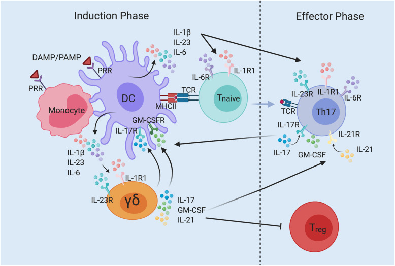
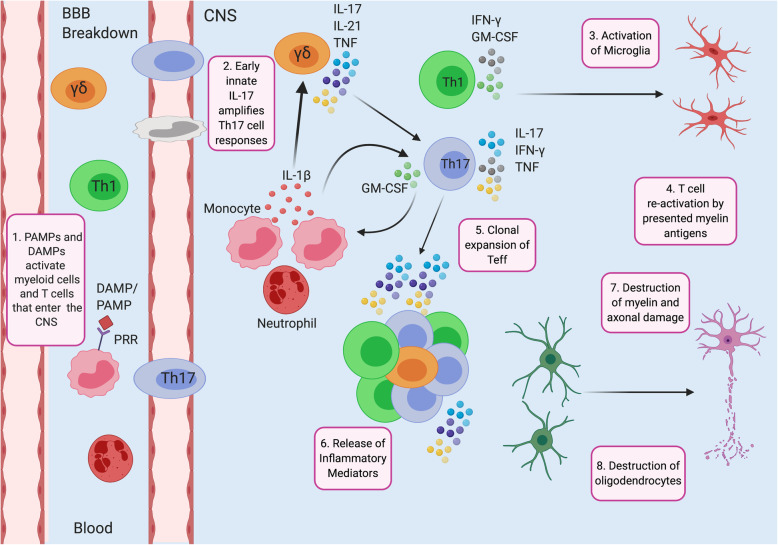
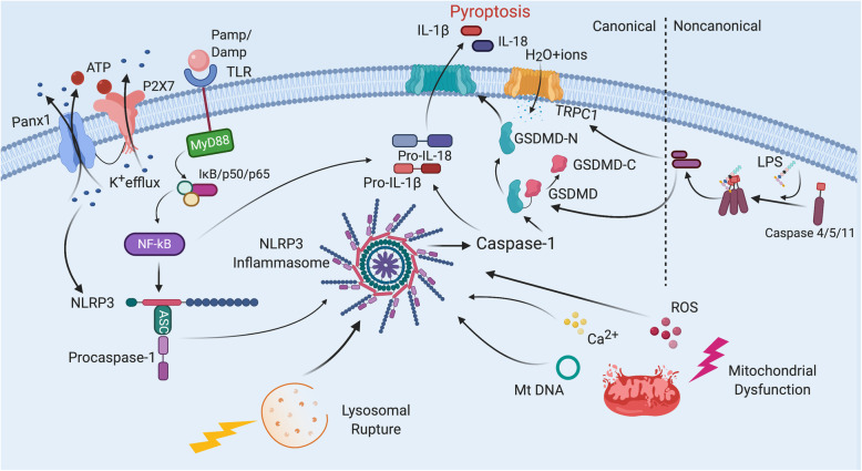
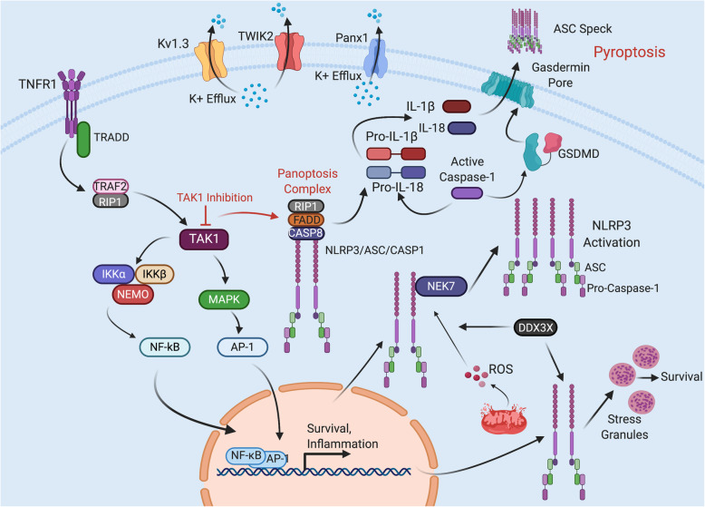
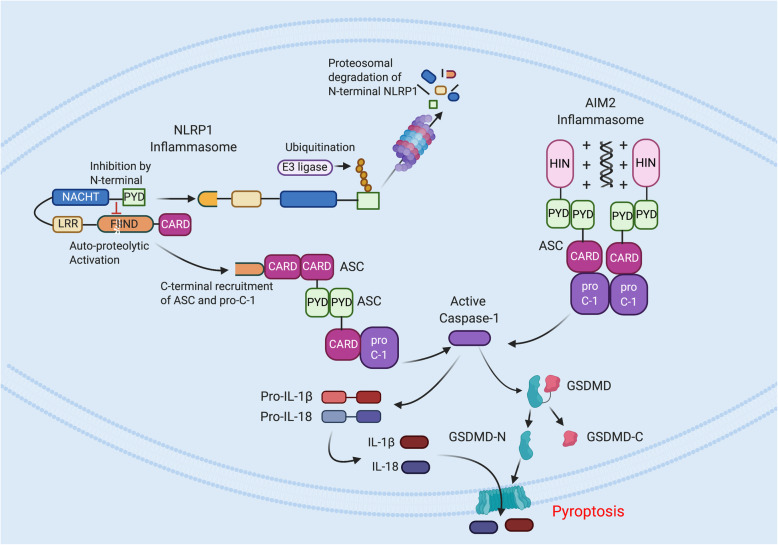
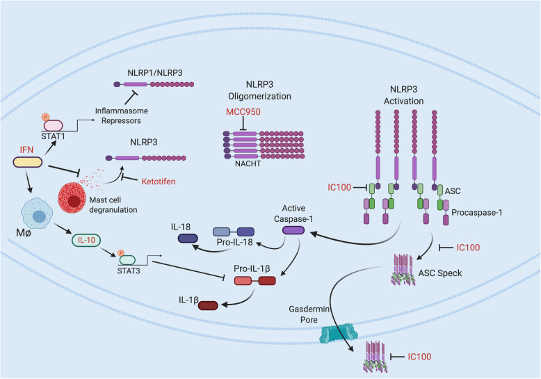
Verification
Claims the paper makes
The claims and statistics of this paper haven’t been checked yet.
Run the check — free, no account needed. It reads the open-access text and checks the sources it cites.
Claims other papers make about this one
Each is checked against this paper’s own text: does it say what the citing paper says it does?
The cited source says what the claim attributes to it.
In the citing paper
“Aberrant activation of the microglial NLRP3 inflammasome has been implicated in the onset and progression of multiple sclerosis (MS), an autoimmune demyelinating disease of the CNS [192, 193].”
From Microglial function, INPP5D/SHIP1 signaling, and NLRP3 inflammasome activation: implications for Alzheimer’s disease (2023) · cites this paper as [192]
Self-check
Percentages, sums and reported differences re-computed from the paper’s own numbers.
The self-check needs the paper’s full text.
Citations
Cites
Pattern Recognition Receptors and Inflammation
Takeuchi, O., & Akira, S. (2010) · Cell · doi:10.1016/j.cell.2010.01.022 · Cited by 8,895
Martinon, F., Burns, K., & Tschopp, J. (2002) · Molecular Cell · doi:10.1016/s1097-2765(02)00599-3 · Cited by 6,066
A role for mitochondria in nlrp3 inflammasome activation
Zhou, R., Yazdi, A. S., Menu, P., & Tschopp, J. (2011) · Nature · doi:10.1038/nature09663 · Cited by 5,602
The NLRP3 inflammasome: molecular activation and regulation to therapeutics
Swanson, K. V., Deng, M., & Ting, J. P.-Y. (2019) · Nature Reviews Immunology · doi:10.1038/s41577-019-0165-0 · Cited by 4,884
Caspase-11 cleaves gasdermin D for non-canonical inflammasome signalling
Kayagaki, N., Stowe, I. B., Lee, B. L., O’Rourke, K., Anderson, K., Warming, S., Cuellar, T., Haley, B., Roose-Girma, M., Phung, Q. T., et al. (2015) · Nature · doi:10.1038/nature15541 · Cited by 3,686
Activation and regulation of the inflammasomes
Latz, E., Xiao, T. S., & Stutz, A. (2013) · Nature Reviews Immunology · doi:10.1038/nri3452 · Cited by 2,884
Muñoz-Planillo, R., Kuffa, P., Martínez-Colón, G. J., Smith, B. L., Rajendiran, T. M., & Núñez, G. (2013) · Immunity · doi:10.1016/j.immuni.2013.05.016 · Cited by 2,167
Differential activation of the inflammasome by caspase-1 adaptors ASC and Ipaf
Mariathasan, S., Newton, K., Monack, D. M., Vucic, D., French, D. M., Lee, W. P., Roose-Girma, M., Erickson, S. L., & Dixit, V. M. (2004) · Nature · doi:10.1038/nature02664 · Cited by 1,864
NLRP3 inflammasome activation: the convergence of multiple signalling pathways on ROS production?
Tschopp, J., & Schroder, K. (2010) · Nature reviews. Immunology · doi:10.1038/nri2725 · Cited by 1,859
Unified Polymerization Mechanism for the Assembly of ASC-Dependent Inflammasomes
Lu, A., Magupalli, V. G., Ruan, J., Yin, Q., Atianand, M. K., Vos, M. R., Schröder, G. F., Fitzgerald, K. A., Wu, H., & Egelman, E. H. (2014) · Cell · doi:10.1016/j.cell.2014.02.008 · Cited by 1,353
Caspase‐1‐induced pyroptotic cell death
Miao, E. A., Rajan, J. V., & Aderem, A. A. (2011) · Immunological Reviews · doi:10.1111/j.1600-065x.2011.01044.x · Cited by 1,162
MCC950 directly targets the NLRP3 ATP-hydrolysis motif for inflammasome inhibition
Coll, R. C., Hill, J. R., Day, C. J., Zamoshnikova, A., Boucher, D., Massey, N. L., Chitty, J. L., Fraser, J. A., Jennings, M. P., Robertson, A. A. B., et al. (2019) · Nature Chemical Biology · doi:10.1038/s41589-019-0277-7 · Cited by 1,041
Pathogen blockade of TAK1 triggers caspase-8–dependent cleavage of gasdermin D and cell death
Ørning, P., Weng, D., Starheim, K. K., Ratner, D., Best, Z., Lee, B. L., Brooks, A., Xia, S., Wu, H., Kelliher, M. A., et al. (2018) · Science · doi:10.1126/science.aau2818 · Cited by 1,039
Caspases in Cell Death, Inflammation, and Pyroptosis
Kesavardhana, S., Malireddi, R. K. S., & Kanneganti, T. (2020) · Annual Review of Immunology · doi:10.1146/annurev-immunol-073119-095439 · Cited by 954
Type I Interferon Inhibits Interleukin-1 Production and Inflammasome Activation
Guarda, G., Braun, M., Staehli, F., Tardivel, A., Mattmann, C., Förster, I., Farlik, M., Decker, T., Pasquier, R. D., Romero, P. J., et al. (2011) · Immunity · doi:10.1016/j.immuni.2011.02.006 · Cited by 924
Caspase-8 induces cleavage of gasdermin D to elicit pyroptosis during Yersinia infection
Sarhan, J., Liu, B. C., Muendlein, H. I., Li, P., Nilson, R., Tang, A. Y., Rongvaux, A., Bunnell, S. C., Shao, F., Green, D. R., et al. (2018) · Proceedings of the National Academy of Sciences · doi:10.1073/pnas.1809548115 · Cited by 901
GsdmD p30 elicited by caspase-11 during pyroptosis forms pores in membranes
Aglietti, R. A., Estevez, A., Gupta, A., Ramirez, M. G., Liu, P. S., Kayagaki, N., Ciferri, C., Dixit, V. M., & Dueber, E. C. (2016) · Proceedings of the National Academy of Sciences · doi:10.1073/pnas.1607769113 · Cited by 898
Microtubule-driven spatial arrangement of mitochondria promotes activation of the NLRP3 inflammasome
Misawa, T., Takahama, M., Kozaki, T., Lee, H., Zou, J., Saitoh, T., & Akira, S. (2013) · Nature Immunology · doi:10.1038/ni.2550 · Cited by 894
Shi, H., Wang, Y., Li, X., Zhan, X., Tang, M., Fina, M., Su, L., Pratt, D., Bu, C. H., Hildebrand, S., et al. (2015) · Nature Immunology · doi:10.1038/ni.3333 · Cited by 771
The cell biology of inflammasomes: Mechanisms of inflammasome activation and regulation
Sharma, D., & Kanneganti, T. (2016) · The Journal of Cell Biology · doi:10.1083/jcb.201602089 · Cited by 754
Reconstituted NALP1 Inflammasome Reveals Two-Step Mechanism of Caspase-1 Activation
Faustin, B., Lartigue, L., Bruey, J., Luciano, F., Sergienko, E. A., Bailly‐Maitre, B., Volkmann, N., Hanein, D., Rouiller, I., & Reed, J. C. (2007) · Molecular Cell · doi:10.1016/j.molcel.2007.01.032 · Cited by 709
The multifaceted role of CD4+ T cells in CD8+ T cell memory
Laidlaw, B. J., Craft, J. E., & Kaech, S. M. (2016) · Nature Reviews Immunology · doi:10.1038/nri.2015.10 · Cited by 657
Progressive multiple sclerosis: from pathogenic mechanisms to treatment
Correale, J. D., Gaitán, M. I., Ysrraelit, M. C., & Fiol, M. P. (2016) · Brain · doi:10.1093/brain/aww258 · Cited by 609
The Pannexin 1 Channel Activates the Inflammasome in Neurons and Astrocytes
Silverman, W. R., Vaccari, J. P. D. R., Locovei, S., Qiu, F., Carlsson, S. K., Scemes, E., Keane, R. W., & Dahl, G. (2009) · Journal of Biological Chemistry · doi:10.1074/jbc.m109.004804 · Cited by 549
Self-citationRedundant roles for inflammasome receptors NLRP3 and NLRC4 in host defense against Salmonella
Broz, P., Newton, K., Lamkanfi, M., Mariathasan, S., Dixit, V. M., & Monack, D. M. (2010) · Journal of Experimental Medicine · doi:10.1084/jem.20100257 · Cited by 549
Memory B Cells Activate Brain-Homing, Autoreactive CD4+ T Cells in Multiple Sclerosis
Jelčić, I., Nimer, F. A., Wang, J., Lentsch, V., Planas, R., Jelčić, I., Madjovski, A., Ruhrmann, S., Faigle, W., Frauenknecht, K. B. M., et al. (2018) · Cell · doi:10.1016/j.cell.2018.08.011 · Cited by 532
T helper 1 immunity requires complement-driven NLRP3 inflammasome activity in CD4 + T cells
Arbore, G., West, E. E., Spolski, R. J., Robertson, A. A. B., Klos, A., Rheinheimer, C., Dutow, P., Woodruff, T. M., Yu, Z., O'Neill, L. A. J., et al. (2016) · Science · doi:10.1126/science.aad1210 · Cited by 513
DDX3X acts as a live-or-die checkpoint in stressed cells by regulating NLRP3 inflammasome
Samir, P., Kesavardhana, S., Patmore, D. M., Gingras, S., Malireddi, R. K. S., Karki, R., Guy, C. S., Briard, B., Place, D. E., Bhattacharya, A., et al. (2019) · Nature · doi:10.1038/s41586-019-1551-2 · Cited by 439
The TWIK2 Potassium Efflux Channel in Macrophages Mediates NLRP3 Inflammasome-Induced Inflammation
Di, A., Xiong, S., Ye, Z., Malireddi, R. K. S., Kometani, S., Zhong, M., Mittal, M., Hong, Z., Kanneganti, T., Rehman, J., et al. (2018) · Immunity · doi:10.1016/j.immuni.2018.04.032 · Cited by 427
Reassessing B cell contributions in multiple sclerosis
Li, R., Patterson, K. R., & Bar‐Or, A. (2018) · Nature Immunology · doi:10.1038/s41590-018-0135-x · Cited by 417
An update on immunopathogenesis, diagnosis, and treatment of multiple sclerosis
Garg, N., & Smith, T. W. (2015) · Brain and Behavior · doi:10.1002/brb3.362 · Cited by 398
NLR members NLRC4 and NLRP3 mediate sterile inflammasome activation in microglia and astrocytes
Freeman, L., Guo, H., David, C. N., Brickey, W. J., Jha, S., & Ting, J. P.-Y. (2017) · Journal of Experimental Medicine · doi:10.1084/jem.20150237 · Cited by 397
Malireddi, R. K. S., Gurung, P., Kesavardhana, S., Samir, P., Burton, A. R., Mummareddy, H., Vogel, P., Pelletier, S. W., Burgula, S., & Kanneganti, T. (2019) · The Journal of Experimental Medicine · doi:10.1084/jem.20191644 · Cited by 391
The DNA-sensing AIM2 inflammasome controls radiation-induced cell death and tissue injury
Hu, B., Jin, C., Li, H., Tong, J., Ouyang, X., Cetinbas, N. M., Zhu, S., Strowig, T., Lam, F. C., Zhao, C. N., et al. (2016) · Science · doi:10.1126/science.aaf7532 · Cited by 384
Classification, diagnosis, and differential diagnosis of multiple sclerosis
Sand, I. K. (2015) · Current Opinion in Neurology · doi:10.1097/wco.0000000000000206 · Cited by 359
A Molecular Platform in Neurons Regulates Inflammation after Spinal Cord Injury
de Rivero Vaccari, J. P., Lotocki, G., Marcillo, A. E., Dietrich, W. D., & Keane, R. W. (2008) · The Journal of Neuroscience · doi:10.1523/jneurosci.0157-08.2008 · Cited by 332
Self-citationDuncan, J. A., & Canna, S. (2017) · Immunological Reviews · doi:10.1111/imr.12607 · Cited by 326
Vaccari, J. P. D. R., Dietrich, W. D., & Keane, R. W. (2014) · Journal of Cerebral Blood Flow & Metabolism · doi:10.1038/jcbfm.2013.227 · Cited by 319
Self-citationde Rivero Vaccari, J. P., Lotocki, G., Alonso, O. F., Bramlett, H. M., Dietrich, W. D., & Keane, R. W. (2009) · Journal of Cerebral Blood Flow & Metabolism · doi:10.1038/jcbfm.2009.46 · Cited by 312
Self-citationPyroptotic Neuronal Cell Death Mediated by the AIM2 Inflammasome
Adamczak, S. E., Vaccari, J. P. D. R., Dale, G. A., Brand, F. J., Nonner, D., Bullock, M. R. R., Dahl, G., Dietrich, W. D., & Keane, R. W. (2014) · Journal of Cerebral Blood Flow & Metabolism · doi:10.1038/jcbfm.2013.236 · Cited by 289
Self-citationInoue, M., Williams, K. L., Gunn, M. D., & Shinohara, M. L. (2012) · Proceedings of the National Academy of Sciences · doi:10.1073/pnas.1201836109 · Cited by 288
Karki, R., Man, S. M., Malireddi, R. K. S., Gurung, P., Vogel, P., Lamkanfi, M., & Kanneganti, T.-D. (2015) · Cell Host & Microbe · doi:10.1016/j.chom.2015.01.006 · Cited by 272
Gasdermin D Restrains Type I Interferon Response to Cytosolic DNA by Disrupting Ionic Homeostasis
Banerjee, I., Behl, B., Mendonça, M. S. A., Shrivastava, G., Russo, A. J., Ménoret, A., Ghosh, A., Vella, A. T., Vanaja, S. K., Sarkar, S. N., et al. (2018) · Immunity · doi:10.1016/j.immuni.2018.07.006 · Cited by 266
More to Life than NF-κB in TNFR1 Signaling
Ting, A. T., & Bertrand, M. J. M. (2016) · Trends in Immunology · doi:10.1016/j.it.2016.06.002 · Cited by 265
Finger, J. N., Lich, J. D., Dare, L. C., Cook, M. N., Brown, K. K., Duraiswami, C., Bertin, J., & Gough, P. J. (2012) · Journal of Biological Chemistry · doi:10.1074/jbc.m112.378323 · Cited by 264
Villar, L. M., Sádaba, M. C., Roldán, E., Masjuán, J., González‐Porqué, P., Villarrubia, N., Espiño, M., García‐Trujillo, J. A., Bootello, A., & Alvarez-Cermeño, J. C. (2005) · Journal of Clinical Investigation · doi:10.1172/jci22833 · Cited by 259
TAK1 restricts spontaneous NLRP3 activation and cell death to control myeloid proliferation
Malireddi, R. K. S., Gurung, P., Mavuluri, J., Dasari, T. K., Klco, J. M., Chi, H., & Kanneganti, T. (2018) · The Journal of Experimental Medicine · doi:10.1084/jem.20171922 · Cited by 249
Opdenbosch, N. V., Gurung, P., Walle, L. V., Fossoul, A., Kanneganti, T., & Lamkanfi, M. (2014) · Nature Communications · doi:10.1038/ncomms4209 · Cited by 235
Caspase-8 as an Effector and Regulator of NLRP3 Inflammasome Signaling
Antonopoulos, C. H., Russo, H. M., Sanadi, C. E. E., Martin, B. N., Li, X., Kaiser, W. J., Mocarski, E. S., & Dubyak, G. (2015) · Journal of Biological Chemistry · doi:10.1074/jbc.m115.652321 · Cited by 232
Grandemange, S., Sanchez, E., Louis‐Plence, P., Mau‐Them, F. T., Bessis, D., Coubes, C., Frouin, É., Seyger, M. M. B., Girard, M., Puechberty, J., et al. (2016) · Annals of the Rheumatic Diseases · doi:10.1136/annrheumdis-2016-210021 · Cited by 231
Syed, S. A., Beurel, E., Loewenstein, D., Lowell, J. A., Craighead, W. E., Dunlop, B. W., Mayberg, H. S., Dhabhar, F. S., Dietrich, W. D., Keane, R. W., et al. (2018) · Neuron · doi:10.1016/j.neuron.2018.08.001 · Cited by 214
Self-citationVajjhala, P. R., Mirams, R. E., & Hill, J. M. (2012) · Journal of Biological Chemistry · doi:10.1074/jbc.m112.381228 · Cited by 205
Holding RIPK1 on the Ubiquitin Leash in TNFR1 Signaling
Peltzer, N., Darding, M., & Walczak, H. (2016) · Trends in Cell Biology · doi:10.1016/j.tcb.2016.01.006 · Cited by 188
Guo, Q., Wu, Y., Hou, Y., Liu, Y., Liu, T., Zhang, H., Fan, C., Guan, H., Li, Y., Shan, Z., et al. (2018) · Frontiers in Immunology · doi:10.3389/fimmu.2018.01197 · Cited by 180
Function and Regulation of Noncanonical Caspase-4/5/11 Inflammasome
Matikainen, S., Nyman, T. A., & Cypryk, W. (2020) · The Journal of Immunology · doi:10.4049/jimmunol.2000373 · Cited by 179
Specific peripheral B cell tolerance defects in patients with multiple sclerosis
Kinnunen, T. T., Chamberlain, N., Morbach, H., Cantaert, T., Lynch, M. T., Preston‐Hurlburt, P., Herold, K. C., Hafler, D. A., O’Connor, K. C., & Meffre, E. (2013) · Journal of Clinical Investigation · doi:10.1172/jci68775 · Cited by 170
Adamczak, S. E., Dale, G. A., Vaccari, J. P. D. R., Bullock, M. R., Dietrich, W. D., & Keane, R. W. (2012) · Journal of neurosurgery · doi:10.3171/2012.9.jns12815 · Cited by 168
Self-citationIL-1β Breaks Tolerance through Expansion of CD25+ Effector T Cells
O’Sullivan, B., Thomas, H. E., Pai, S., Santamaría, P., Iwakura, Y., Steptoe, R. J., Kay, T. W. H., & Thomas, R. (2006) · The Journal of Immunology · doi:10.4049/jimmunol.176.12.7278 · Cited by 164
T cell–intrinsic ASC critically promotes TH17-mediated experimental autoimmune encephalomyelitis
Martin, B. N., Wang, C., Zhang, C.-J., Kang, Z., Gulen, M. F., Zepp, J. A., Zhao, J., Bian, G., Do, J., Min, B., et al. (2016) · Nature Immunology · doi:10.1038/ni.3389 · Cited by 159
Proteolytic Processing of Nlrp1b Is Required for Inflammasome Activity
Frew, B. C., Joag, V., & Mogridge, J. (2012) · PLoS Pathogens · doi:10.1371/journal.ppat.1002659 · Cited by 156
How Inflammasomes Inform Adaptive Immunity
Evavold, C. L., & Kagan, J. C. (2017) · Journal of Molecular Biology · doi:10.1016/j.jmb.2017.09.019 · Cited by 155
Malhotra, S., Costa, C., Eixarch, H., Keller, C. W., Amann, L., Martínez‐Banaclocha, H., Midaglia, L. S., Sarró, E., Machín-Díaz, I., Villar, L. M., et al. (2020) · Brain · doi:10.1093/brain/awaa084 · Cited by 138
Radiation Exposure Induces Inflammasome Pathway Activation in Immune Cells
Stoecklein, V. M., Osuka, A., Ishikawa, S., Lederer, M. R., Wanke-Jellinek, L., & Lederer, J. A. (2014) · The Journal of Immunology · doi:10.4049/jimmunol.1303051 · Cited by 137
The Diagnosis and Treatment of Optic Neuritis
Wilhelm, H. M., & Schabet, M. (2015) · Deutsches Ärzteblatt international · doi:10.3238/arztebl.2015.0616 · Cited by 136
Th17 cells, γδ T cells and their interplay in EAE and multiple sclerosis
McGinley, A. M., Edwards, S. C., Raverdeau, M., & Mills, K. H. G. (2018) · Journal of Autoimmunity · doi:10.1016/j.jaut.2018.01.001 · Cited by 130
Inflammasome Proteins As Biomarkers of Multiple Sclerosis
Keane, R. W., Dietrich, W. D., & Vaccari, J. P. D. R. (2018) · Frontiers in Neurology · doi:10.3389/fneur.2018.00135 · Cited by 130
Self-citationYao, Y. F., Vent‐Schmidt, J., McGeough, M. D., Wong, M. Q., Hoffman, H. M., Steiner, T. S., & Levings, M. K. (2015) · The Journal of Immunology · doi:10.4049/jimmunol.1403225 · Cited by 128
Sun, Y. E., Ma, J., Li, D., Li, P., Zhou, X., Li, Y., He, Z., Qin, L., Liang, L., & Luo, X. (2019) · Journal of Neuroinflammation · doi:10.1186/s12974-019-1452-1 · Cited by 123
Inflammasome proteins as biomarkers of traumatic brain injury
Kerr, N. A., Lee, S. W., Pérez‐Bárcena, J., Crespí, C., Ibáñez, J., Bullock, M. R., Dietrich, W. D., Keane, R. W., & Vaccari, J. P. D. R. (2018) · PLoS ONE · doi:10.1371/journal.pone.0210128 · Cited by 122
Self-citationde Jong, B. A., Huizinga, T. W. J., Bollen, E. L. E. M., Uitdehaag, B. M. J., Bosma, G. P. T., van Buchem, M. A., Remarque, E. J., Burgmans, A. C. S., Kalkers, N. F., Polman, C. H., et al. (2002) · Journal of Neuroimmunology · doi:10.1016/s0165-5728(02)00056-5 · Cited by 119
Lee, S. W., Vaccari, J. P. D. R., Truettner, J. S., Dietrich, W. D., & Keane, R. W. (2019) · Journal of Neuroinflammation · doi:10.1186/s12974-019-1423-6 · Cited by 115
Self-citationNLRP3 inflammasome is associated with the response to IFN-β in patients with multiple sclerosis
Malhotra, S., Río, J., Urcelay, E., Nurtdinov, R., Bustamante, M. F., Fernández, Ó., Oliver‐Martos, B., Zettl, U. K., Brassat, D., Killestein, J., et al. (2015) · Brain · doi:10.1093/brain/awu388 · Cited by 111
The NAIP/NLRC4 inflammasome in infection and pathology
Bauer, R., & Rauch, I. (2020) · Molecular Aspects of Medicine · doi:10.1016/j.mam.2020.100863 · Cited by 107
Inflammasome Proteins in Serum and Serum-Derived Extracellular Vesicles as Biomarkers of Stroke
Kerr, N. A., García-Contreras, M., Abbassi, S., Mejias, N. H., Desousa, B. R., Ricordi, C., Dietrich, W. D., Keane, R. W., & Vaccari, J. P. D. R. (2018) · Frontiers in Molecular Neuroscience · doi:10.3389/fnmol.2018.00309 · Cited by 102
Self-citationInoue, M., & Shinohara, M. L. (2013) · Immunology · doi:10.1111/imm.12081 · Cited by 91
Microglial Inflammasome Activation in Penetrating Ballistic-Like Brain Injury
Lee, S. W., Gajavelli, S., Spurlock, M. S., Andreoni, C. B., Vaccari, J. P. D. R., Bullock, M. R., Keane, R. W., & Dietrich, W. D. (2018) · Journal of Neurotrauma · doi:10.1089/neu.2017.5530 · Cited by 89
Self-citationKhan, N., Kuo, A., Brockman, D. A., Cooper, M. A., & Smith, M. T. (2017) · Inflammopharmacology · doi:10.1007/s10787-017-0401-9 · Cited by 88
Desu, H. L., Plastini, M. J., Illiano, P., Bramlett, H. M., Dietrich, W. D., Vaccari, J. P. D. R., Brambilla, R., & Keane, R. W. (2020) · Journal of Neuroinflammation · doi:10.1186/s12974-020-01826-0 · Cited by 83
Self-citationMicroglial NLRP3 inflammasome activation in multiple sclerosis
(2020) · Advances in Protein Chemistry and Structural Biology · doi:10.1016/bs.apcsb.2019.08.007 · Cited by 83
Gov, L., Schneider, C. A., de Lima, T. S., Pandori, W., & Lodoen, M. B. (2017) · The Journal of Immunology · doi:10.4049/jimmunol.1700245 · Cited by 82
Soares, J. L. S., de Oliveira, E. M. L., & Pontillo, A. (2019) · Multiple Sclerosis and Related Disorders · doi:10.1016/j.msard.2019.01.023 · Cited by 81
Vilariño‐Güell, C., Zimprich, A. A., Boneschi, F. M., Herculano, B., Wang, Z., Matesanz, F., Urcelay, E., Vandenbroeck, K., Leyva, L., Gris, D., et al. (2019) · PLoS Genetics · doi:10.1371/journal.pgen.1008180 · Cited by 79
Identification of rare genetic variation of NLRP1 gene in familial multiple sclerosis
Maver, A., Lavtar, P., Ristič, S., Stopinšek, S., Simčič, S., Hočevar, K., Sepčić, J., Drulovic, J. S., Pekmezovic, T. D., Novaković, I. V., et al. (2017) · Scientific Reports · doi:10.1038/s41598-017-03536-9 · Cited by 66
Xu, L., Zhang, C., Jiang, N., He, D., Bai, Y., & Xin, Y. (2018) · Journal of Cellular Biochemistry · doi:10.1002/jcb.27792 · Cited by 65
Vladimirova, O. V., Lu, F. M., Shawver, L., & Kálmán, B. (1999) · Inflammation Research · doi:10.1007/s000110050480 · Cited by 58
Imani, D., Azimi, A., Salehi, Z., Rezaei, N., Emamnejad, R., Sadr, M., & Izad, M. H. (2018) · International Journal of Immunogenetics · doi:10.1111/iji.12401 · Cited by 50
Vidmar, L., Maver, A., Drulovic, J. S., Sepčić, J., Novaković, I. V., Ristič, S., Šega, S., & Peterlin, B. (2019) · Scientific Reports · doi:10.1038/s41598-019-45598-x · Cited by 49
Noroozi, S., Ebrahimi, H. A., Arababadi, M. K., Nakhaee, N., & Asadikaram, G. (2016) · Molecular Neurobiology · doi:10.1007/s12035-016-9864-8 · Cited by 49
The effects of interferon‐β on interleukin‐10 in multiple sclerosis patients
Ersoy, E., Kuş, C. N., Sener, U. T., Çoker, I., & Zorlu, Y. (2005) · European Journal of Neurology · doi:10.1111/j.1468-1331.2004.00986.x · Cited by 48
Reubold, T. F., Hahne, G., Wohlgemuth, S., & Eschenburg, S. (2014) · FEBS Letters · doi:10.1016/j.febslet.2014.07.017 · Cited by 45
Role of the HIN Domain in Regulation of Innate Immune Responses
Shaw, N., & Liu, Z. (2013) · Molecular and Cellular Biology · doi:10.1128/mcb.00857-13 · Cited by 44
Markowitz, C. (2013) · PubMed · Cited by 43
To protect or adversely affect? The dichotomous role of the NLRP1 inflammasome in human disease
Tupik, J. D., Nagai-Singer, M. A., & Allen, I. C. (2020) · Molecular Aspects of Medicine · doi:10.1016/j.mam.2020.100858 · Cited by 35
Zhu, J., Yang, Y. F., Hu, S.-G., Zhang, Q., Yu, J., & Zhang, Y.-M. (2017) · Experimental and Therapeutic Medicine · doi:10.3892/etm.2017.4490 · Cited by 33
Calming Down Mast Cells with Ketotifen: A Potential Strategy for Multiple Sclerosis Therapy?
Pinke, K. H., Zorzella-Pezavento, S. F. G., Fraga‐Silva, T. F. D. C., Mimura, L. A. N., de Oliveira, L. R. C., Ishikawa, L. L. W., Fernandes, A. A. H., Lara, V. S., & Sartori, A. (2019) · Neurotherapeutics · doi:10.1007/s13311-019-00775-8 · Cited by 28
Yu, P., Chen, J., Dai, Y., Jiang, Y., Qiu, W., Gu, Y., & Wang, H. (2019) · Multiple Sclerosis and Related Disorders · doi:10.1016/j.msard.2019.101888 · Cited by 26
CARD ‐ and pyrin‐only proteins regulating inflammasome activation and immunity
Matusiak, M., Opdenbosch, N. V., & Lamkanfi, M. (2015) · Immunological Reviews · doi:10.1111/imr.12282 · Cited by 26
Hagman, S., Kolasa, M., Basnyat, P., Helminen, M., Kähönen, M. A. P., Dastidar, P., Lehtimäki, T. J., & Elovaara, I. (2015) · Journal of Neuroimmunology · doi:10.1016/j.jneuroim.2015.02.006 · Cited by 19
Analysis of NOD-like receptor NLRP1 in multiple sclerosis families
Bernales, C. Q., Encarnacion, M. J., Criscuoli, M. G., Yee, I. M. L., Traboulsee, A. L., Sadovnick, A. D., & Vilariño‐Güell, C. (2017) · Immunogenetics · doi:10.1007/s00251-017-1034-2 · Cited by 19
Attig, F., Spitzbarth, I., Kalkuhl, A., Deschl, U., Puff, C., Baumgärtner, W., & Ulrich, R. (2019) · International Journal of Molecular Sciences · doi:10.3390/ijms20133217 · Cited by 15
NLRP1 – One NLR to guard them all
Lacey, C. A., & Miao, E. A. (2019) · The EMBO Journal · doi:10.15252/embj.2019102494 · Cited by 13
NLRP3 polymorphisms and response to interferon-beta in multiple sclerosis patients
Malhotra, S., Sorosina, M., Río, J., Peroni, S., Midaglia, L. S., Villar, L. M., Alvarez-Cermeño, J. C., Schröder, I., Esposito, F., Clarelli, F., et al. (2017) · Multiple Sclerosis Journal · doi:10.1177/1352458517739137 · Cited by 12
[NLRP3 inflammasome and multiple sclerosis/EAE].
Inoue, M., & Shinohara, M. L. (2015) · PubMed · Cited by 3
Research works printed but not linked: 1
- Kesavardhana S, RKS M, Kanneganti T-D. Caspases in cell death, inflammation, and pyroptosis, vol. 38; 2020. p. 567–95.
Cited by
Sorted by citations
Inflammation and immune dysfunction in Parkinson disease
Tansey, M. G., Wallings, R. L., Houser, M. C., Herrick, M. K., Keating, C. E., & Joers, V. (2022) · Nature Reviews Immunology · doi:10.1038/s41577-022-00684-6 · Cited by 1,382
Anti-Inflammatory Effects of GLP-1 Receptor Activation in the Brain in Neurodegenerative Diseases
Diz-Chaves, Y., Mastoor, Z., Spuch, C., González-Matías, L. C., & Mallo, F. (2022) · International Journal of Molecular Sciences · doi:10.3390/ijms23179583 · Cited by 148
Terzioglu, G., & Young-Pearse, T. L. (2023) · Molecular Neurodegeneration · doi:10.1186/s13024-023-00674-9 · Cited by 91
Huang, S., Chen, Z., Fan, B., Chen, Y., Zhou, L., Jiang, B., Long, H., Zhong, W., Li, X., & Li, Y. (2021) · Journal of Neuroimmunology · doi:10.1016/j.jneuroim.2021.577543 · Cited by 80
Inflammasome activation in traumatic brain injury and Alzheimer's disease
Johnson, N. H., Vaccari, J. P. D. R., Bramlett, H. M., Keane, R. W., & Dietrich, W. D. (2022) · Translational research · doi:10.1016/j.trsl.2022.08.014 · Cited by 74
Self-citation
Metadata
- Authors
- Govindarajan, V., de Rivero Vaccari, J. P., & Keane, R. W.
- Affiliations
- University of Miami (US)
- Corresponding author
- Keane, Robert W.
- Venue
- Journal of Neuroinflammation
- Published
- 2020-09-02
- Volume
- 17
- Issue
- 1
- Pages
- 260-260
- ISSN
- 1742-2094 (ISSN-L)
- PMC
- PMC7469327
- OpenAlex
- W3082497244
- Cited by
- 110
- References
- 103
- Citations by year
- 2026: 12 · 2025: 15 · 2024: 23 · 2023: 26 · 2022: 21 · 2021: 10 · 2020: 3
- FWCI
- 1.44
- Type
- journal article
- Language
- en
- Open access
- gold
- doi.org
- Locations
- Journal of Neuroinflammation (publishedVersion, cc-by)
- PubMed Central (publishedVersion, cc-by)
- PubMed (publishedVersion)
- DOAJ (DOAJ: Directory of Open Access Journals) (submittedVersion, cc-by-sa)
- Funders
- National Institute of Neurological Disorders and Stroke (R01NS113969-01)
- Topics
- Life Sciences › Biochemistry, Genetics and Molecular Biology › Molecular Biology › Inflammasome and immune disorders
- Health Sciences › Medicine › Clinical Neurology › Multiple Sclerosis Research Studies
- Life Sciences › Neuroscience › Neurology › Neuroinflammation and Neurodegeneration Mechanisms
- Keywords
- multiple sclerosis, inflammasome, caspase-1, IL-1β, IL-18, pyroptosis, demyelination, autoimmune T cells, innate immunity, neuroinflammation, therapeutic target
- MeSH terms
- Animals, Humans, Immunologic Factors, Multiple Sclerosis, Signal Transduction, Cytokines, Caspases, Inflammasomes
About these checks
Registry, retraction, Cites and statistics results are computed, not judged, with one exception: a printed reference that no index lists is read once by a model before it counts as a citation error. Claims are extracted by a model and kept only when their quote appears verbatim in the text; each cited source is then checked, with a model’s help, for whether it says what the claim relies on. Those verdicts can be wrong — read the claim’s quote and the cited source before relying on one.
Statistics and arithmetic are recomputed, not judged.
Confidence bands are shown only for fields calibrated on our annotated test set. None is calibrated yet, so no band is shown.
What counts as a claim ›How we measure confidence ›How we check this ›
Are you an author, or do you see a problem with this page? Request a review
