

Functional brain-specific microvessels from iPSC-derived human brain microvascular endothelial cells: the role of matrix composition on monolayer formation
Open accessThe DOI matches its Crossref record; 71 of the 71 references it cites were found in a registry or an index.
Abstract
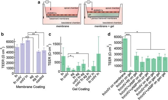
BACKGROUND: Transwell-based models of the blood-brain barrier (BBB) incorporating monolayers of human brain microvascular endothelial cells (dhBMECs) derived from induced pluripotent stem cells show many of the key features of the BBB, including expression of transporters and efflux pumps, expression of tight junction proteins, and physiological values of transendothelial electrical resistance. The fabrication of 3D BBB models using dhBMECs has so far been unsuccessful due to the poor adhesion and survival of these cells on matrix materials commonly used in tissue engineering. METHODS: To address this issue, we systematically screened a wide range of matrix materials (collagen I, hyaluronic acid, and fibrin), compositions (laminin/entactin), protein coatings (fibronectin, laminin, collagen IV, perlecan, and agrin), and soluble factors (ROCK inhibitor and cyclic adenosine monophosphate) in 2D culture to assess cell adhesion, spreading, and barrier function. RESULTS: collagen I gels coated with basement membrane proteins (fibronectin, collagen IV, and laminin), cell coverage was high but did not reliably reach confluence. The transendothelial electrical resistance (TEER) on collagen I gels coated with basement membrane proteins was lower than on coated transwell membranes. Agrin, a heparin sulfate proteoglycan found in basement membranes of the brain, promoted monolayer formation but resulted in a significant decrease in transendothelial electrical resistance (TEER). However, the addition of ROCK inhibitor, cAMP, or cross-linking the gels to increase stiffness, resulted in a significant improvement of TEER values and enabled the formation of confluent monolayers. CONCLUSIONS: Having identified matrix compositions that promote monolayer formation and barrier function, we successfully fabricated dhBMEC microvessels in cross-linked collagen I gels coated with fibronectin and collagen IV, and treated with ROCK inhibitor and cAMP. We measured apparent permeability values for Lucifer yellow, comparable to values obtained in the transwell assay. During these experiments we observed no focal leaks, suggesting the formation of tight junctions that effectively block paracellular transport.
Overview
The claims and statistics of this paper haven’t been checked yet.
Free: it reads the open-access text and checks the first 5 claims that cite a source we can check. Create a free account to check every claim.
Integrity signals
Registry
Cites
Statistics
Figures



Verification
Claims the paper makes
The claims and statistics of this paper haven’t been checked yet.
Free: it reads the open-access text and checks the first 5 claims that cite a source we can check. Create a free account to check every claim.
Claims other papers make about this one
Each is checked against this paper’s own text: does it say what the citing paper says it does?
The source backs part of the claim, not all of it — or backs it for a different population, condition or comparison.
In the citing paper
“dhBMEC monolayers express a wide range of BBB markers, including tight junction (TJ) proteins (Fig. 1b), transporters, and efflux pumps, and typically attain transendothelial electrical resistance (TEER) > 1500 Ω cm2 [27, 28, 31, 34–37].”
From Role of iPSC-derived pericytes on barrier function of iPSC-derived brain microvascular endothelial cells in 2D and 3D (2019) · cites this paper as [27]
Experimental — accuracy not yet measured
Self-check
Percentages, sums and reported differences re-computed from the paper’s own numbers.
The self-check needs the paper’s full text.
Citations
Cites
Tissue Cells Feel and Respond to the Stiffness of Their Substrate
Discher, D. E., Janmey, P., & Wang, Y.-L. (2005) · Science · doi:10.1126/science.1116995 · Cited by 6,382
Astrocyte-endothelial interactions at the blood-brain barrier
Abbott, N. J., Rönnbäck, L., & Hansson, E. (2006) · Nature Reviews Neuroscience · doi:10.1038/nrn1824 · Cited by 5,595
Cerebral organoids model human brain development and microcephaly
Lancaster, M. A., Renner, M., Martin, C.-A., Wenzel, D., Bicknell, L. S., Hurles, M. E., Homfray, T., Penninger, J. M., Jackson, A. P., & Knoblich, J. A. (2013) · Nature · doi:10.1038/nature12517 · Cited by 5,372 · cited 1×
“The laminin concentrations were based on previous studies of neuronal outgrowth and cerebral organoid development [35, 36].”
MethodNew Perspectives in Cell Adhesion: RGD and Integrins
Ruoslahti, E., & Pierschbacher, M. D. (1987) · Science · doi:10.1126/science.2821619 · Cited by 4,865
Cell Adhesion: The Molecular Basis of Tissue Architecture and Morphogenesis
Gumbiner, B. M. (1996) · Cell · doi:10.1016/s0092-8674(00)81279-9 · Cited by 3,471
Effects of substrate stiffness on cell morphology, cytoskeletal structure, and adhesion
Yeung, T., Georges, P. C., Flanagan, L. A., Marg, B., Ortiz, M., Funaki, M., Zahir, N., Ming, W., Weaver, V., & Janmey, P. A. (2005) · Cell Motility and the Cytoskeleton · doi:10.1002/cm.20041 · Cited by 2,325
A ROCK inhibitor permits survival of dissociated human embryonic stem cells
Watanabe, K., Ueno, M., Kamiya, D., Nishiyama, A., Matsumura, M., Wataya, T., Takahashi, J. B., Nishikawa, S., Nishikawa, S.-I., Muguruma, K., et al. (2007) · Nature Biotechnology · doi:10.1038/nbt1310 · Cited by 2,055
MDCK (Madin-Darby Canine Kidney) Cells: A Tool for Membrane Permeability Screening
Irvine, J. D., Takahashi, L., Lockhart, K., Cheong, J., Tolan, J. W., Selick, H. E., & Grove, J. R. (1999) · Journal of Pharmaceutical Sciences · doi:10.1021/js9803205 · Cited by 980
In vitro microvessels for the study of angiogenesis and thrombosis
Zheng, Y., Chen, J., Craven, M., Choi, N. W., Totorica, S., Diaz-Santana, A., Kermani, P., Hempstead, B., Fischbach-Teschl, C., López, J. A., et al. (2012) · Proceedings of the National Academy of Sciences · doi:10.1073/pnas.1201240109 · Cited by 894
Derivation of blood-brain barrier endothelial cells from human pluripotent stem cells
Lippmann, E. S., Azarin, S. M., Kay, J. E., Nessler, R. A., Wilson, H. K., Al-Ahmad, A., Palecek, S. P., & Shusta, E. V. (2012) · Nature Biotechnology · doi:10.1038/nbt.2247 · Cited by 800
Research works printed but not linked: 1
- Yeung T, Georges PC, Flanagan LA, Marg B, Ortiz M, Funaki M, Zahir N, Ming W, Weaver V, Janmey PA. Effects of substrate stiffness on cell morphology, cytoskeletal structure, and adhesion. Cell Motil Cytoskelet. 2005;60:24–34. · DOI · Paper page
Cited by
Two new Later Stone Age sites from the Final Pleistocene in the Falémé Valley, eastern Senegal
Ndiaye, M., Lespez, L., Tribolo, C., Rassé, M., Hadjas, I., Davidoux, S., Huysecom, É., & Douze, K. (2024) · Monash University Research Portal (Monash University) · doi:10.3929/ethz-b-000667478 · Cited by 23,868
Park, T.-E., Mustafaoglu, N., Herland, A., Hasselkus, R., Mannix, R., FitzGerald, E. A., Prantil-Baun, R., Watters, A., Henry, O., Benz, M., et al. (2019) · Nature Communications · doi:10.1038/s41467-019-10588-0 · Cited by 594
Wevers, N. R., Kasi, D. G., Gray, T., Wilschut, K. J., Smith, B., van Vught, R., Shimizu, F., Sano, Y., Kanda, T., Marsh, G., et al. (2018) · Fluids and Barriers of the CNS · doi:10.1186/s12987-018-0108-3 · Cited by 353
In Vitro Models of the Blood-Brain Barrier
Neuhaus, W. (2020) · Handbook of Experimental Pharmacology · doi:10.1007/164_2020_370 · Cited by 236
Linville, R. M., DeStefano, J. G., Sklar, M. B., Xu, Z., Farrell, A. M., Bogorad, M. I., Chu, C., Walczak, P., Cheng, L., Mahairaki, V., et al. (2019) · Biomaterials · doi:10.1016/j.biomaterials.2018.10.023 · Cited by 199
Self-citation
Metadata
- Authors
- Katt, M. E., Linville, R. M., Mayo, L. N., Xu, Z. S., & Searson, P. C.
- Affiliations
- Johns Hopkins University (US)
- Corresponding author
- Katt, Moriah E.
- Venue
- Fluids and Barriers of the CNS
- Published
- 2018-02-20
- Volume
- 15
- Issue
- 1
- Pages
- 7-7
- ISSN
- 2045-8118 (ISSN-L)
- PMC
- PMC5819713
- OpenAlex
- W2795689570
- Cited by
- 102
- References
- 71
- Citations by year
- 2026: 5 · 2025: 11 · 2024: 8 · 2023: 12 · 2022: 15 · 2021: 11 · 2020: 18 · 2019: 15 · 2018: 7
- FWCI
- 5.74
- Type
- journal article
- Language
- en
- Open access
- gold
- doi.org
- Licence
- cc-by
- creativecommons.org/licenses/by/4.0/ (tdm)
- creativecommons.org/licenses/by/4.0/ (vor)
- Locations
- Fluids and Barriers of the CNS (publishedVersion, cc-by)
- Europe PMC (PubMed Central) (publishedVersion, cc-by)
- PubMed Central (publishedVersion, cc-by)
- PubMed (publishedVersion)
- DOAJ (DOAJ: Directory of Open Access Journals) (submittedVersion, cc-by-sa)
- Funders
- Defense Threat Reduction Agency (HDTRA1-15-1-0046)
- Topics
- Life Sciences › Neuroscience › Neurology › Barrier Structure and Function Studies
- Physical Sciences › Engineering › Biomedical Engineering › 3D Printing in Biomedical Research
- Life Sciences › Biochemistry, Genetics and Molecular Biology › Cell Biology › Cellular Mechanics and Interactions
- Keywords
- blood-brain barrier, human brain microvascular endothelial cells, iPSC-derived endothelial cells, collagen I gel, basement membrane proteins, fibronectin, type IV collagen, agrin, ROCK inhibitor, cyclic AMP, transendothelial electrical resistance, cell adhesion, tight junctions, paracellular permeability, heparan sulfate proteoglycans, extracellular matrix composition
- MeSH terms
- Brain, Capillary Permeability, Cell Adhesion, Cell Line, Extracellular Matrix, Fibronectins, Fluorescent Antibody Technique, Humans, Electric Impedance, Cell Culture Techniques, Tight Junctions, Tissue Engineering, Fibril-Associated Collagens, Endothelial Cells, Green Fluorescent Proteins, Tissue Scaffolds, Microvessels, Induced Pluripotent Stem Cells
About these checks
Registry, retraction, Cites and statistics results are computed, not judged, with one exception: a printed reference that no index lists is read once by a model before it counts as a citation error. Claims are extracted by a model and kept only when their quote appears verbatim in the text; each cited source is then checked, with a model’s help, for whether it says what the claim relies on. Those verdicts can be wrong — read the claim’s quote and the cited source before relying on one.
Statistics and arithmetic are recomputed, not judged.
Confidence bands are shown only for fields calibrated on our annotated test set. None is calibrated yet, so no band is shown.
What counts as a claim ›How we measure confidence ›How we check this ›
Are you an author, or do you see a problem with this page? Request a review
How to cite
Cite CoChat for the checks on this page, and the paper for its own findings.
- This page
- CoChat. (2026). Citation check: Functional brain-specific microvessels from iPSC-derived human brain microvascular endothelial cells: the role of matrix composition on monolayer formation. https://app.cochat.ai/papers/10.1186/s12987-018-0092-7
- The paper
- Katt, M. E., Linville, R. M., Mayo, L. N., Xu, Z. S., & Searson, P. C. (2018). Functional brain-specific microvessels from iPSC-derived human brain microvascular endothelial cells: the role of matrix composition on monolayer formation. Fluids and Barriers of the CNS. https://doi.org/10.1186/s12987-018-0092-7
Data sources
Bibliographic data: Semantic Scholar, OpenAlex, Crossref.
Contains information from the Semantic Scholar Academic Graph Datasets, made available under the ODC Attribution License (ODC-BY 1.0).
Semantic Scholar’s data is described in: Kinney, R., et al. (2023). The Semantic Scholar Open Data Platform. arXiv:2301.10140.
