Cited by 24Interstitial Lung Diseases and Idiopathic Pulmonary FibrosisOccupational and environmental lung diseasesNanoparticles: synthesis and applications

The DOI matches its Crossref record; 2 of the 2 references it cites were found in a registry or an index.

MMP-3-mediated cleavage of OPN is involved in copper oxide nanoparticle-induced activation of fibroblasts

Open access
Zhang, Yuanbao, Mo, Yiqun, Zhang, Yue, Yuan, Jiali, Zhang, Qunwei·2023·Particle and Fibre Toxicology

Abstract

Background: Copper oxide nanoparticles (Nano-CuO) are one of the most produced and used nanomaterials. Previous studies have shown that exposure to Nano-CuO caused acute lung injury, inflammation, and fibrosis. However, the mechanisms underlying Nano-CuO-induced lung fibrosis are still unclear. Here, we hypothesized that exposure of human lung epithelial cells and macrophages to Nano-CuO would upregulate MMP-3, which cleaved osteopontin (OPN), resulting in fibroblast activation and lung fibrosis. Methods: A triple co-culture model was established to explore the mechanisms underlying Nano-CuO-induced fibroblast activation. Cytotoxicity of Nano-CuO on BEAS-2B, U937* macrophages, and MRC-5 fibroblasts were determined by alamarBlue and MTS assays. The expression or activity of MMP-3, OPN, and fibrosis-associated proteins was determined by Western blot or zymography assay. Migration of MRC-5 fibroblasts was evaluated by wound healing assay. MMP-3 siRNA and an RGD-containing peptide, GRGDSP, were used to explore the role of MMP-3 and cleaved OPN in fibroblast activation. Results: Exposure to non-cytotoxic doses of Nano-CuO (0.5 and 1 µg/mL) caused increased expression and activity of MMP-3 in the conditioned media of BEAS-2B and U937* cells, but not MRC-5 fibroblasts. Nano-CuO exposure also caused increased production of cleaved OPN fragments, which was abolished by MMP-3 siRNA transfection. Conditioned media from Nano-CuO-exposed BEAS-2B, U937*, or the co-culture of BEAS-2B and U937* caused activation of unexposed MRC-5 fibroblasts. However, direct exposure of MRC-5 fibroblasts to Nano-CuO did not induce their activation. In a triple co-culture system, exposure of BEAS-2B and U937* cells to Nano-CuO caused activation of unexposed MRC-5 fibroblasts, while transfection of MMP-3 siRNA in BEAS-2B and U937* cells significantly inhibited the activation and migration of MRC-5 fibroblasts. In addition, pretreatment with GRGDSP peptide inhibited Nano-CuO-induced activation and migration of MRC-5 fibroblasts in the triple co-culture system. Conclusions: Our results demonstrated that Nano-CuO exposure caused increased production of MMP-3 from lung epithelial BEAS-2B cells and U937* macrophages, which cleaved OPN, resulting in the activation of lung fibroblasts MRC-5. These results suggest that MMP-3-cleaved OPN may play a key role in Nano-CuO-induced activation of lung fibroblasts. More investigations are needed to confirm whether these effects are due to the nanoparticles themselves and/or Cu ions.

Overview

The claims and statistics of this paper haven’t been checked yet.

Run the check — free, no account needed. It reads the open-access text and checks the sources it cites.

A few free checks a day · papers already verified open instantly · results are public

Integrity signals

Registry

Crossref · 10.1186/s12989-023-00532-y

Cites

2 of 2 references verified0 citation errors · 0 not checkable

Statistics

Figures

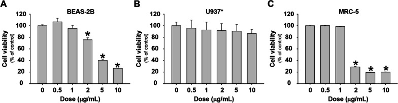
Fig. 1 Cytotoxicity of Nano-CuO on BEAS-2B cells, U937-derived macrophages (U937*), and MRC-5 fibroblasts. BEAS-2B (a), U937* macrophages (b), and MRC-5 fibroblasts (c) were seeded into 96-well plates and treated with 0.5, 1, 2, 5, and 10 µg/mL of Nano-CuO for 24 h (a and b) and 48 h (c), respectively. Cells without treatment were used as control. The cytotoxicity was determined by alamarBlue assay. Data represent mean ± SE (n = 6). * Significant difference as compared to the control group, p < 0.05
Fig. 1 Cytotoxicity of Nano-CuO on BEAS-2B cells, U937-derived macrophages (U937*), and MRC-5 fibroblasts. BEAS-2B (a), U937* macrophages (b), and MRC-5 fibroblasts (c) were seeded into 96-well plates and treated with 0.5, 1, 2, 5, and 10 µg/mL of Nano-CuO for 24 h (a and b) and 48 h (c), respectively. Cells without treatment were used as control. The cytotoxicity was determined by alamarBlue assay. Data represent mean ± SE (n = 6). * Significant difference as compared to the control group, p < 0.05CC BY · PubMed Central
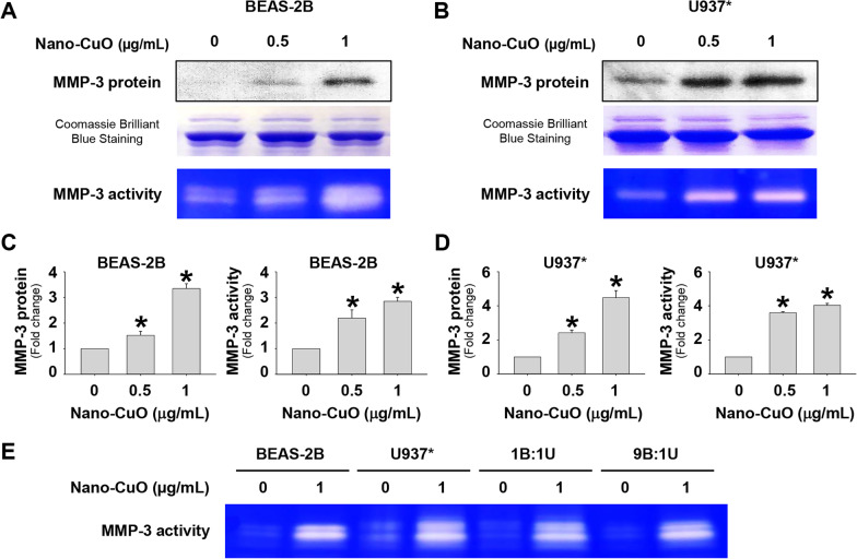
Fig. 2 Increased expression and activity of MMP-3 in the conditioned media of BEAS-2B cells and U937* macrophages exposed to Nano-CuO. BEAS-2B cells (a and c) and U937* macrophages (b and d) were exposed to 0.5 and 1 µg/mL of Nano-CuO for 12 h. After treatment, the conditioned media were collected. Cells without treatment were used as control. MMP-3 protein level in the conditioned media was detected by Western blot, while its activity was detected by β-casein zymography. Equal protein loading was verified by Coomassie Brilliant Blue staining. The activity of MMP-3 was also determined in the conditioned media from co-culture system (e). a, b, and e were the results of a single experiment, while c and d were normalized band densitometry readings averaged from three independent experiments. Data represent mean ± SE (n = 3). * Significant difference as compared to the control group, p < 0.05
Fig. 2 Increased expression and activity of MMP-3 in the conditioned media of BEAS-2B cells and U937* macrophages exposed to Nano-CuO. BEAS-2B cells (a and c) and U937* macrophages (b and d) were exposed to 0.5 and 1 µg/mL of Nano-CuO for 12 h. After treatment, the conditioned media were collected. Cells without treatment were used as control. MMP-3 protein level in the conditioned media was detected by Western blot, while its activity was detected by β-casein zymography. Equal protein loading was verified by Coomassie Brilliant Blue staining. The activity of MMP-3 was also determined in the conditioned media from co-culture system (e). a, b, and e were the results of a single experiment, while c and d were normalized band densitometry readings averaged from three independent experiments. Data represent mean ± SE (n = 3). * Significant difference as compared to the control group, p < 0.05CC BY · PubMed Central
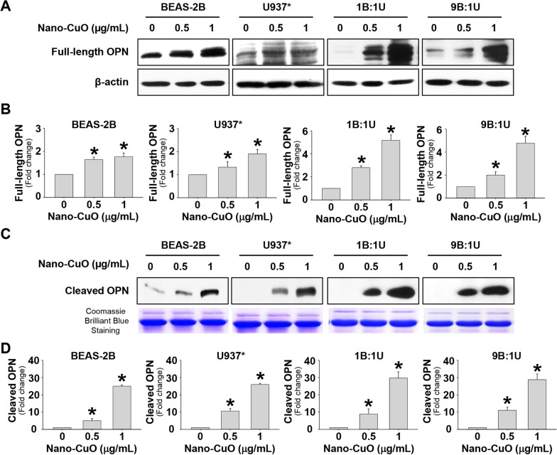
Fig. 3 Increased expression of OPN in BEAS-2B cells and U937* macrophages exposed to Nano-CuO. BEAS-2B cells, U937* macrophages, or co-culture of BEAS-2B and U937* macrophages at a ratio of 1:1 or 9:1 (BEAS-2B:U937*) were exposed to 0.5 and 1 µg/mL of Nano-CuO for 12 h. After treatment, the cells and conditioned media were collected. Cells without treatment were used as control. Full-length OPN (66 kDa) in the cells and cleaved OPN (40 kDa N-terminal fragment) in the conditioned media were detected by Western blot. Equal protein loading was verified by β-actin expression (a and b) or Coomassie Brilliant Blue staining (c and d). a and c were the results of a single experiment. b and d were results of three independent experiments. Data represent mean ± SE (n = 3). * Significant difference as compared to the control group, p < 0.05
Fig. 3 Increased expression of OPN in BEAS-2B cells and U937* macrophages exposed to Nano-CuO. BEAS-2B cells, U937* macrophages, or co-culture of BEAS-2B and U937* macrophages at a ratio of 1:1 or 9:1 (BEAS-2B:U937*) were exposed to 0.5 and 1 µg/mL of Nano-CuO for 12 h. After treatment, the cells and conditioned media were collected. Cells without treatment were used as control. Full-length OPN (66 kDa) in the cells and cleaved OPN (40 kDa N-terminal fragment) in the conditioned media were detected by Western blot. Equal protein loading was verified by β-actin expression (a and b) or Coomassie Brilliant Blue staining (c and d). a and c were the results of a single experiment. b and d were results of three independent experiments. Data represent mean ± SE (n = 3). * Significant difference as compared to the control group, p < 0.05CC BY · PubMed Central
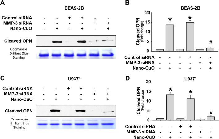
Fig. 4 The role of MMP-3 on Nano-CuO-induced production of cleaved OPN in BEAS-2B cells and U937* macrophages. BEAS-2B cells (a and b) and U937* macrophages (c and d) were transfected with 30 nM of MMP-3 siRNA or Negative Control No. 2 siRNA. After transfection, the cells were exposed to 1 µg/mL of Nano-CuO for 12 h. The conditioned media were collected to detect the level of cleaved OPN (40 kDa N-terminal fragment) by Western blot. Equal protein loading was verified by Coomassie Brilliant Blue staining. a and c were the results of a single experiment. b and d were normalized band densitometry readings averaged from three independent experiments. Data represent mean ± SE (n = 3). * Significant difference as compared to the control group, p < 0.05; # Significant difference as compared to the Nano-CuO-treated group transfected with Negative Control No. 2 siRNA, p < 0.05
Fig. 4 The role of MMP-3 on Nano-CuO-induced production of cleaved OPN in BEAS-2B cells and U937* macrophages. BEAS-2B cells (a and b) and U937* macrophages (c and d) were transfected with 30 nM of MMP-3 siRNA or Negative Control No. 2 siRNA. After transfection, the cells were exposed to 1 µg/mL of Nano-CuO for 12 h. The conditioned media were collected to detect the level of cleaved OPN (40 kDa N-terminal fragment) by Western blot. Equal protein loading was verified by Coomassie Brilliant Blue staining. a and c were the results of a single experiment. b and d were normalized band densitometry readings averaged from three independent experiments. Data represent mean ± SE (n = 3). * Significant difference as compared to the control group, p < 0.05; # Significant difference as compared to the Nano-CuO-treated group transfected with Negative Control No. 2 siRNA, p < 0.05CC BY · PubMed Central
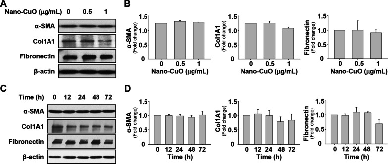
Fig. 5 Direct Nano-CuO exposure had no effects on activation of MRC-5 fibroblasts. MRC-5 cells were treated with 0.5 and 1 µg/mL of Nano-CuO for 48 h (a and b) or with 1 µg/mL of Nano-CuO for 12, 24, 48, and 72 h (c and d). Cells without treatment were used as control. Proteins were isolated from the cells to detect the expression of α-SMA, Col1A1, and fibronectin. a and c were the results of a single Western blot experiment. b and d were the average expression levels of α-SMA, Col1A1, and fibronectin normalized to β-actin from three independent experiments. Data represent mean ± SE (n = 3)
Fig. 5 Direct Nano-CuO exposure had no effects on activation of MRC-5 fibroblasts. MRC-5 cells were treated with 0.5 and 1 µg/mL of Nano-CuO for 48 h (a and b) or with 1 µg/mL of Nano-CuO for 12, 24, 48, and 72 h (c and d). Cells without treatment were used as control. Proteins were isolated from the cells to detect the expression of α-SMA, Col1A1, and fibronectin. a and c were the results of a single Western blot experiment. b and d were the average expression levels of α-SMA, Col1A1, and fibronectin normalized to β-actin from three independent experiments. Data represent mean ± SE (n = 3)CC BY · PubMed Central
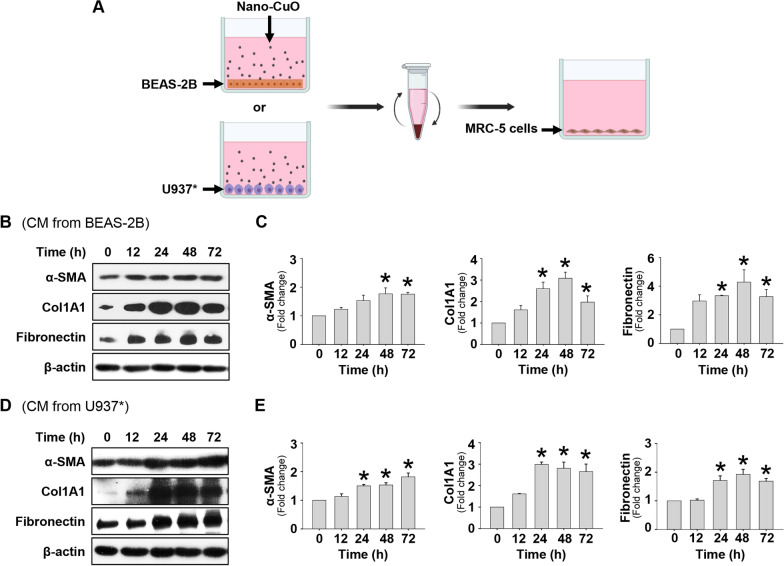
Fig. 6 Activation of MRC-5 fibroblasts cultured in the conditioned media from Nano-CuO-exposed BEAS-2B or U937* cells. a is the experimental protocol. BEAS-2B or U937* cells were treated with 1 µg/mL of Nano-CuO for 12 h and the conditioned media (CM) were collected to culture MRC-5 fibroblasts. After 0, 12, 24, 48, and 72 h culture, the MRC-5 fibroblasts were collected for protein isolation and Western blot. b and d were the results of a single Western blot experiment. c and e were the average expression levels of α-SMA, Col1A1, and fibronectin normalized to β-actin from three independent experiments. Data represent mean ± SE (n = 3). * Significant difference as compared to the control group, p < 0.05. Graphics in a were created with BioRender (https://BioRender.com)
Fig. 6 Activation of MRC-5 fibroblasts cultured in the conditioned media from Nano-CuO-exposed BEAS-2B or U937* cells. a is the experimental protocol. BEAS-2B or U937* cells were treated with 1 µg/mL of Nano-CuO for 12 h and the conditioned media (CM) were collected to culture MRC-5 fibroblasts. After 0, 12, 24, 48, and 72 h culture, the MRC-5 fibroblasts were collected for protein isolation and Western blot. b and d were the results of a single Western blot experiment. c and e were the average expression levels of α-SMA, Col1A1, and fibronectin normalized to β-actin from three independent experiments. Data represent mean ± SE (n = 3). * Significant difference as compared to the control group, p < 0.05. Graphics in a were created with BioRender (https://BioRender.com)CC BY · PubMed Central
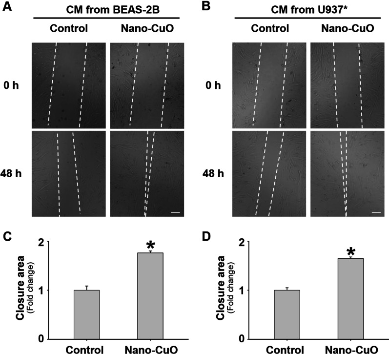
Fig. 7 Conditioned media from Nano-CuO-exposed BEAS-2B or U937* cells enhanced migration of unexposed MRC-5 fibroblasts. BEAS-2B or U937* cells were treated with 0 or 1 µg/mL of Nano-CuO for 12 h and the conditioned media (CM) were collected to culture MRC-5 fibroblasts for 48 h. Migration of MRC-5 cells was detected by wound healing assay. a and b were representative images. c and d were the average closure area from three independent experiments. Scale bars in a and b represent 200 µm. Data represent mean ± SE (n = 3). * Significant difference as compared to the control group, p < 0.05
Fig. 7 Conditioned media from Nano-CuO-exposed BEAS-2B or U937* cells enhanced migration of unexposed MRC-5 fibroblasts. BEAS-2B or U937* cells were treated with 0 or 1 µg/mL of Nano-CuO for 12 h and the conditioned media (CM) were collected to culture MRC-5 fibroblasts for 48 h. Migration of MRC-5 cells was detected by wound healing assay. a and b were representative images. c and d were the average closure area from three independent experiments. Scale bars in a and b represent 200 µm. Data represent mean ± SE (n = 3). * Significant difference as compared to the control group, p < 0.05CC BY · PubMed Central
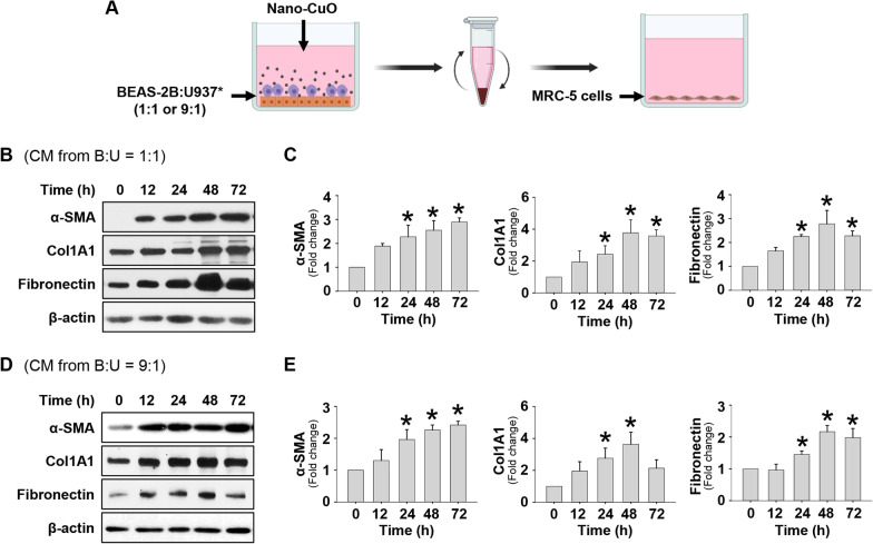
Fig. 8 Cultured in the conditioned media from Nano-CuO-exposed co-culture of BEAS-2B and U937* cells caused activation of unexposed MRC-5 fibroblasts. a is the experimental protocol of the co-culture experiments. Co-culture of BEAS-2B and U937* cells with ratio of 1:1 or 9:1 (BEAS-2B:U937*) was treated with 1 µg/mL of Nano-CuO for 12 h and the conditioned media (CM) were collected. MRC-5 fibroblasts were then cultured in the conditioned media for 0, 12, 24, 48, and 72 h. Proteins from MRC-5 cells were isolated to detect the expression of α-SMA, Col1A1, and fibronectin. b and d were the results of a single Western blot experiment. c and e were the average expression levels of α-SMA, Col1A1, and fibronectin normalized to β-actin from three independent experiments. Data represent mean ± SE (n = 3). * Significant difference as compared to the control group, p < 0.05. Graphics in a were created with BioRender (https://BioRender.com)
Fig. 8 Cultured in the conditioned media from Nano-CuO-exposed co-culture of BEAS-2B and U937* cells caused activation of unexposed MRC-5 fibroblasts. a is the experimental protocol of the co-culture experiments. Co-culture of BEAS-2B and U937* cells with ratio of 1:1 or 9:1 (BEAS-2B:U937*) was treated with 1 µg/mL of Nano-CuO for 12 h and the conditioned media (CM) were collected. MRC-5 fibroblasts were then cultured in the conditioned media for 0, 12, 24, 48, and 72 h. Proteins from MRC-5 cells were isolated to detect the expression of α-SMA, Col1A1, and fibronectin. b and d were the results of a single Western blot experiment. c and e were the average expression levels of α-SMA, Col1A1, and fibronectin normalized to β-actin from three independent experiments. Data represent mean ± SE (n = 3). * Significant difference as compared to the control group, p < 0.05. Graphics in a were created with BioRender (https://BioRender.com)CC BY · PubMed Central
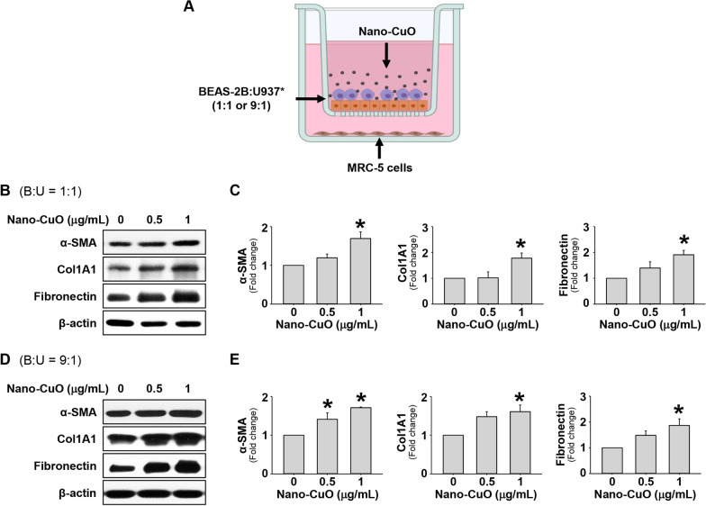
Fig. 9 Indirect exposure to Nano-CuO caused activation of MRC-5 fibroblasts in a triple co-culture system. a is the experimental protocol for the triple co-culture experiments. BEAS-2B cells and U937* cells were seeded at the ratio of 1:1 (b and c) or 9:1 (d and e) (BEAS-2B:U937*) in the inserts of Transwell® units and MRC-5 cells were seeded in the lower chambers of the units. After establishment of the triple co-culture system, BEAS-2B and U937* cells were exposed to 0, 0.5 and 1 µg/mL of Nano-CuO for 12 h, then the inserts were taken out. MRC-5 cells in the lower chamber were incubated for another 36 h and proteins were isolated to detect the expression of α-SMA, Col1A1, and fibronectin. b and d were the results of a single Western blot experiment. c and e were the average expression levels of α-SMA, Col1A1, and fibronectin normalized to β-actin from three independent experiments. Data represent mean ± SE (n = 3). * Significant difference as compared to the control group, p < 0.05. Graphics in a were created with BioRender (https://BioRender.com)
Fig. 9 Indirect exposure to Nano-CuO caused activation of MRC-5 fibroblasts in a triple co-culture system. a is the experimental protocol for the triple co-culture experiments. BEAS-2B cells and U937* cells were seeded at the ratio of 1:1 (b and c) or 9:1 (d and e) (BEAS-2B:U937*) in the inserts of Transwell® units and MRC-5 cells were seeded in the lower chambers of the units. After establishment of the triple co-culture system, BEAS-2B and U937* cells were exposed to 0, 0.5 and 1 µg/mL of Nano-CuO for 12 h, then the inserts were taken out. MRC-5 cells in the lower chamber were incubated for another 36 h and proteins were isolated to detect the expression of α-SMA, Col1A1, and fibronectin. b and d were the results of a single Western blot experiment. c and e were the average expression levels of α-SMA, Col1A1, and fibronectin normalized to β-actin from three independent experiments. Data represent mean ± SE (n = 3). * Significant difference as compared to the control group, p < 0.05. Graphics in a were created with BioRender (https://BioRender.com)CC BY · PubMed Central
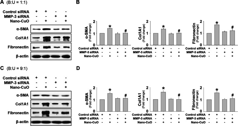
Fig. 10 The role of MMP-3 on Nano-CuO-induced activation of MRC-5 fibroblasts in the triple co-culture system. BEAS-2B and U937* cells were seeded at the ratio of 1:1 (a and b) or 9:1 (c and d) (BEAS-2B:U937*) in the inserts of Transwell® units and transfected with 30 nM of MMP-3 siRNA or Negative Control No. 2 siRNA. After transfection, the cells were exposed to 1 µg/mL of Nano-CuO for 12 h. After exposure, the inserts were taken out and the MRC-5 fibroblasts seeded in the lower chamber were incubated for another 36 h. The proteins isolated from MRC-5 fibroblasts were used to detect the expression levels of α-SMA, Col1A1, and fibronectin by Western blot. a and c were the results of a single Western blot experiment. b and d were the average expression level of α-SMA, Col1A1, and fibronectin normalized to β-actin from three independent experiments. Data represent mean ± SE (n = 3). * Significant difference as compared to the control group, p < 0.05; # Significant difference as compared to the Nano-CuO-treated group transfected with Negative Control No. 2 siRNA, p < 0.05
Fig. 10 The role of MMP-3 on Nano-CuO-induced activation of MRC-5 fibroblasts in the triple co-culture system. BEAS-2B and U937* cells were seeded at the ratio of 1:1 (a and b) or 9:1 (c and d) (BEAS-2B:U937*) in the inserts of Transwell® units and transfected with 30 nM of MMP-3 siRNA or Negative Control No. 2 siRNA. After transfection, the cells were exposed to 1 µg/mL of Nano-CuO for 12 h. After exposure, the inserts were taken out and the MRC-5 fibroblasts seeded in the lower chamber were incubated for another 36 h. The proteins isolated from MRC-5 fibroblasts were used to detect the expression levels of α-SMA, Col1A1, and fibronectin by Western blot. a and c were the results of a single Western blot experiment. b and d were the average expression level of α-SMA, Col1A1, and fibronectin normalized to β-actin from three independent experiments. Data represent mean ± SE (n = 3). * Significant difference as compared to the control group, p < 0.05; # Significant difference as compared to the Nano-CuO-treated group transfected with Negative Control No. 2 siRNA, p < 0.05CC BY · PubMed Central
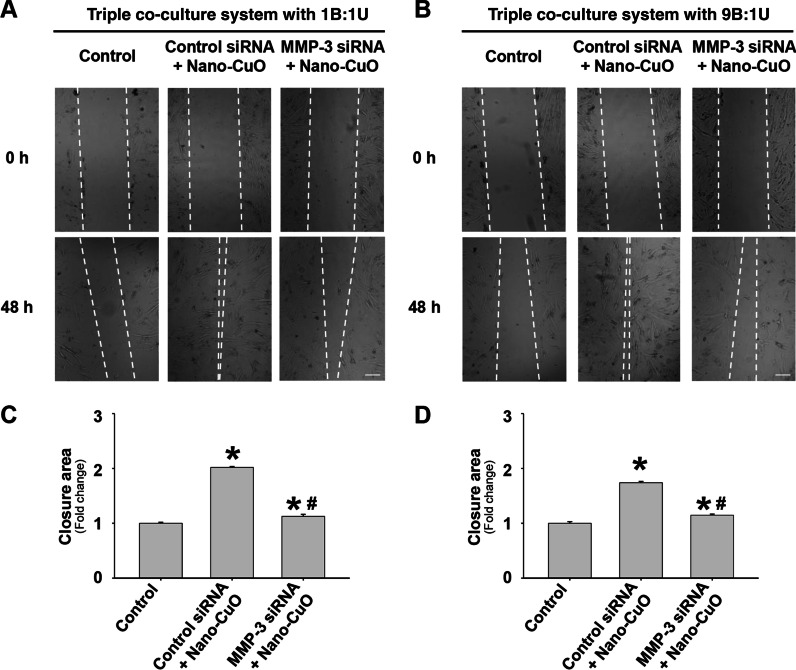
Fig. 11 MMP-3 siRNA transfection attenuated Nano-CuO-induced migration of MRC-5 fibroblasts in the triple co-culture system. BEAS-2B and U937* cells were seeded at a ratio of 1:1 (a and c) or 9:1 (b and d) (BEAS-2B:U937*) in the inserts of Transwell® units and transfected with 30 nM of MMP-3 siRNA or Negative Control No. 2 siRNA. After transfection, the cells were exposed to 1 µg/mL of Nano-CuO for 12 h. After exposure, the inserts were taken out and the MRC-5 fibroblasts seeded in the lower chamber were incubated for another 36 h. Migration of MRC-5 cells was detected by wound healing assay. Cells without any treatments were used as the control. a and b were representative images. c and d were the average closure area from three independent experiments. Scale bars in a and b represent 200 µm. Data represent mean ± SE (n = 3). * Significant difference as compared to the control group, p < 0.05; # Significant difference as compared to the Nano-CuO-treated group with control siRNA transfection, p < 0.05
Fig. 11 MMP-3 siRNA transfection attenuated Nano-CuO-induced migration of MRC-5 fibroblasts in the triple co-culture system. BEAS-2B and U937* cells were seeded at a ratio of 1:1 (a and c) or 9:1 (b and d) (BEAS-2B:U937*) in the inserts of Transwell® units and transfected with 30 nM of MMP-3 siRNA or Negative Control No. 2 siRNA. After transfection, the cells were exposed to 1 µg/mL of Nano-CuO for 12 h. After exposure, the inserts were taken out and the MRC-5 fibroblasts seeded in the lower chamber were incubated for another 36 h. Migration of MRC-5 cells was detected by wound healing assay. Cells without any treatments were used as the control. a and b were representative images. c and d were the average closure area from three independent experiments. Scale bars in a and b represent 200 µm. Data represent mean ± SE (n = 3). * Significant difference as compared to the control group, p < 0.05; # Significant difference as compared to the Nano-CuO-treated group with control siRNA transfection, p < 0.05CC BY · PubMed Central
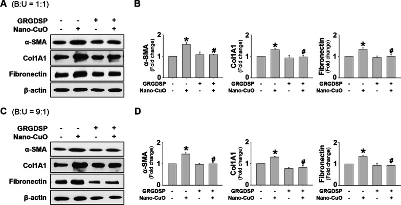
Fig. 12 The role of MMP-3-cleaved OPN in Nano-CuO-induced activation of MRC-5 fibroblasts in the triple co-culture system. BEAS-2B and U937* cells were seeded at the ratio of 1:1 (a and b) or 9:1 (c and d) (BEAS-2B:U937*) in the inserts, and MRC-5 fibroblasts were seeded in the lower chambers of Transwell® units. MRC-5 fibroblasts were pretreated with 200 µg/mL of GRGDSP peptides, then BEAS-2B and U937* cells were exposed to 1 µg/mL of Nano-CuO for 12 h. After exposure, the inserts were taken out and the MRC-5 fibroblasts were incubated for another 36 h. The proteins from MRC-5 fibroblasts were isolated to determine the activation of MRC-5 fibroblasts by Western blot. a and c were the results of a single Western blot experiment. b and d were the average expression levels of α-SMA, Col1A1, and fibronectin normalized to β-actin from three independent experiments. Data represent mean ± SE (n = 3). * Significant difference as compared to the control group, p < 0.05; # Significant difference as compared to the Nano-CuO-treated alone group, p < 0.05
Fig. 12 The role of MMP-3-cleaved OPN in Nano-CuO-induced activation of MRC-5 fibroblasts in the triple co-culture system. BEAS-2B and U937* cells were seeded at the ratio of 1:1 (a and b) or 9:1 (c and d) (BEAS-2B:U937*) in the inserts, and MRC-5 fibroblasts were seeded in the lower chambers of Transwell® units. MRC-5 fibroblasts were pretreated with 200 µg/mL of GRGDSP peptides, then BEAS-2B and U937* cells were exposed to 1 µg/mL of Nano-CuO for 12 h. After exposure, the inserts were taken out and the MRC-5 fibroblasts were incubated for another 36 h. The proteins from MRC-5 fibroblasts were isolated to determine the activation of MRC-5 fibroblasts by Western blot. a and c were the results of a single Western blot experiment. b and d were the average expression levels of α-SMA, Col1A1, and fibronectin normalized to β-actin from three independent experiments. Data represent mean ± SE (n = 3). * Significant difference as compared to the control group, p < 0.05; # Significant difference as compared to the Nano-CuO-treated alone group, p < 0.05CC BY · PubMed Central
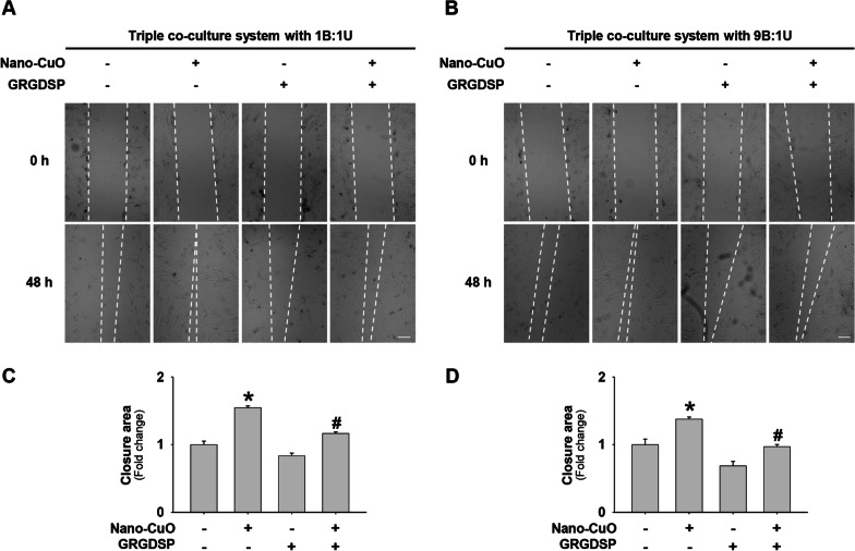
Fig. 13 Pretreatment of GRGDSP peptides inhibited Nano-CuO-induced migration of MRC-5 fibroblasts in the triple co-culture system. BEAS-2B and U937* cells were seeded at the ratio of 1:1 (a and c) or 9:1 (b and d) (BEAS-2B:U937*) in the inserts and MRC-5 fibroblasts were seeded in the lower chambers of Transwell® units. MRC-5 fibroblasts were pretreated with 200 µg/mL of GRGDSP peptides, then BEAS-2B and U937* cells were exposed to 1 µg/mL of Nano-CuO for 12 h. After exposure, the inserts were taken out and the MRC-5 fibroblasts were incubated for another 36 h. Migration of MRC-5 cells was detected by wound healing assay. a and b were representative images. c and d were the average closure area from three independent experiments. Scale bars in a and b represent 200 µm. Data represent mean ± SE (n = 3). * Significant difference as compared to the control group, p < 0.05; # Significant difference as compared to the Nano-CuO-treated alone group, p < 0.05
Fig. 13 Pretreatment of GRGDSP peptides inhibited Nano-CuO-induced migration of MRC-5 fibroblasts in the triple co-culture system. BEAS-2B and U937* cells were seeded at the ratio of 1:1 (a and c) or 9:1 (b and d) (BEAS-2B:U937*) in the inserts and MRC-5 fibroblasts were seeded in the lower chambers of Transwell® units. MRC-5 fibroblasts were pretreated with 200 µg/mL of GRGDSP peptides, then BEAS-2B and U937* cells were exposed to 1 µg/mL of Nano-CuO for 12 h. After exposure, the inserts were taken out and the MRC-5 fibroblasts were incubated for another 36 h. Migration of MRC-5 cells was detected by wound healing assay. a and b were representative images. c and d were the average closure area from three independent experiments. Scale bars in a and b represent 200 µm. Data represent mean ± SE (n = 3). * Significant difference as compared to the control group, p < 0.05; # Significant difference as compared to the Nano-CuO-treated alone group, p < 0.05CC BY · PubMed Central
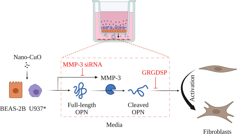
Fig. 14 Schematic diagram of potential mechanisms of Nano-CuO-induced activation of fibroblasts. Exposure of lung epithelial BEAS-2B cells and U937* macrophages to Nano-CuO caused increased expression and activity of MMP-3 and increased production of MMP-3-cleaved OPN, leading to the activation of fibroblasts. Graphics were created with BioRender (https://BioRender.com)
Fig. 14 Schematic diagram of potential mechanisms of Nano-CuO-induced activation of fibroblasts. Exposure of lung epithelial BEAS-2B cells and U937* macrophages to Nano-CuO caused increased expression and activity of MMP-3 and increased production of MMP-3-cleaved OPN, leading to the activation of fibroblasts. Graphics were created with BioRender (https://BioRender.com)CC BY · PubMed Central

Verification

Claims the paper makes

The claims and statistics of this paper haven’t been checked yet.

Run the check — free, no account needed. It reads the open-access text and checks the sources it cites.

A few free checks a day · papers already verified open instantly · results are public

Claims other papers make about this one

Each is checked against this paper’s own text: does it say what the citing paper says it does?

Self-check

Percentages, sums and reported differences re-computed from the paper’s own numbers.

The self-check needs the paper’s full text.

Citations

Cites

Show all 2 references

Research works printed but not linked: 80

  1. Colvin VL. The potential environmental impact of engineered nanomaterials. Nat Biotechnol. 2003;21(10):1166–70. · DOI
  2. Niska K, Zielinska E, Radomski MW, et al. Metal nanoparticles in dermatology and cosmetology: interactions with human skin cells. Chem Biol Interact. 2018;295:38–51. · DOI
  3. Stankic S, Suman S, Haque F, et al. Pure and multi metal oxide nanoparticles: synthesis, antibacterial and cytotoxic properties. J Nanobiotechnol. 2016;14(1):73. · DOI
  4. Bhaumik A, Shearin AM, Patel R, et al. Significant enhancement of optical absorption through nano-structuring of copper based oxide semiconductors: possible future materials for solar energy applications. Phys Chem Chem Phys. 2014;16(22):11054–66. · DOI
  5. Longano D, Ditaranto N, Cioffi N, et al. Analytical characterization of laser-generated copper nanoparticles for antibacterial composite food packaging. Anal Bioanal Chem. 2012;403(4):1179–86. · DOI
  6. Ren G, Hu D, Cheng EW, et al. Characterisation of copper oxide nanoparticles for antimicrobial applications. Int J Antimicrob Agents. 2009;33(6):587–90. · DOI
  7. Naz S, Gul A, Zia M. Toxicity of copper oxide nanoparticles: a review study. IET Nanobiotechnol. 2020;14(1):1–13. · DOI
  8. Costa PM, Gosens I, Williams A, et al. Transcriptional profiling reveals gene expression changes associated with inflammation and cell proliferation following short-term inhalation exposure to copper oxide nanoparticles. J Appl Toxicol. 2018;38(3):385–97. · DOI
  9. Juganson K, Ivask A, Blinova I, et al. NanoE-Tox: new and in-depth database concerning ecotoxicity of nanomaterials. Beilstein J Nanotechnol. 2015;6:1788–804. · DOI
  10. Karlsson HL, Cronholm P, Gustafsson J, et al. Copper oxide nanoparticles are highly toxic: a comparison between metal oxide nanoparticles and carbon nanotubes. Chem Res Toxicol. 2008;21(9):1726–32. · DOI · Paper page
  11. Ko JW, Shin NR, Park JW, et al. Copper oxide nanoparticles induce collagen deposition via TGF-beta1/Smad3 signaling in human airway epithelial cells. Nanotoxicology. 2018;12(3):239–50. · DOI
  12. Lai X, Zhao H, Zhang Y, et al. Intranasal delivery of copper oxide nanoparticles induces pulmonary toxicity and fibrosis in C57BL/6 mice. Sci Rep. 2018;8(1):4499. · DOI
  13. Zhang Y, Mo Y, Yuan J, et al. MMP-3 activation is involved in copper oxide nanoparticle-induced Epithelial–mesenchymal transition in human lung epithelial cells. Nanotoxicology. 2021;15(10):1380–402. · DOI
  14. Yamashita CM, Dolgonos L, Zemans RL, et al. Matrix metalloproteinase 3 is a mediator of pulmonary fibrosis. Am J Pathol. 2011;179(4):1733–45. · DOI
  15. Darby IA, Hewitson TD. Fibroblast differentiation in wound healing and fibrosis. Int Rev Cytol. 2007;257:143–79. · DOI
  16. Dong J, Ma Q. Osteopontin enhances multi-walled carbon nanotube-triggered lung fibrosis by promoting TGF-beta1 activation and myofibroblast differentiation. Part Fibre Toxicol. 2017;14(1):18. · DOI
  17. Bonner JC. Mesenchymal cell survival in airway and interstitial pulmonary fibrosis. Fibrogenesis Tissue Repair. 2010;3:15. · DOI
  18. Dong J, Ma Q. Advances in mechanisms and signaling pathways of carbon nanotube toxicity. Nanotoxicology. 2015;9(5):658–76. · DOI
  19. Dong J, Ma Q. Myofibroblasts and lung fibrosis induced by carbon nanotube exposure. Part Fibre Toxicol. 2016;13(1):60. · DOI
  20. Hussain S, Sangtian S, Anderson SM, et al. Inflammasome activation in airway epithelial cells after multi-walled carbon nanotube exposure mediates a profibrotic response in lung fibroblasts. Part Fibre Toxicol. 2014;11:28. · DOI
  21. Kabadi PK, Rodd AL, Simmons AE, et al. A novel human 3D lung microtissue model for nanoparticle-induced cell-matrix alterations. Part Fibre Toxicol. 2019;16(1):15. · DOI
  22. Ryman-Rasmussen JP, Tewksbury EW, Moss OR, et al. Inhaled multiwalled carbon nanotubes potentiate airway fibrosis in murine allergic asthma. Am J Respir Cell Mol Biol. 2009;40(3):349–58. · DOI
  23. Snyder-Talkington BN, Schwegler-Berry D, Castranova V, et al. Multi-walled carbon nanotubes induce human microvascular endothelial cellular effects in an alveolar-capillary co-culture with small airway epithelial cells. Part Fibre Toxicol. 2013;10:35. · DOI
  24. Venter C, Niesler C. A triple co-culture method to investigate the effect of macrophages and fibroblasts on myoblast proliferation and migration. Biotechniques. 2018;64(2):52–8. · DOI
  25. Wang P, Nie X, Wang Y, et al. Multiwall carbon nanotubes mediate macrophage activation and promote pulmonary fibrosis through TGF-beta/Smad signaling pathway. Small. 2013;9(22):3799–811. · DOI
  26. Xanthou G, Alissafi T, Semitekolou M, et al. Osteopontin has a crucial role in allergic airway disease through regulation of dendritic cell subsets. Nat Med. 2007;13(5):570–8. · DOI
  27. Ophascharoensuk V, Giachelli CM, Gordon K, et al. Obstructive uropathy in the mouse: role of osteopontin in interstitial fibrosis and apoptosis. Kidney Int. 1999;56(2):571–80. · DOI
  28. Rangaswami H, Bulbule A, Kundu GC. Osteopontin: role in cell signaling and cancer progression. Trends Cell Biol. 2006;16(2):79–87. · DOI
  29. Wang KX, Denhardt DT. Osteopontin: role in immune regulation and stress responses. Cytokine Growth Factor Rev. 2008;19(5–6):333–45. · DOI · Paper page
  30. Pardo A, Gibson K, Cisneros J, et al. Up-regulation and profibrotic role of osteopontin in human idiopathic pulmonary fibrosis. PLoS Med. 2005;2(9): e251. · DOI
  31. Samitas K, Zervas E, Vittorakis S, et al. Osteopontin expression and relation to disease severity in human asthma. Eur Respir J. 2011;37(2):331–41. · DOI
  32. Simoes DC, Xanthou G, Petrochilou K, et al. Osteopontin deficiency protects against airway remodeling and hyperresponsiveness in chronic asthma. Am J Respir Crit Care Med. 2009;179(10):894–902. · DOI
  33. Takahashi F, Takahashi K, Okazaki T, et al. Role of osteopontin in the pathogenesis of bleomycin-induced pulmonary fibrosis. Am J Respir Cell Mol Biol. 2001;24(3):264–71. · DOI
  34. Khaliullin TO, Kisin ER, Murray AR, et al. Mediation of the single-walled carbon nanotubes induced pulmonary fibrogenic response by osteopontin and TGF-beta1. Exp Lung Res. 2017;43(8):311–26. · DOI
  35. Denhardt DT, Noda M, O’Regan AW, et al. Osteopontin as a means to cope with environmental insults: regulation of inflammation, tissue remodeling, and cell survival. J Clin Invest. 2001;107(9):1055–61. · DOI
  36. Senger DR, Perruzzi CA. Cell migration promoted by a potent GRGDS-containing thrombin-cleavage fragment of osteopontin. Biochim Biophys Acta. 1996;1314(1–2):13–24. · DOI
  37. Takahashi K, Takahashi F, Tanabe KK, et al. The carboxyl-terminal fragment of osteopontin suppresses arginine-glycine-asparatic acid-dependent cell adhesion. Biochem Mol Biol Int. 1998;46(6):1081–92.
  38. Agnihotri R, Crawford HC, Haro H, et al. Osteopontin, a novel substrate for matrix metalloproteinase-3 (stromelysin-1) and matrix metalloproteinase-7 (matrilysin). J Biol Chem. 2001;276(30):28261–7. · DOI
  39. Szalay G, Sauter M, Haberland M, et al. Osteopontin: a fibrosis-related marker molecule in cardiac remodeling of enterovirus myocarditis in the susceptible host. Circ Res. 2009;104(7):851–9. · DOI
  40. Yu M, Mo Y, Wan R, et al. Regulation of plasminogen activator inhibitor-1 expression in endothelial cells with exposure to metal nanoparticles. Toxicol Lett. 2010;195(1):82–9. · DOI
  41. Mo Y, Wan R, Wang J, et al. Diabetes is associated with increased sensitivity of alveolar macrophages to urban particulate matter exposure. Toxicology. 2009;262(2):130–7. · DOI
  42. Akerlund E, Islam MS, McCarrick S, et al. Inflammation and (secondary) genotoxicity of Ni and NiO nanoparticles. Nanotoxicology. 2019;13(8):1060–72. · DOI
  43. Barosova H, Karakocak BB, Septiadi D, et al. An in vitro lung system to assess the proinflammatory hazard of carbon nanotube aerosols. Int J Mol Sci. 2020. https://doi.org/10.3390/ijms21155335. · DOI
  44. Rothen-Rutishauser BM, Kiama SG, Gehr P. A three-dimensional cellular model of the human respiratory tract to study the interaction with particles. Am J Respir Cell Mol Biol. 2005;32(4):281–9. · DOI
  45. Xia T, Hamilton RF, Bonner JC, et al. Interlaboratory evaluation of in vitro cytotoxicity and inflammatory responses to engineered nanomaterials: the NIEHS Nano GO Consortium. Environ Health Perspect. 2013;121(6):683–90. · DOI
  46. Verhoeckx K, Cotter P, Lopez-Exposito I, et al. The impact of food bioactives on health: in vitro and ex vivo models. Cham; 2015. · DOI
  47. Cappellini F, Di Bucchianico S, Karri V, et al. Dry generation of CeO2 nanoparticles and deposition onto a co-culture of A549 and THP-1 cells in air-liquid interface-dosimetry considerations and comparison to submerged exposure. Nanomaterials (Basel). 2020. https://doi.org/10.3390/nano10040618. · DOI
  48. Crapo JD, Barry BE, Gehr P, et al. Cell number and cell characteristics of the normal human lung. Am Rev Respir Dis. 1982;126(2):332–7.
  49. García-de-Alba C, Becerril C, Ruiz V, et al. Expression of matrix metalloproteases by fibrocytes: possible role in migration and homing. Am J Respir Crit Care Med. 2010;182(9):1144–52. · DOI
  50. Gomez DE, Alonso DF, Yoshiji H, et al. Tissue inhibitors of metalloproteinases: structure, regulation and biological functions. Eur J Cell Biol. 1997;74(2):111–22.
  51. Jantzen K, Roursgaard M, Desler C, et al. Oxidative damage to DNA by diesel exhaust particle exposure in co-cultures of human lung epithelial cells and macrophages. Mutagenesis. 2012;27(6):693–701. · DOI
  52. Loret T, Peyret E, Dubreuil M, et al. Air-liquid interface exposure to aerosols of poorly soluble nanomaterials induces different biological activation levels compared to exposure to suspensions. Part Fibre Toxicol. 2016;13(1):58. · DOI
  53. Fundamental inhalation toxicology. In: Derelanko MJ, Hollinger MA, editor. Handbook of toxicology. Boca Raton: CRC Press; 2001.
  54. Klein SG, Hennen J, Serchi T, et al. Potential of coculture in vitro models to study inflammatory and sensitizing effects of particles on the lung. Toxicol In Vitro. 2011;25(8):1516–34. · DOI
  55. Muller L, Riediker M, Wick P, et al. Oxidative stress and inflammation response after nanoparticle exposure: differences between human lung cell monocultures and an advanced three-dimensional model of the human epithelial airways. J R Soc Interface. 2010;7(Suppl 1):S27-40.
  56. Rothen-Rutishauser B, Blank F, Muhlfeld C, et al. In vitro models of the human epithelial airway barrier to study the toxic potential of particulate matter. Expert Opin Drug Metab Toxicol. 2008;4(8):1075–89. · DOI
  57. Wottrich R, Diabate S, Krug HF. Biological effects of ultrafine model particles in human macrophages and epithelial cells in mono- and co-culture. Int J Hyg Environ Health. 2004;207(4):353–61. · DOI
  58. Kasper J, Hermanns MI, Bantz C, et al. Inflammatory and cytotoxic responses of an alveolar-capillary coculture model to silica nanoparticles: comparison with conventional monocultures. Part Fibre Toxicol. 2011;8(1):6. · DOI
  59. Snyder-Talkington BN, Dong C, Zhao X, et al. Multi-walled carbon nanotube-induced gene expression in vitro: concordance with in vivo studies. Toxicology. 2015;328:66–74. · DOI
  60. Frohlich E, Mercuri A, Wu S, et al. Measurements of deposition, lung surface area and lung fluid for simulation of inhaled compounds. Front Pharmacol. 2016;7:181. · DOI
  61. Asgharian B, Price OT, Oldham M, et al. Computational modeling of nanoscale and microscale particle deposition, retention and dosimetry in the mouse respiratory tract. Inhal Toxicol. 2014;26(14):829–42. · DOI
  62. Hu G, Christman JW. Editorial: alveolar macrophages in lung inflammation and resolution. Front Immunol. 2019;10:2275. · DOI
  63. Mori R, Shaw TJ, Martin P. Molecular mechanisms linking wound inflammation and fibrosis: knockdown of osteopontin leads to rapid repair and reduced scarring. J Exp Med. 2008;205(1):43–51. · DOI
  64. O’Regan A. The role of osteopontin in lung disease. Cytokine Growth Factor Rev. 2003;14(6):479–88. · DOI
  65. Fujita K, Fukuda M, Fukui H, et al. Intratracheal instillation of single-wall carbon nanotubes in the rat lung induces time-dependent changes in gene expression. Nanotoxicology. 2015;9(3):290–301. · DOI
  66. Mangum J, Bermudez E, Sar M, et al. Osteopontin expression in particle-induced lung disease. Exp Lung Res. 2004;30(7):585–98. · DOI
  67. Thompson EA, Sayers BC, Glista-Baker EE, et al. Innate immune responses to nanoparticle exposure in the lung. J Environ Immunol Toxicol. 2014;1(3):150–6.
  68. Christensen B, Sorensen ES. Osteopontin is highly susceptible to cleavage in bovine milk and the proteolytic fragments bind the alphaVbeta(3)-integrin receptor. J Dairy Sci. 2014;97(1):136–46. · DOI
  69. Kon S, Nakayama Y, Matsumoto N, et al. A novel cryptic binding motif, LRSKSRSFQVSDEQY, in the C-terminal fragment of MMP-3/7-cleaved osteopontin as a novel ligand for alpha9beta1 integrin is involved in the anti-type II collagen antibody-induced arthritis. PLoS ONE. 2014;9(12): e116210. · DOI
  70. Leitner L, Schuch K, Jurets A, et al. Immunological blockade of adipocyte inflammation caused by increased matrix metalloproteinase-cleaved osteopontin in obesity. Obesity (Silver Spring). 2015;23(4):779–85. · DOI
  71. Lindsey ML, Zouein FA, Tian Y, et al. Osteopontin is proteolytically processed by matrix metalloproteinase 9. Can J Physiol Pharmacol. 2015;93(10):879–86. · DOI
  72. Tan TK, Zheng G, Hsu TT, et al. Matrix metalloproteinase-9 of tubular and macrophage origin contributes to the pathogenesis of renal fibrosis via macrophage recruitment through osteopontin cleavage. Lab Invest. 2013;93(4):434–49. · DOI
  73. Apte SS, Parks WC. Metalloproteinases: a parade of functions in matrix biology and an outlook for the future. Matrix Biol. 2015;44–46:1–6. · DOI
  74. Li N, Wang L, Shi F, et al. Silica nanoparticle induces pulmonary fibroblast transdifferentiation via macrophage route: potential mechanism revealed by proteomic analysis. Toxicol In Vitro. 2021;76: 105220. · DOI
  75. Mo Y, Zhang Y, Wan R, et al. miR-21 mediates nickel nanoparticle-induced pulmonary injury and fibrosis. Nanotoxicology. 2020;14(9):1175–97. · DOI
  76. Mangum JB, Turpin EA, Antao-Menezes A, et al. Single-walled carbon nanotube (SWCNT)-induced interstitial fibrosis in the lungs of rats is associated with increased levels of PDGF mRNA and the formation of unique intercellular carbon structures that bridge alveolar macrophages in situ. Part Fibre Toxicol. 2006;3:15. · DOI
  77. Hatipoglu OF, Uctepe E, Opoku G, et al. Osteopontin silencing attenuates bleomycin-induced murine pulmonary fibrosis by regulating epithelial–mesenchymal transition. Biomed Pharmacother. 2021;139: 111633. · DOI · Paper page
  78. Henson TE, Navratilova J, Tennant AH, et al. In vitro intestinal toxicity of copper oxide nanoparticles in rat and human cell models. Nanotoxicology. 2019;13(6):795–811. · DOI
  79. He H, Zou Z, Wang B, et al. Copper oxide nanoparticles induce oxidative DNA damage and cell death via copper ion-mediated p38 MAPK activation in vascular endothelial cells. Int J Nanomed. 2020;15:3291–302. · DOI
  80. Strauch BM, Niemand RK, Winkelbeiner NL, et al. Comparison between micro- and nanosized copper oxide and water soluble copper chloride: interrelationship between intracellular copper concentrations, oxidative stress and DNA damage response in human lung cells. Part Fibre Toxicol. 2017;14(1):28. · DOI

Cited by

Metadata

Authors
Zhang, Y., Mo, Y., Zhang, Y., Yuan, J., & Zhang, Q.
Affiliations
  • Northwestern University (US)
  • University of Louisville (US)
Corresponding author
Zhang, Qunwei
Venue
Particle and Fibre Toxicology
Published
2023-05-22
Volume
20
Issue
1
Pages
22-22
ISSN
1743-8977 (ISSN-L)
OpenAlex
W4377291875
Cited by
24
References
2
Citations by year
2026: 5 · 2025: 10 · 2024: 9
FWCI
4.42
Type
journal article
Language
en
Open access
Locations
  • Particle and Fibre Toxicology (publishedVersion, cc-by)
  • PubMed Central (publishedVersion, cc-by)
  • PubMed (publishedVersion)
  • DOAJ (DOAJ: Directory of Open Access Journals) (submittedVersion, cc-by-sa)
Funders
National Institutes of Health (ES023696)
Topics
  • Health Sciences › Medicine › Pulmonary and Respiratory Medicine › Interstitial Lung Diseases and Idiopathic Pulmonary Fibrosis
  • Health Sciences › Medicine › Pulmonary and Respiratory Medicine › Occupational and environmental lung diseases
  • Physical Sciences › Materials Science › Materials Chemistry › Nanoparticles: synthesis and applications
Keywords
copper oxide nanoparticles, MMP-3, fibroblast activation, pulmonary fibrosis, nanoparticle toxicity, macrophages, lung epithelial cells, fibroblast migration, RGD peptide, lung injury, GRGDSP peptide, GRGDS peptide
MeSH terms
Cell Line, Copper, Epithelial Cells, Fibroblasts, Humans, Lung, Macrophages, Coculture Techniques, Matrix Metalloproteinase 3, Osteopontin, Metal Nanoparticles

About these checks

Registry, retraction, Cites and statistics results are computed, not judged, with one exception: a printed reference that no index lists is read once by a model before it counts as a citation error. Claims are extracted by a model and kept only when their quote appears verbatim in the text; each cited source is then checked, with a model’s help, for whether it says what the claim relies on. Those verdicts can be wrong — read the claim’s quote and the cited source before relying on one.

Statistics and arithmetic are recomputed, not judged.

Confidence bands are shown only for fields calibrated on our annotated test set. None is calibrated yet, so no band is shown.

What counts as a claim ›How we measure confidence ›How we check this ›

Are you an author, or do you see a problem with this page? Request a review