

The DOI matches its Crossref record; 1 of the 1 references it cites were found in a registry or an index.
CircASH2L facilitates tumor-like biologic behaviours and inflammation of fibroblast-like synoviocytes via miR-129-5p/HIPK2 axis in rheumatoid arthritis
Abstract
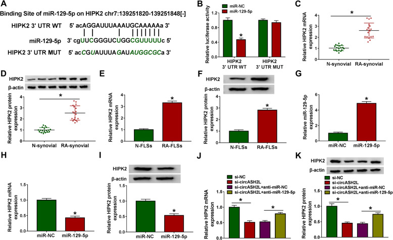
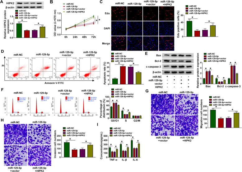
Background: Previous study showed that circular RNA Absent-Small-Homeotic-2--Like protein (circASH2L) was higher in rheumatoid arthritis (RA) patients. However, the roles and mechanisms of circASH2L in RA progression remain unclear. Methods: Levels analysis was conducted using western blot and qRT-PCR. The proliferation, apoptosis, cell cycle progression, migration, invasiveness, and inflammation of RA fibroblast-like synoviocytes (RA-FLSs) were determined via MTT, flow cytometry, western blot, transwell, and ELISA assays. Results: CircASH2L knockdown in RA-FLSs suppressed cell proliferative, migratory, and invasive capacities, triggered cell cycle arrest, promoted apoptosis, and inhibited inflammation. Mechanistically, circASH2L targeted miR-129-5p, and repression of miR-129-5p abolished the functions of circASH2L silencing on the growth, motility, and inflammation of RA-FLSs. Besides, miR-129-5p was found to directly target HIPK2, and suppressed the tumor-like biologic behaviors and inflammation of RA-FLSs via regulating HIPK2. Importantly, we proved that circASH2L could modulate HIPK2 expression via miR-129-5p. Conclusion: CircASH2L promoted RA-FLS growth, motility, and inflammation through miR-129-5p/HIPK2 axis.
Overview
The claims and statistics of this paper haven’t been checked yet.
Run the check — free, no account needed. It reads the open-access text and checks the sources it cites.
Integrity signals
Registry
Cites
Statistics
Figures
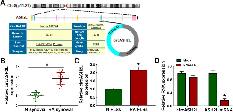
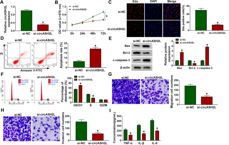
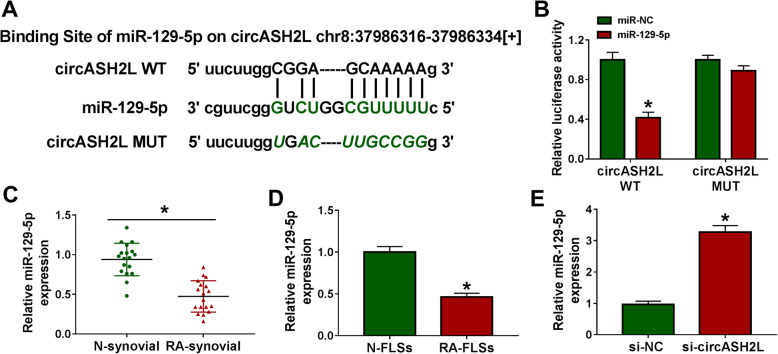
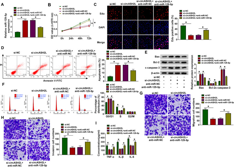
Verification
Claims the paper makes
The claims and statistics of this paper haven’t been checked yet.
Run the check — free, no account needed. It reads the open-access text and checks the sources it cites.
Claims other papers make about this one
Each is checked against this paper’s own text: does it say what the citing paper says it does?
In the citing paper
“In line with these observations, decreased levels of miR-129-5p have been associated with the induction of inflammatory processes in various other diseases such as kidney injury or rheumatoid arthritis [59, 84].”
From A role for astrocytic miR-129-5p in frontotemporal dementia (2025) · cites this paper as [84]
Self-check
Percentages, sums and reported differences re-computed from the paper’s own numbers.
The self-check needs the paper’s full text.
Citations
Cites
From the RNA world to the clinic
Sullenger, B. A., & Nair, S. (2016) · Science · doi:10.1126/science.aad8709 · Cited by 274
Research works printed but not linked: 29
- McInnes IB, Schett G. Pathogenetic insights from the treatment of rheumatoid arthritis. Lancet (London, England). 2017;389(10086):2328–37. · DOI
- Covic T, Cumming SR, Pallant JF, Manolios N, Emery P, Conaghan PG, et al. Depression and anxiety in patients with rheumatoid arthritis: prevalence rates based on a comparison of the Depression, Anxiety and Stress Scale (DASS) and the hospital, Anxiety and Depression Scale (HADS). BMC psychiatry. 2012;12(1):6. https://doi.org/10.1186/1471-244X-12-6. · DOI
- Croia C, Bursi R, Sutera D, Petrelli F, Alunno A, Puxeddu I. One year in review 2019: pathogenesis of rheumatoid arthritis. Clinical and experimental rheumatology. 2019;37(3):347–57.
- Neumann E, Lefèvre S, Zimmermann B, Gay S, Müller-Ladner U. Rheumatoid arthritis progression mediated by activated synovial fibroblasts. Trends in molecular medicine. 2010;16(10):458–68. https://doi.org/10.1016/j.molmed.2010.07.004. · DOI
- Bottini N, Firestein GS. Duality of fibroblast-like synoviocytes in RA: passive responders and imprinted aggressors. Nature reviews Rheumatology. 2013;9(1):24–33. https://doi.org/10.1038/nrrheum.2012.190. · DOI
- Lefevre S, Meier FM, Neumann E, Muller-Ladner U. Role of synovial fibroblasts in rheumatoid arthritis. Current pharmaceutical design. 2015;21(2):130–41. https://doi.org/10.2174/1381612820666140825122036. · DOI
- Rybak-Wolf A, Stottmeister C, Glažar P, Jens M, Pino N, Giusti S, et al. Circular RNAs in the mammalian brain are highly abundant, conserved, and dynamically expressed. Molecular cell. 2015;58(5):870–85. https://doi.org/10.1016/j.molcel.2015.03.027. · DOI · Paper page
- Memczak S, Jens M, Elefsinioti A, Torti F, Krueger J, Rybak A, et al. Circular RNAs are a large class of animal RNAs with regulatory potency. Nature. 2013;495(7441):333–8. https://doi.org/10.1038/nature11928. · DOI · Paper page
- Zhou R, Wu Y, Wang W, Su W, Liu Y, Wang Y, et al. Circular RNAs (circRNAs) in cancer. Cancer letters. 2018;425:134–42. https://doi.org/10.1016/j.canlet.2018.03.035. · DOI
- Yu CY, Kuo HC. The emerging roles and functions of circular RNAs and their generation. Journal of biomedical science. 2019;26(1):29. https://doi.org/10.1186/s12929-019-0523-z. · DOI
- Marques-Rocha JL, Samblas M, Milagro FI, Bressan J, Martínez JA, Marti A. Noncoding RNAs, cytokines, and inflammation-related diseases. FASEB journal : official publication of the Federation of American Societies for Experimental Biology. 2015;29(9):3595–611. https://doi.org/10.1096/fj.14-260323. · DOI
- Vo JN, Cieslik M, Zhang Y, Shukla S, Xiao L, Zhang Y, et al. The landscape of circular RNA in cancer. Cell. 2019;176(4):869–81 e813. · DOI
- Altesha MA, Ni T, Khan A, Liu K, Zheng X. Circular RNA in cardiovascular disease. Journal of cellular physiology. 2019;234(5):5588–600. https://doi.org/10.1002/jcp.27384. · DOI
- Chen X, Yang T, Wang W, Xi W, Zhang T, Li Q, et al. Circular RNAs in immune responses and immune diseases. Theranostics. 2019;9(2):588–607. https://doi.org/10.7150/thno.29678. · DOI
- Li B, Li N, Zhang L, Li K, Xie Y, Xue M, et al. Hsa_circ_0001859 regulates ATF2 expression by functioning as an MiR-204/211 sponge in human rheumatoid arthritis. J Immunology Res. 2018;2018:9412387.
- Tang X, Wang J, Xia X, Tian J, Rui K, Xu H, et al. Elevated expression of ciRS-7 in peripheral blood mononuclear cells from rheumatoid arthritis patients. Diagnostic pathology. 2019;14(1):11. https://doi.org/10.1186/s13000-019-0783-7. · DOI
- Zheng F, Yu X, Huang J, Dai Y. Circular RNA expression profiles of peripheral blood mononuclear cells in rheumatoid arthritis patients, based on microarray chip technology. Molecular medicine reports. 2017;16(6):8029–36. https://doi.org/10.3892/mmr.2017.7638. · DOI
- Bartok B, Firestein GS. Fibroblast-like synoviocytes: key effector cells in rheumatoid arthritis. Immunological reviews. 2010;233(1):233–55. https://doi.org/10.1111/j.0105-2896.2009.00859.x. · DOI
- Jeck WR, Sharpless NE. Detecting and characterizing circular RNAs. Nature biotechnology. 2014;32(5):453–61. https://doi.org/10.1038/nbt.2890. · DOI
- Jin Y, Shenoy AK, Doernberg S, Chen H, Luo H, Shen H, et al. FBXO11 promotes ubiquitination of the Snail family of transcription factors in cancer progression and epidermal development. Cancer letters. 2015;362(1):70–82. https://doi.org/10.1016/j.canlet.2015.03.037. · DOI
- Luo Q, Liu J, Fu B, Zhang L, Guo Y, Huang Z, et al. Circular RNAs Hsa_circ_0002715 and Hsa_circ_0035197 in peripheral blood are novel potential biomarkers for new-onset rheumatoid arthritis. Disease markers. 2019;2019:2073139.
- Luo Q, Zhang L, Li X, Fu B, Deng Z, Qing C, et al. Identification of circular RNAs hsa_circ_0044235 in peripheral blood as novel biomarkers for rheumatoid arthritis. Clinical and experimental immunology. 2018;194(1):118–24. https://doi.org/10.1111/cei.13181. · DOI
- Ouyang Q, Wu J, Jiang Z, Zhao J, Wang R, Lou A, et al. Microarray expression profile of circular RNAs in peripheral blood mononuclear cells from rheumatoid arthritis patients. Cellular physiology and biochemistry : international journal of experimental cellular physiology, biochemistry, and pharmacology. 2017;42(2):651–9. https://doi.org/10.1159/000477883. · DOI
- Hansen TB, Jensen TI, Clausen BH, Bramsen JB, Finsen B, Damgaard CK, et al. Natural RNA circles function as efficient microRNA sponges. Nature. 2013;495(7441):384–8. https://doi.org/10.1038/nature11993. · DOI · Paper page
- Chien Y, Tsai PH, Lai YH, Lu KH, Liu CY, Lin HF, et al. CircularRNA as novel biomarkers in liver diseases. Journal of the Chinese Medical Association : JCMA. 2020;83(1):15–7. https://doi.org/10.1097/JCMA.0000000000000230. · DOI
- Zhang Y, Yan N, Wang X, Chang Y, Wang Y. MiR-129-5p regulates cell proliferation and apoptosis via IGF-1R/Src/ERK/Egr-1 pathway in RA-fibroblast-like synoviocytes. Bioscience Rep. 2019;39:12.
- Saul VV, Schmitz ML. Posttranslational modifications regulate HIPK2, a driver of proliferative diseases. J Mol Med (Berlin, Germany). 2013;91(9):1051–8. · DOI
- Blaquiere JA, Verheyen EM. Homeodomain-interacting protein kinases: diverse and complex roles in development and disease. Curr Topics Dev Biol. 2017;123:73–103. https://doi.org/10.1016/bs.ctdb.2016.10.002. · DOI
- Li Y, Lai-Han Leung E, Pan H, Yao X, Huang Q, Wu M, et al. Identification of potential genetic causal variants for rheumatoid arthritis by whole-exome sequencing. Oncotarget. 2017;8(67):111119–29. https://doi.org/10.18632/oncotarget.22630. · DOI
Cited by
Sorted by citations
A role for astrocytic miR-129-5p in frontotemporal dementia
Kaurani, L., Pradhan, R., Schröder, S., Burkhardt, S., Schuetz, A.-L., Krüger, D. M., Pena, T., Heutink, P., Sananbenesi, F., & Fischer, A. (2025) · Translational Psychiatry · doi:10.1038/s41398-025-03338-y · Cited by 10
Metadata
- Authors
- Li, X., Qu, M., Zhang, J., Chen, K., & Ma, X.
- Venue
- Journal of Orthopaedic Surgery and Research
- Published
- 2021-05-08
- Volume
- 16
- Issue
- 1
- ISSN
- 1749-799X
- PMC
- PMC8106127
- Cited by
- 18
- References
- 1
- Language
- en
About these checks
Registry, retraction, Cites and statistics results are computed, not judged, with one exception: a printed reference that no index lists is read once by a model before it counts as a citation error. Claims are extracted by a model and kept only when their quote appears verbatim in the text; each cited source is then checked, with a model’s help, for whether it says what the claim relies on. Those verdicts can be wrong — read the claim’s quote and the cited source before relying on one.
Statistics and arithmetic are recomputed, not judged.
Confidence bands are shown only for fields calibrated on our annotated test set. None is calibrated yet, so no band is shown.
What counts as a claim ›How we measure confidence ›How we check this ›
Are you an author, or do you see a problem with this page? Request a review
How to cite
Cite CoChat for the checks on this page, and the paper for its own findings.
- This page
- CoChat. (2026). Citation check: CircASH2L facilitates tumor-like biologic behaviours and inflammation of fibroblast-like synoviocytes via miR-129-5p/HIPK2 axis in rheumatoid arthritis. https://app.cochat.ai/papers/10.1186/s13018-021-02432-3
- The paper
- Li, X., Qu, M., Zhang, J., Chen, K., & Ma, X. (2021). CircASH2L facilitates tumor-like biologic behaviours and inflammation of fibroblast-like synoviocytes via miR-129-5p/HIPK2 axis in rheumatoid arthritis. Journal of Orthopaedic Surgery and Research. https://doi.org/10.1186/s13018-021-02432-3
