Student or educator? Sign up with your school email and get 6 months of Pro free.
Cited by 179

The DOI matches its Crossref record; 42 of the 42 references it cites were found in a registry or an index.

Exosomes derived from microRNA-138-5p-overexpressing bone marrow-derived mesenchymal stem cells confer neuroprotection to astrocytes following ischemic stroke via inhibition of LCN2

Deng, Yiming, Chen, Duanduan, Gao, Feng, Lv, Hong, Zhang, Guojun, Sun, Xuan, Liu, Lian, Mo, Dapeng, Ma, Ning, Song, Ligang, Huo, Xiaochuan, Yan, Tianyi, Zhang, Jingbo, Miao, Zhongrong·2019·Journal of Biological Engineering

Abstract

Background: MicroRNAs (miRNAs) are implicated in the progression of ischemic stroke (IS) and bone marrow-derived mesenchymal stem cells (BMSCs)-derived exosomes play a role in IS therapy. Herein we hypothesized that the BMSCs-derived exosomes containing overexpressed miR-138-5p could protect the astrocytes following IS involved with lipocalin 2 (LCN2). Methods: The differentially expressed gene related to IS was initially identified by bioinformatics analysis. miR-138-5p was predicted to regulate LCN2. The expression of miR-138-5p and LCN2 was altered in the oxygen-glucose deprivation (OGD)-induced astrocytes. Furthermore, the cell behaviors and inflammatory responses were evaluated both in astrocytes alone and astrocytes co-cultured with exosomes derived from BMSCs overexpressing miR-138-5p to explore the involvement of miR-138-5p and LCN2 in IS. Besides, middle cerebral artery occlusion (MCAO) mouse model was established to explore the effect of BMSCs-derived exosomal miR-138-5p in IS in vivo. Results: LCN2 was highly expressed in IS. Besides, LCN2 was a target gene of miR-138-5p. BMSCs-derived exosomes could be endocytosed by astrocytes via co-culture. Overexpression of miR-138-5p promoted the proliferation and inhibited apoptosis of astrocytes injured by OGD, accompanied by the reduced expression of inflammatory factors, which was achieved by down-regulating LCN2. More importantly, BMSCs delivered miR-138-5p to the astrocytes via exosomes and BMSCs-derived exosomal miR-138-5p alleviated neuron injury in IS mice. Conclusion: BMSCs-derived exosomal miR-138-5p reduces neurological impairment by promoting proliferation and inhibiting inflammatory responses of astrocytes following IS by targeting LCN2, which may provide a novel target for IS treatment.

Overview

The claims and statistics of this paper haven’t been checked yet.

Run the check — free, no account needed. It reads the open-access text and checks the sources it cites.

A few free checks a day · papers already verified open instantly · results are public

Integrity signals

Registry

Crossref · 10.1186/s13036-019-0193-0

Cites

42 of 42 references verified0 citation errors · 0 not checkable

Statistics

Figures

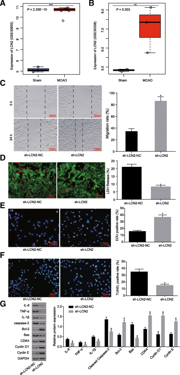
Fig. 1 LCN2 is highly expressed in IS and may affect the proliferation and apoptosis of OGD-induced astrocytes. a the expression of LCN2 in the GSE30655 chip; *** p < 0.001. b, the expression of LCN2 in the GSE35338 chip; ** p < 0.01. c, the ability of cell repair determined by scratch test (scale bar = 100 μm); d, LIVE/DEAD staining (scale bar = 50 μm) and quantitative analysis for LDH release rate; e, cell proliferation detected by EdU staining (scale bar = 50 μm); f, TUNEL staining for astrocyte apoptosis (scale bar = 50 μm); g, Western blot analysis for protein bands and levels of inflammatory factors, proliferation and apoptosis marker proteins. The data were all expressed as mean ± standard deviation. The comparison between two groups was analyzed by non-paired t-test. The experiment was repeated three times. *, p < 0.05 vs. sh-LCN2-NC. IS, ischemic stroke; MCAO, middle cerebral artery occlusion model; GAPDH, glyceraldehyde-3-phosphate dehydrogenase; Bax, Bcl-2-associated X protein; Bcl-2, B-cell CLL/Lymphoma 2; IL-6, interleukin-6; IL-1β, interleukin-1β; TNF-α, tumor necrosis factor-α; sh-LCN2-NC, astrocytes transfected with sh-LCN2-NC plasmid; sh-LCN2, astrocytes transfected with sh-LCN2
Fig. 1 LCN2 is highly expressed in IS and may affect the proliferation and apoptosis of OGD-induced astrocytes. a the expression of LCN2 in the GSE30655 chip; *** p < 0.001. b, the expression of LCN2 in the GSE35338 chip; ** p < 0.01. c, the ability of cell repair determined by scratch test (scale bar = 100 μm); d, LIVE/DEAD staining (scale bar = 50 μm) and quantitative analysis for LDH release rate; e, cell proliferation detected by EdU staining (scale bar = 50 μm); f, TUNEL staining for astrocyte apoptosis (scale bar = 50 μm); g, Western blot analysis for protein bands and levels of inflammatory factors, proliferation and apoptosis marker proteins. The data were all expressed as mean ± standard deviation. The comparison between two groups was analyzed by non-paired t-test. The experiment was repeated three times. *, p < 0.05 vs. sh-LCN2-NC. IS, ischemic stroke; MCAO, middle cerebral artery occlusion model; GAPDH, glyceraldehyde-3-phosphate dehydrogenase; Bax, Bcl-2-associated X protein; Bcl-2, B-cell CLL/Lymphoma 2; IL-6, interleukin-6; IL-1β, interleukin-1β; TNF-α, tumor necrosis factor-α; sh-LCN2-NC, astrocytes transfected with sh-LCN2-NC plasmid; sh-LCN2, astrocytes transfected with sh-LCN2CC BY · PubMed Central
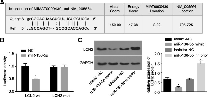
Fig. 2 LCN2 is a target gene of miR-138-5p. a, predicted binding sites of miR-138-5p in 3’UTR of LCN2; b, luciferase activity of HEK293 cells detected by dual luciferase reporter gene assay; c, protein bands and quantitative analysis for LCN2 expression in astrocytes. The comparison among multiple groups was analyzed by one-way analysis of variance and the comparison between two groups was analyzed by non-paired t-test. The experiment was repeated three times. *, p < 0.05 vs. NC and mimic-NC. NC, negative control; WT, wide type; MUT, mutant type; GAPDH, glyceraldehyde-3-phosphate dehydrogenase; mimic-NC, astrocytes transfected with mimic-NC; miR-138-5p mimic, astrocytes transfected with miR-138-5p mimic; inhibitor-NC, astrocytes transfected with inhibitor-NC; miR-138-5p inhibitor, astrocytes transfected with miR-138-5p inhibitor
Fig. 2 LCN2 is a target gene of miR-138-5p. a, predicted binding sites of miR-138-5p in 3’UTR of LCN2; b, luciferase activity of HEK293 cells detected by dual luciferase reporter gene assay; c, protein bands and quantitative analysis for LCN2 expression in astrocytes. The comparison among multiple groups was analyzed by one-way analysis of variance and the comparison between two groups was analyzed by non-paired t-test. The experiment was repeated three times. *, p < 0.05 vs. NC and mimic-NC. NC, negative control; WT, wide type; MUT, mutant type; GAPDH, glyceraldehyde-3-phosphate dehydrogenase; mimic-NC, astrocytes transfected with mimic-NC; miR-138-5p mimic, astrocytes transfected with miR-138-5p mimic; inhibitor-NC, astrocytes transfected with inhibitor-NC; miR-138-5p inhibitor, astrocytes transfected with miR-138-5p inhibitorCC BY · PubMed Central
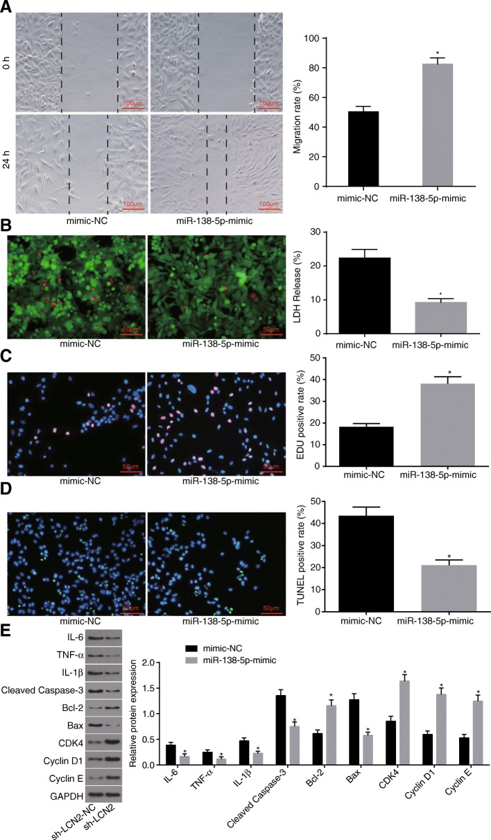
Fig. 3 Overexpression of miR-138-5p inhibits apoptosis of astrocytes injured by OGD. a, the repair ability of damages in cells determined by scratch test (scale bar = 100 μm), and quantitative analysis for cell migration and the number of exosomes; b, LIVE / DEAD staining for LDH release rate (scale bar = 50 μm); c, cell proliferation detected by EdU staining and quantitative analysis for EdU positive expression (scale bar = 50 μm); d, TUNEL staining (scale bar = 50 μm) and quantitative analysis for TUNEL positive rate; e, protein bands and quantitative analysis for inflammatory factors, proliferation and apoptosis marker proteins determined by Western blot analysis. The data were all expressed as mean ± standard deviation. The comparison between two groups was analyzed by non-paired t-test. The experiment was repeated three times. *, p < 0.05 vs. mimic-NC. OGD, oxygen-glucose deprivation; LDH, lactate dehydrogenase; EdU, 5-Ethynyl-2′-deoxyuridine; TUNEL, terminal deoxynucleotidyl transferase-mediated dUTP nick-end labeling; GAPDH, glyceraldehyde-3-phosphate dehydrogenase; Bax, Bcl-2-associated X protein; Bcl-2, B-cell CLL/Lymphoma 2; IL-6, interleukin-6; IL-1β, interleukin-1β; TNF-α, tumor necrosis factor-α; mimic-NC, astrocytes transfected with mimic-NC plasmid; miR-138-5p-mimic, astrocytes transfected with miR-138-5p-mimic plasmid
Fig. 3 Overexpression of miR-138-5p inhibits apoptosis of astrocytes injured by OGD. a, the repair ability of damages in cells determined by scratch test (scale bar = 100 μm), and quantitative analysis for cell migration and the number of exosomes; b, LIVE / DEAD staining for LDH release rate (scale bar = 50 μm); c, cell proliferation detected by EdU staining and quantitative analysis for EdU positive expression (scale bar = 50 μm); d, TUNEL staining (scale bar = 50 μm) and quantitative analysis for TUNEL positive rate; e, protein bands and quantitative analysis for inflammatory factors, proliferation and apoptosis marker proteins determined by Western blot analysis. The data were all expressed as mean ± standard deviation. The comparison between two groups was analyzed by non-paired t-test. The experiment was repeated three times. *, p < 0.05 vs. mimic-NC. OGD, oxygen-glucose deprivation; LDH, lactate dehydrogenase; EdU, 5-Ethynyl-2′-deoxyuridine; TUNEL, terminal deoxynucleotidyl transferase-mediated dUTP nick-end labeling; GAPDH, glyceraldehyde-3-phosphate dehydrogenase; Bax, Bcl-2-associated X protein; Bcl-2, B-cell CLL/Lymphoma 2; IL-6, interleukin-6; IL-1β, interleukin-1β; TNF-α, tumor necrosis factor-α; mimic-NC, astrocytes transfected with mimic-NC plasmid; miR-138-5p-mimic, astrocytes transfected with miR-138-5p-mimic plasmidCC BY · PubMed Central
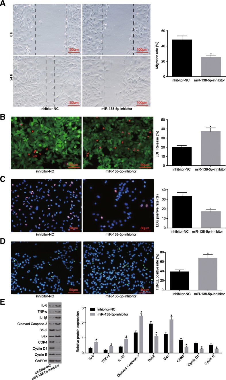
Fig. 4 Downregulation of miR-138-5p promotes apoptosis of astrocytes injured by OGD. a, the repair ability of damages in cells determined by scratch test (scale bar = 100 μm), and quantitative analysis for cell migration and the number of exosomes; b, LIVE / DEAD staining for LDH release rate (scale bar = 50 μm); c, cell proliferation detected by EdU staining and quantitative analysis for EDU positive expression (scale bar = 50 μm); d, TUNEL staining (scale bar = 50 μm) and quantitative analysis for TUNEL positive rate; e, expression of inflammatory factors, proliferation and apoptosis marker proteins determined by Western blot analysis. The data were all expressed as mean ± standard deviation. The comparison between two groups was analyzed by non-paired t-test. The experiment was repeated three times. *, p < 0.05 vs. inhibitor-NC. OGD, oxygen-glucose deprivation; LDH, lactate dehydrogenase; EdU, 5-Ethynyl-2′-deoxyuridine; TUNEL, terminal deoxynucleotidyl transferase-mediated dUTP nick-end labeling; GAPDH, glyceraldehyde-3-phosphate dehydrogenase; Bax, Bcl-2-associated X protein; Bcl-2, B-cell CLL/Lymphoma 2; IL-6, interleukin-6; IL-1β, interleukin-1β; TNF-α, tumor necrosis factor-α; inhibitor-NC, astrocytes transfected with inhibitor-NC plasmid; miR-138-5p-inhibitor, astrocytes transfected with miR-138-5p-inhibitor plasmid
Fig. 4 Downregulation of miR-138-5p promotes apoptosis of astrocytes injured by OGD. a, the repair ability of damages in cells determined by scratch test (scale bar = 100 μm), and quantitative analysis for cell migration and the number of exosomes; b, LIVE / DEAD staining for LDH release rate (scale bar = 50 μm); c, cell proliferation detected by EdU staining and quantitative analysis for EDU positive expression (scale bar = 50 μm); d, TUNEL staining (scale bar = 50 μm) and quantitative analysis for TUNEL positive rate; e, expression of inflammatory factors, proliferation and apoptosis marker proteins determined by Western blot analysis. The data were all expressed as mean ± standard deviation. The comparison between two groups was analyzed by non-paired t-test. The experiment was repeated three times. *, p < 0.05 vs. inhibitor-NC. OGD, oxygen-glucose deprivation; LDH, lactate dehydrogenase; EdU, 5-Ethynyl-2′-deoxyuridine; TUNEL, terminal deoxynucleotidyl transferase-mediated dUTP nick-end labeling; GAPDH, glyceraldehyde-3-phosphate dehydrogenase; Bax, Bcl-2-associated X protein; Bcl-2, B-cell CLL/Lymphoma 2; IL-6, interleukin-6; IL-1β, interleukin-1β; TNF-α, tumor necrosis factor-α; inhibitor-NC, astrocytes transfected with inhibitor-NC plasmid; miR-138-5p-inhibitor, astrocytes transfected with miR-138-5p-inhibitor plasmidCC BY · PubMed Central
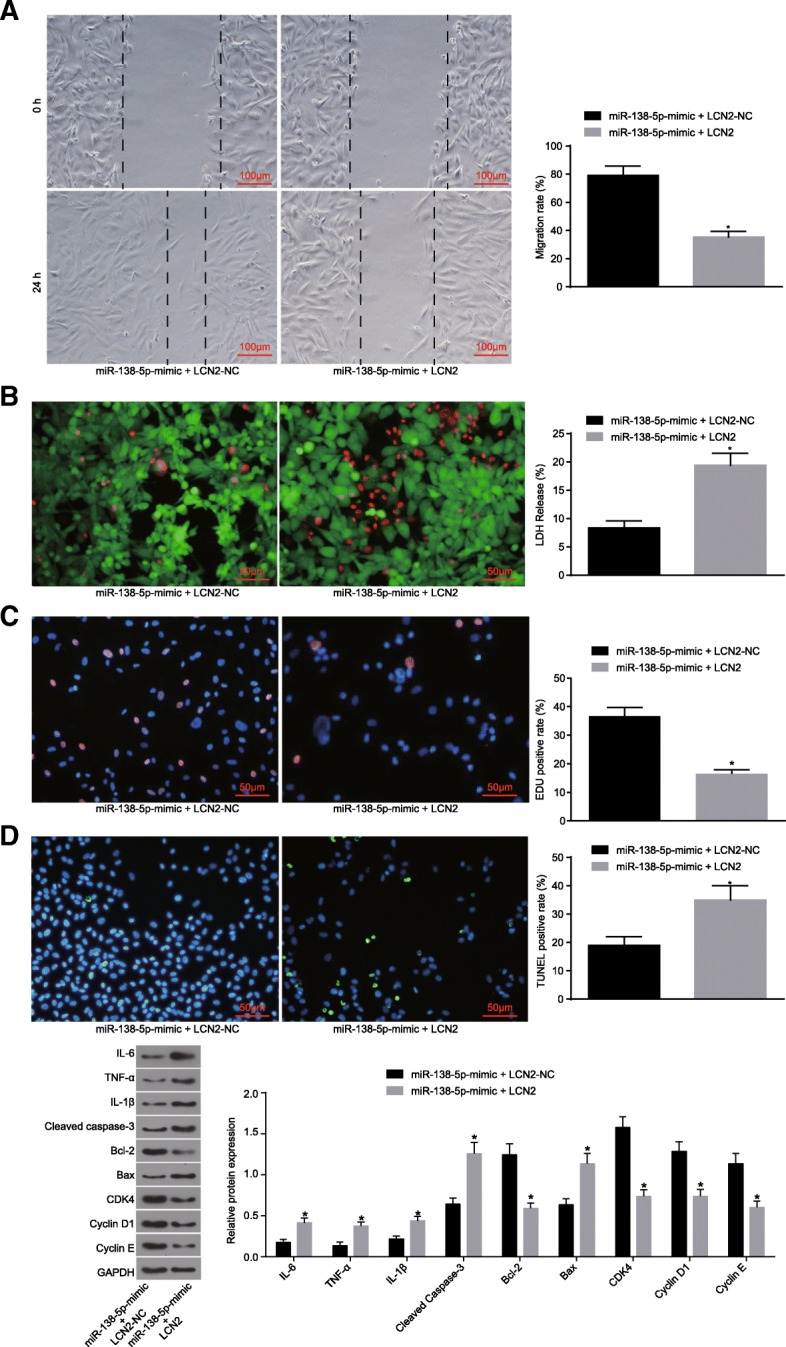
Fig. 5 miR-138-5p plays a role in proliferation, apoptosis and inflammation of astrocytes by regulating LCN2 gene. a, the repair ability of damages in cells determined by scratch test (scale bar = 100 μm), and quantitative analysis for cell migration and the number of exosomes; b, LIVE/DEAD staining for LDH release rate (scale bar = 50 μm); b, cell proliferation detected by EdU staining and quantitative analysis for EdU positive expression (scale bar = 50 μm); d, TUNEL staining (scale bar = 50 μm) and quantitative analysis for TUNEL positive rate; e, expression of inflammatory factors, proliferation and apoptosis marker proteins determined by Western blot analysis. The data were all expressed as mean ± standard deviation. The comparison between two groups was analyzed by non-paired t-test. The experiment was repeated three times. *, p < 0.05 vs. miR-138-5p -mimic + LCN2-NC. OGD, oxygen-glucose deprivation; LDH, lactate dehydrogenase; EdU, 5-Ethynyl-2′-deoxyuridine; TUNEL, terminal deoxynucleotidyl transferase-mediated dUTP nick-end labeling; GAPDH, glyceraldehyde-3-phosphate dehydrogenase; Bax, Bcl-2-associated X protein; Bcl-2, B-cell CLL/Lymphoma 2; IL-6, interleukin-6; IL-1β, interleukin-1β; TNF-α, tumor necrosis factor-α; miR-138-5p-mimic + LCN2-NC, astrocytes transfected with miR-138-5p-mimic + LCN2-NC plasmid; miR-138-5p-mimic + LCN2, astrocytes transfected with miR-138-5p-mimic + LCN2
Fig. 5 miR-138-5p plays a role in proliferation, apoptosis and inflammation of astrocytes by regulating LCN2 gene. a, the repair ability of damages in cells determined by scratch test (scale bar = 100 μm), and quantitative analysis for cell migration and the number of exosomes; b, LIVE/DEAD staining for LDH release rate (scale bar = 50 μm); b, cell proliferation detected by EdU staining and quantitative analysis for EdU positive expression (scale bar = 50 μm); d, TUNEL staining (scale bar = 50 μm) and quantitative analysis for TUNEL positive rate; e, expression of inflammatory factors, proliferation and apoptosis marker proteins determined by Western blot analysis. The data were all expressed as mean ± standard deviation. The comparison between two groups was analyzed by non-paired t-test. The experiment was repeated three times. *, p < 0.05 vs. miR-138-5p -mimic + LCN2-NC. OGD, oxygen-glucose deprivation; LDH, lactate dehydrogenase; EdU, 5-Ethynyl-2′-deoxyuridine; TUNEL, terminal deoxynucleotidyl transferase-mediated dUTP nick-end labeling; GAPDH, glyceraldehyde-3-phosphate dehydrogenase; Bax, Bcl-2-associated X protein; Bcl-2, B-cell CLL/Lymphoma 2; IL-6, interleukin-6; IL-1β, interleukin-1β; TNF-α, tumor necrosis factor-α; miR-138-5p-mimic + LCN2-NC, astrocytes transfected with miR-138-5p-mimic + LCN2-NC plasmid; miR-138-5p-mimic + LCN2, astrocytes transfected with miR-138-5p-mimic + LCN2CC BY · PubMed Central
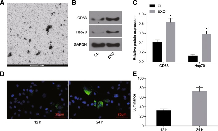
Fig. 6 Endocytosis, intracellular sorting, and processing of BMSCs-derived exosomes by astrocytes. a, electron microscopic examination of exosomes (scale bar = 200 nm); b, protein bands of surface makers of BMSCs-derived exosomes determined by Western blot analysis (CL, BMSC lysate; EXO, exosomes); c, quantitative analysis regarding protein expression of surface makers of BMSCs-derived exosomes determined by Western blot analysis; d, exosome surface fluorescence (scale bar = 25 μm); e, quantitative analysis for endocytosis at different time points. The data were all expressed as mean ± standard deviation. The comparison between two groups was analyzed by non-paired t-test. The experiment was repeated three times. *, p < 0.05 vs. CL or endocytosis at 12 h. BMSCs, bone marrow-derived mesenchymal stem cells
Fig. 6 Endocytosis, intracellular sorting, and processing of BMSCs-derived exosomes by astrocytes. a, electron microscopic examination of exosomes (scale bar = 200 nm); b, protein bands of surface makers of BMSCs-derived exosomes determined by Western blot analysis (CL, BMSC lysate; EXO, exosomes); c, quantitative analysis regarding protein expression of surface makers of BMSCs-derived exosomes determined by Western blot analysis; d, exosome surface fluorescence (scale bar = 25 μm); e, quantitative analysis for endocytosis at different time points. The data were all expressed as mean ± standard deviation. The comparison between two groups was analyzed by non-paired t-test. The experiment was repeated three times. *, p < 0.05 vs. CL or endocytosis at 12 h. BMSCs, bone marrow-derived mesenchymal stem cellsCC BY · PubMed Central
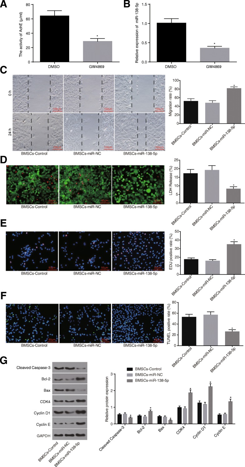
Fig. 7 BMSCs deliver miR-138-5p to the astrocytes via the exosomes. a, quantitative analysis for the number of exosomes; b, the expression of the miR-138-5p in the co-culture system detected by RT-qPCR; c, repair ability of damages in cells determined by scratch test (scale bar = 100 μm), and quantitative analysis for cell migration and the number of exosomes; d, LIVE / DEAD staining for LDH release rate (scale bar = 50 μm); e, cell proliferation detected by EdU staining and quantitative analysis for EDU positive expression (scale bar = 50 μm); f, TUNEL staining (scale bar = 50 μm) and quantitative analysis for TUNEL positive rate; g, protein bands and quantitative analysis for expression of inflammatory factors, proliferation and apoptosis marker proteins determined by Western blot analysis. The data were all expressed as mean ± standard deviation. The comparison among multiple groups was analyzed by one-way analysis of variance. The experiment was repeated three times. *, p < 0.05 vs. DMSO or BMSCs-Control. LDH, lactate dehydrogenase; EdU, 5-Ethynyl-2′-deoxyuridine; TUNEL, terminal deoxynucleotidyl transferase-mediated dUTP nick-end labeling; GAPDH, glyceraldehyde-3-phosphate dehydrogenase; Bax, Bcl-2-associated X protein; Bcl-2, B-cell CLL/Lymphoma 2; IL-6, interleukin-6; IL-1β, interleukin-1β; TNF-α, tumor necrosis factor-α; BMSCs-control, astrocytes co-cultured with BMSCs-derived exosomes without any treatment; BMSCs-miR-NC, astrocytes co-cultured with BMSCs-derived exosomes infected with miR-NC; BMSCs-miR-138-5p, astrocytes co-cultured with BMSCs-derived exosomes infected with miR-138-5p overexpression; BMSCs, bone marrow-derived mesenchymal stem cells
Fig. 7 BMSCs deliver miR-138-5p to the astrocytes via the exosomes. a, quantitative analysis for the number of exosomes; b, the expression of the miR-138-5p in the co-culture system detected by RT-qPCR; c, repair ability of damages in cells determined by scratch test (scale bar = 100 μm), and quantitative analysis for cell migration and the number of exosomes; d, LIVE / DEAD staining for LDH release rate (scale bar = 50 μm); e, cell proliferation detected by EdU staining and quantitative analysis for EDU positive expression (scale bar = 50 μm); f, TUNEL staining (scale bar = 50 μm) and quantitative analysis for TUNEL positive rate; g, protein bands and quantitative analysis for expression of inflammatory factors, proliferation and apoptosis marker proteins determined by Western blot analysis. The data were all expressed as mean ± standard deviation. The comparison among multiple groups was analyzed by one-way analysis of variance. The experiment was repeated three times. *, p < 0.05 vs. DMSO or BMSCs-Control. LDH, lactate dehydrogenase; EdU, 5-Ethynyl-2′-deoxyuridine; TUNEL, terminal deoxynucleotidyl transferase-mediated dUTP nick-end labeling; GAPDH, glyceraldehyde-3-phosphate dehydrogenase; Bax, Bcl-2-associated X protein; Bcl-2, B-cell CLL/Lymphoma 2; IL-6, interleukin-6; IL-1β, interleukin-1β; TNF-α, tumor necrosis factor-α; BMSCs-control, astrocytes co-cultured with BMSCs-derived exosomes without any treatment; BMSCs-miR-NC, astrocytes co-cultured with BMSCs-derived exosomes infected with miR-NC; BMSCs-miR-138-5p, astrocytes co-cultured with BMSCs-derived exosomes infected with miR-138-5p overexpression; BMSCs, bone marrow-derived mesenchymal stem cellsCC BY · PubMed Central
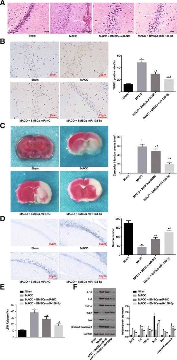
Fig. 8 BMSCs-derived exosomal miR-138-5p reduces neuron injury following IS in vivo. a, HE staining for hippocampal tissues of MCAO mouse models (scale bar = 25 μm); b, TUNEL staining to detect neuron apoptosis (scale bar = 25 μm) and quantitative analysis for the apoptotic rate; c, TTC staining showing the volume changes of cerebral infarction in mice and the corresponding quantitative analysis; d, Nissl staining (scale bar = 50 μm) showing the number of neurons and the corresponding quantitative analysis; e, quantitative analysis for LDH content; f, protein expression of inflammatory factors, proliferation and apoptosis marker proteins determined by Western blot analysis. The data were all measurement data, expressed as mean ± standard deviation. The comparison among multiple groups was analyzed by one-way analysis of variance. The experiment was repeated three times. *, p < 0.05 vs. sham. #, p < 0.05 vs. MCAO. n = 10. LDH, lactate dehydrogenase; TUNEL, terminal deoxynucleotidyl transferase-mediated dUTP nick-end labeling; MCAO, middle cerebral artery occlusion; GAPDH, glyceraldehyde-3-phosphate dehydrogenase; Bax, Bcl-2-associated X protein; Bcl-2, B-cell CLL/Lymphoma 2; IL-6, interleukin-6; IL-1β, interleukin-1β; TNF-α, tumor necrosis factor-α; BMSCs-control, MCAO mice without any treatment; BMSCs-miR-NC, MCAO mice injected with BMSCs-miR-NC exosomes; BMSCs-miR-138-5p, MCAO mice injected with BMSCs-miR-138-5p exosomes
Fig. 8 BMSCs-derived exosomal miR-138-5p reduces neuron injury following IS in vivo. a, HE staining for hippocampal tissues of MCAO mouse models (scale bar = 25 μm); b, TUNEL staining to detect neuron apoptosis (scale bar = 25 μm) and quantitative analysis for the apoptotic rate; c, TTC staining showing the volume changes of cerebral infarction in mice and the corresponding quantitative analysis; d, Nissl staining (scale bar = 50 μm) showing the number of neurons and the corresponding quantitative analysis; e, quantitative analysis for LDH content; f, protein expression of inflammatory factors, proliferation and apoptosis marker proteins determined by Western blot analysis. The data were all measurement data, expressed as mean ± standard deviation. The comparison among multiple groups was analyzed by one-way analysis of variance. The experiment was repeated three times. *, p < 0.05 vs. sham. #, p < 0.05 vs. MCAO. n = 10. LDH, lactate dehydrogenase; TUNEL, terminal deoxynucleotidyl transferase-mediated dUTP nick-end labeling; MCAO, middle cerebral artery occlusion; GAPDH, glyceraldehyde-3-phosphate dehydrogenase; Bax, Bcl-2-associated X protein; Bcl-2, B-cell CLL/Lymphoma 2; IL-6, interleukin-6; IL-1β, interleukin-1β; TNF-α, tumor necrosis factor-α; BMSCs-control, MCAO mice without any treatment; BMSCs-miR-NC, MCAO mice injected with BMSCs-miR-NC exosomes; BMSCs-miR-138-5p, MCAO mice injected with BMSCs-miR-138-5p exosomesCC BY · PubMed Central

Verification

Claims the paper makes

The claims and statistics of this paper haven’t been checked yet.

Run the check — free, no account needed. It reads the open-access text and checks the sources it cites.

A few free checks a day · papers already verified open instantly · results are public

Claims other papers make about this one

Each is checked against this paper’s own text: does it say what the citing paper says it does?

Self-check

Percentages, sums and reported differences re-computed from the paper’s own numbers.

The self-check needs the paper’s full text.

Citations

Cites

Show all 42 references

Cited by

See all 179

Metadata

Authors
Deng, Y., Chen, D., Gao, F., Lv, H., Zhang, G., Sun, X., Liu, L., Mo, D., Ma, N., Song, L., Huo, X., Yan, T., Zhang, J., & Miao, Z.
Venue
Journal of Biological Engineering
Published
2019-08-28
Volume
13
Issue
1
ISSN
1754-1611
Cited by
179
References
42
Language
en
Licence
Funders
  • the Beijing Municipal Administration of Hospitals’ Youth Program (QML20180506)
  • National Natural Science Foundation of China (81671776, 81371290 and 81502391)
  • Beijing Municipal Science & Technology Commission (Z161100001116122)

About these checks

Registry, retraction, Cites and statistics results are computed, not judged, with one exception: a printed reference that no index lists is read once by a model before it counts as a citation error. Claims are extracted by a model and kept only when their quote appears verbatim in the text; each cited source is then checked, with a model’s help, for whether it says what the claim relies on. Those verdicts can be wrong — read the claim’s quote and the cited source before relying on one.

Statistics and arithmetic are recomputed, not judged.

Confidence bands are shown only for fields calibrated on our annotated test set. None is calibrated yet, so no band is shown.

What counts as a claim ›How we measure confidence ›How we check this ›

Are you an author, or do you see a problem with this page? Request a review

How to cite

Cite CoChat for the checks on this page, and the paper for its own findings.

This page
CoChat. (2026). Citation check: Exosomes derived from microRNA-138-5p-overexpressing bone marrow-derived mesenchymal stem cells confer neuroprotection to astrocytes following ischemic stroke via inhibition of LCN2. https://app.cochat.ai/papers/10.1186/s13036-019-0193-0
The paper
Deng, Y., Chen, D., Gao, F., Lv, H., Zhang, G., Sun, X., Liu, L., Mo, D., Ma, N., Song, L., Huo, X., Yan, T., Zhang, J., & Miao, Z. (2019). Exosomes derived from microRNA-138-5p-overexpressing bone marrow-derived mesenchymal stem cells confer neuroprotection to astrocytes following ischemic stroke via inhibition of LCN2. Journal of Biological Engineering. https://doi.org/10.1186/s13036-019-0193-0

Data sources

Bibliographic data: Semantic Scholar, OpenAlex, Crossref.

Contains information from the Semantic Scholar Academic Graph Datasets, made available under the ODC Attribution License (ODC-BY 1.0).

Semantic Scholar’s data is described in: Kinney, R., et al. (2023). The Semantic Scholar Open Data Platform. arXiv:2301.10140.