Cited by 61

The DOI matches its Crossref record; 5 of the 5 references it cites were found in a registry or an index.

Lentivirus-mediated expression of human secreted amyloid precursor protein-alpha prevents development of memory and plasticity deficits in a mouse model of Alzheimer’s disease.

Tan, Valerie T. Y., Mockett, Bruce G., Ohline, Shane M., Parfitt, Karen D., Wicky, Hollie E., Peppercorn, Katie, Schoderboeck, Lucia, Yahaya, Mohamad Fairuz bin, Tate, Warren P., Hughes, Stephanie M., Abraham, Wickliffe C.·2018·Molecular Brain

Overview

The claims and statistics of this paper haven’t been checked yet.

Run the check — free, no account needed. It reads the open-access text and checks the sources it cites.

A few free checks a day · papers already verified open instantly · results are public

Integrity signals

Registry

Crossref · 10.1186/s13041-018-0348-9

Cites

5 of 5 references verified0 citation errors · 0 not checkable

Statistics

Figures

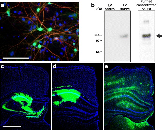
Fig. 1 In vitro and in vivo lentiviral transduction of hippocampal neurons with sAPPα-copGFP. a The transduction marker copGFP (green) was expressed in mouse primary hippocampal neurons co-labelled with the neuronal marker MAP2 (red) and the nuclear marker DAPI (blue). Scale bar, 50 μm. b Detection of sAPPα in media from primary mouse neural cultures transduced with LV-sAPPα, but not LV-control. The right-hand lane of the western blot illustrates the band from a sample fortified with purified sAPPα [22]. c-e Examples of in vivo copGFP expression (green) showing transduction of hippocampal regions containing LV-transduced neurons in slices from wild-type (c), transgenic-control (d) and transgenic-sAPPα (e) mice. Scale bar: 1 mm
Fig. 1 In vitro and in vivo lentiviral transduction of hippocampal neurons with sAPPα-copGFP. a The transduction marker copGFP (green) was expressed in mouse primary hippocampal neurons co-labelled with the neuronal marker MAP2 (red) and the nuclear marker DAPI (blue). Scale bar, 50 μm. b Detection of sAPPα in media from primary mouse neural cultures transduced with LV-sAPPα, but not LV-control. The right-hand lane of the western blot illustrates the band from a sample fortified with purified sAPPα [22]. c-e Examples of in vivo copGFP expression (green) showing transduction of hippocampal regions containing LV-transduced neurons in slices from wild-type (c), transgenic-control (d) and transgenic-sAPPα (e) mice. Scale bar: 1 mmCC BY · PubMed Central
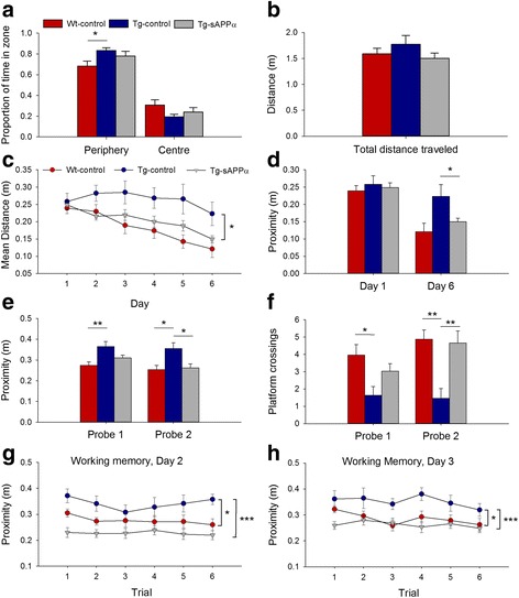
Fig. 2 sAPPα transduction rescued spatial learning and memory in the APP/PS1 mice. a In the open field all groups preferred the periphery (p < 0.001), but this preference was stronger in Tg-controls (n = 11) compared to WT-control (n = 22, p < 0.05, Students t-test). The Tg-sAPPa group showed a trend toward reduced time in the periphery (n = 23, p = 0.084) compared to Tg-controls. b Mean distances travelled by each group were not significantly different. c For spatial reference memory acquisition in the water maze, there was a significant group main effect (F(1,54) = 3.33, p = 0.043) for proximity to the platform, with a strong trend for Tg-controls to be impaired relative to WT-control animals (Tukey p = 0.071), while Tg-sAPPα animals performed significantly better than Tg-control animals (p = 0.046). d There was a significant group main effect (F(1,54) = 3.33, p = 0.043) with a strong trend for impaired performance on day 6 by the Tg-control group compared to WT-controls (p = 0.068) and by enhanced performance of the Tg-sAPPα group compared to Tg-controls (p = 0.018). e, f Tg-control mice had significantly poorer memory for the platform position than WT-controls (Probe 1: proximity p = 0.004, crossings p = 0.037; Probe 2: proximity p = 0.016, crossings p = 0.006). Tg-sAPPα mice exhibited a partial rescue in Probe 1 and a complete rescue in Probe 2 (proximity p = 0.022, crossings p = 0.006 compared to Tg-control). g, h Spatial working memory testing revealed a significant group main effect (G, Day 2: p < 0.001; H, Day 3: p = 0.004). On both days, Tg-controls performed significantly worse than WT-controls (Day 1: p = 0.025; Day 2: p = 0.034) while Tg-sAPPα animals were significantly better than Tg-controls (Day 1: p < 0.001; Day 2: p = 0.003). *p < 0.05, **p < 0.01, ***p < 0.001
Fig. 2 sAPPα transduction rescued spatial learning and memory in the APP/PS1 mice. a In the open field all groups preferred the periphery (p < 0.001), but this preference was stronger in Tg-controls (n = 11) compared to WT-control (n = 22, p < 0.05, Students t-test). The Tg-sAPPa group showed a trend toward reduced time in the periphery (n = 23, p = 0.084) compared to Tg-controls. b Mean distances travelled by each group were not significantly different. c For spatial reference memory acquisition in the water maze, there was a significant group main effect (F(1,54) = 3.33, p = 0.043) for proximity to the platform, with a strong trend for Tg-controls to be impaired relative to WT-control animals (Tukey p = 0.071), while Tg-sAPPα animals performed significantly better than Tg-control animals (p = 0.046). d There was a significant group main effect (F(1,54) = 3.33, p = 0.043) with a strong trend for impaired performance on day 6 by the Tg-control group compared to WT-controls (p = 0.068) and by enhanced performance of the Tg-sAPPα group compared to Tg-controls (p = 0.018). e, f Tg-control mice had significantly poorer memory for the platform position than WT-controls (Probe 1: proximity p = 0.004, crossings p = 0.037; Probe 2: proximity p = 0.016, crossings p = 0.006). Tg-sAPPα mice exhibited a partial rescue in Probe 1 and a complete rescue in Probe 2 (proximity p = 0.022, crossings p = 0.006 compared to Tg-control). g, h Spatial working memory testing revealed a significant group main effect (G, Day 2: p < 0.001; H, Day 3: p = 0.004). On both days, Tg-controls performed significantly worse than WT-controls (Day 1: p = 0.025; Day 2: p = 0.034) while Tg-sAPPα animals were significantly better than Tg-controls (Day 1: p < 0.001; Day 2: p = 0.003). *p < 0.05, **p < 0.01, ***p < 0.001CC BY · PubMed Central
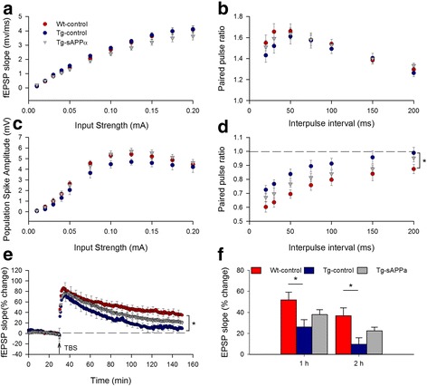
Fig. 3 Expression of sAPPα partially restored hippocampal LTP in transgenic mice. a, b There were no significant genotype or treatment effects on the I/O curves in CA1 for either the (a) EPSP slope, (p = 0.17 and 0.14, respectively) or (b) population spike amplitude (p = 0.14 and 0.63, respectively, indicating that basal synaptic transmission and cell excitability were unaffected in Tg mice and sAPPα over-expression. c There were no group differences in paired-pulse facilitation of the fEPSP (WT-control: n = 22; Tg-control: n = 12; Tg-sAPPα: n = 17, F(2,48) = 0.32, p = 0.73), nor a group x interval interaction (F(2,48) = 0.96, p = 0.39), indicating no difference in basal transmitter release probability. (d) Paired-pulse inhibition was reduced in Tg-controls compared to WT-controls (WT-control: n = 16; Tg-control: n = 10; F(2,38) = 4.18, p = 0.023), indicating an impairment of recurrent inhibition. A partial rescue was produced by sAPPα expression. (e, f) TBS delivered to the Schaffer collaterals induced robust potentiation in all groups but which decayed at different rates between groups. Significant group differences were observed at both 1 h (F(2,49) = 4.20, p = 0.021) and 2 h post-TBS (F(2,49) = 5.44, p = 0.007) with Tg-controls significantly impaired (26.1 ± 6.9%, n = 11) compared to WT-controls (51.9 ± 7.4%, n = 21; p < 0.05) after 1 h. LTP in the Tg-sAPPα indicating a partial recovery of LTP expression. The partial recovery was still observed 2 h post-TBS (WT-controls: 36.8 ± 7.5%, n = 21; Tg-controls: 9.7 ± 6.2%, n = 11; Tg-sAPPα: 22.5 ± 3.5%, n = 18). *p < 0.05
Fig. 3 Expression of sAPPα partially restored hippocampal LTP in transgenic mice. a, b There were no significant genotype or treatment effects on the I/O curves in CA1 for either the (a) EPSP slope, (p = 0.17 and 0.14, respectively) or (b) population spike amplitude (p = 0.14 and 0.63, respectively, indicating that basal synaptic transmission and cell excitability were unaffected in Tg mice and sAPPα over-expression. c There were no group differences in paired-pulse facilitation of the fEPSP (WT-control: n = 22; Tg-control: n = 12; Tg-sAPPα: n = 17, F(2,48) = 0.32, p = 0.73), nor a group x interval interaction (F(2,48) = 0.96, p = 0.39), indicating no difference in basal transmitter release probability. (d) Paired-pulse inhibition was reduced in Tg-controls compared to WT-controls (WT-control: n = 16; Tg-control: n = 10; F(2,38) = 4.18, p = 0.023), indicating an impairment of recurrent inhibition. A partial rescue was produced by sAPPα expression. (e, f) TBS delivered to the Schaffer collaterals induced robust potentiation in all groups but which decayed at different rates between groups. Significant group differences were observed at both 1 h (F(2,49) = 4.20, p = 0.021) and 2 h post-TBS (F(2,49) = 5.44, p = 0.007) with Tg-controls significantly impaired (26.1 ± 6.9%, n = 11) compared to WT-controls (51.9 ± 7.4%, n = 21; p < 0.05) after 1 h. LTP in the Tg-sAPPα indicating a partial recovery of LTP expression. The partial recovery was still observed 2 h post-TBS (WT-controls: 36.8 ± 7.5%, n = 21; Tg-controls: 9.7 ± 6.2%, n = 11; Tg-sAPPα: 22.5 ± 3.5%, n = 18). *p < 0.05CC BY · PubMed Central
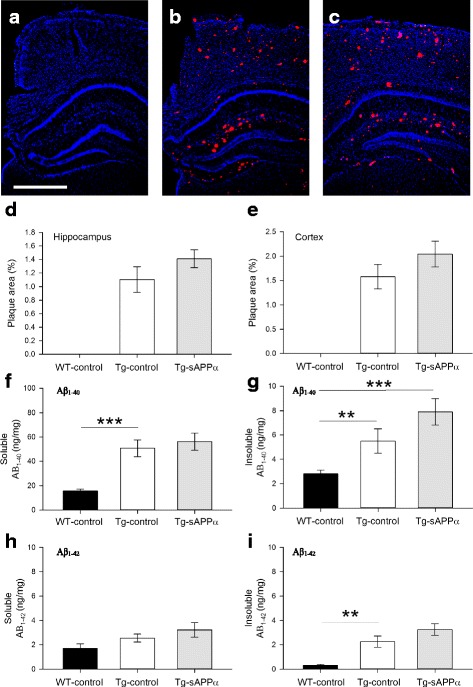
Fig. 4 Effect of sAPPα expression on the development of amyloid pathology. a-c Congo red staining of coronal brain sections from (a) WT-control, (b) Tg-sAPPα and (c) Tg-control animals revealed an absence of amyloid plaques in WT-control brains, but extensive plaque formation in Tg-sAPPα and Tg-control brains. d-e No significant differences in plaque density were observed between Tg- sAPPα and Tg-control in the hippocampus (p = 0.316, d) or overlying neocortex (p = 0.297, E). f-i Human amyloid-β load in the hippocampus. f Soluble Aβ1–40 levels differed between groups (one-way ANOVA F(2,27) = 17.40, p < 0.001), with higher levels in in Tg-control animals compared to wildtypes (WT-control: 15.6 ± 1.6 ng/mg, n = 12; Tg-control: 50.7 ± 6.9 ng/mg, n = 6, post-hoc Tukey p = 0.001), but no effect of sAPPα treatment compared to Tg-control (Tg-sAPPα: 56.1 ± 7.1 ng/mg, n = 12, Tukey = 0.820). g Insoluble Aβ1–40 levels showed a significant overall group effect (F(2,28 = 10.70) and while there was no significant difference between WT-control and Tg-control (WT-control, 2.8 ± 0.3 ng/mg, n = 13, Tg-control, 5.5 ± 1.0 ng/mg, n = 6, Tukey p = 0.145), the levels were elevated in the Tg-sAPPα group compared to WT-control (Tg-sAPPα 7.9 ± 1.1 ng/mg, Tukey p < 0.001) and they were not different to Tg-control (Tukey p + 0.194). (H) Soluble Aβ1–42 levels did not show any differences between groups. i For the insoluble fraction, there was an overall group effect (F(2,28) = 20.47, p < 0.001, whereby Tg-controls showed the expected greater Aβ1–42 load (2.26 ± 0.46 ng/mg, n = 6) compared to WT-controls (0.31 ± 0.077 ng/mg, n = 13; Tukey p = 0.006) and this level was not significantly affected by sAPPα over-expression (3.25 ± 0.48 ng/mg, n = 12, Tukey p = 0.220). **p < 0.01, ***p < 0.001, Scale bar: 1 mm
Fig. 4 Effect of sAPPα expression on the development of amyloid pathology. a-c Congo red staining of coronal brain sections from (a) WT-control, (b) Tg-sAPPα and (c) Tg-control animals revealed an absence of amyloid plaques in WT-control brains, but extensive plaque formation in Tg-sAPPα and Tg-control brains. d-e No significant differences in plaque density were observed between Tg- sAPPα and Tg-control in the hippocampus (p = 0.316, d) or overlying neocortex (p = 0.297, E). f-i Human amyloid-β load in the hippocampus. f Soluble Aβ1–40 levels differed between groups (one-way ANOVA F(2,27) = 17.40, p < 0.001), with higher levels in in Tg-control animals compared to wildtypes (WT-control: 15.6 ± 1.6 ng/mg, n = 12; Tg-control: 50.7 ± 6.9 ng/mg, n = 6, post-hoc Tukey p = 0.001), but no effect of sAPPα treatment compared to Tg-control (Tg-sAPPα: 56.1 ± 7.1 ng/mg, n = 12, Tukey = 0.820). g Insoluble Aβ1–40 levels showed a significant overall group effect (F(2,28 = 10.70) and while there was no significant difference between WT-control and Tg-control (WT-control, 2.8 ± 0.3 ng/mg, n = 13, Tg-control, 5.5 ± 1.0 ng/mg, n = 6, Tukey p = 0.145), the levels were elevated in the Tg-sAPPα group compared to WT-control (Tg-sAPPα 7.9 ± 1.1 ng/mg, Tukey p < 0.001) and they were not different to Tg-control (Tukey p + 0.194). (H) Soluble Aβ1–42 levels did not show any differences between groups. i For the insoluble fraction, there was an overall group effect (F(2,28) = 20.47, p < 0.001, whereby Tg-controls showed the expected greater Aβ1–42 load (2.26 ± 0.46 ng/mg, n = 6) compared to WT-controls (0.31 ± 0.077 ng/mg, n = 13; Tukey p = 0.006) and this level was not significantly affected by sAPPα over-expression (3.25 ± 0.48 ng/mg, n = 12, Tukey p = 0.220). **p < 0.01, ***p < 0.001, Scale bar: 1 mmCC BY · PubMed Central
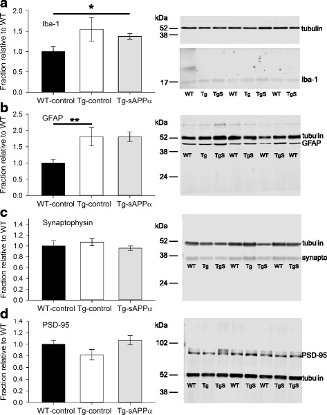
Fig. 5 Cellular markers for neuronal and non-neuronal cells. a Western blot analysis of the microglial marker Iba-1 (insoluble fraction), normalised to WT-control, showed an overall group effect (one-way ANOVA F(2,28) = p = 0.041) with a trend toward higher levels in Tg mice (WT-control: 1.0 ± 0.083, n = 12; Tg-control: 1.37 ± 0.299, n = 6), that was even more evident in the Tg-sAPPα group (Tg-sAPPα: 1.39 ± 0.065, n = 13; post-hoc Tukey p = 0.046). b GFAP levels (insoluble fraction) showed a significant overall group effect (F(2,27) = 10.92, p < 0.001), whereby there was significantly higher levels in the Tg-control group compared to the WT-control group (WT-control: 0.998 ± 0.105, n = 13; Tg-control: 1.81 ± 0.284, n = 6, Tukey p = 0.007) and expression of sAPPα did not alter this higher expression level (Tg-sAPPα: 1.86 ± 0.23, n = 11, Tukey p = 0.974). c Neither genotype (WT-control: 1 ± 0.02, n = 13; Tg-control: 1.07 ± 0.674, n = 6) nor sAPPα treatment (Tg-sAPPα: 0.96 ± 0.043, n = 12) affected the presynaptic marker synaptophysin (soluble fraction). d Levels of the postsynaptic marker PSD-95 (insoluble fraction) were also not affected by the APP/PS1 genotype (WT-control: 1.00 ± 0.072, n = 13; Tg-control: 0.82 ± 0.086, n = 5), although the PSD-95 levels tended to be higher for the Tg-sAPPα group compared to Tg-controls (Tg- sAPPα: 1.066 ± 0.082, n = 12, t(17) = − 1.90, p = 0.074. Representative western blots are presented. Note that in the case of Iba-1, its illustration and that of tubulin are from the same blot, but at different exposures. Tubulin was used as the loading control. *p < 0.05, **p < 0.01; synapto: synaptophysin; Lane labels: WT, WT-control; Tg, Tg-control; TgS, Tg-sAPPα
Fig. 5 Cellular markers for neuronal and non-neuronal cells. a Western blot analysis of the microglial marker Iba-1 (insoluble fraction), normalised to WT-control, showed an overall group effect (one-way ANOVA F(2,28) = p = 0.041) with a trend toward higher levels in Tg mice (WT-control: 1.0 ± 0.083, n = 12; Tg-control: 1.37 ± 0.299, n = 6), that was even more evident in the Tg-sAPPα group (Tg-sAPPα: 1.39 ± 0.065, n = 13; post-hoc Tukey p = 0.046). b GFAP levels (insoluble fraction) showed a significant overall group effect (F(2,27) = 10.92, p < 0.001), whereby there was significantly higher levels in the Tg-control group compared to the WT-control group (WT-control: 0.998 ± 0.105, n = 13; Tg-control: 1.81 ± 0.284, n = 6, Tukey p = 0.007) and expression of sAPPα did not alter this higher expression level (Tg-sAPPα: 1.86 ± 0.23, n = 11, Tukey p = 0.974). c Neither genotype (WT-control: 1 ± 0.02, n = 13; Tg-control: 1.07 ± 0.674, n = 6) nor sAPPα treatment (Tg-sAPPα: 0.96 ± 0.043, n = 12) affected the presynaptic marker synaptophysin (soluble fraction). d Levels of the postsynaptic marker PSD-95 (insoluble fraction) were also not affected by the APP/PS1 genotype (WT-control: 1.00 ± 0.072, n = 13; Tg-control: 0.82 ± 0.086, n = 5), although the PSD-95 levels tended to be higher for the Tg-sAPPα group compared to Tg-controls (Tg- sAPPα: 1.066 ± 0.082, n = 12, t(17) = − 1.90, p = 0.074. Representative western blots are presented. Note that in the case of Iba-1, its illustration and that of tubulin are from the same blot, but at different exposures. Tubulin was used as the loading control. *p < 0.05, **p < 0.01; synapto: synaptophysin; Lane labels: WT, WT-control; Tg, Tg-control; TgS, Tg-sAPPαCC BY · PubMed Central
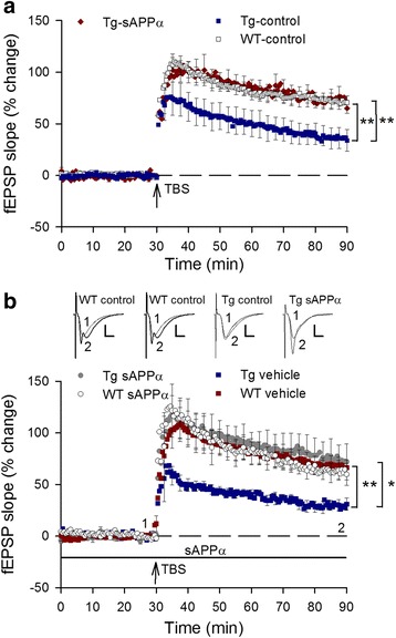
Fig. 6 Delivery of sAPPα, either in vivo or in vitro, after plaque development, completely rescued the impaired LTP in Tg-controls. a Chronically administered sAPPα by lentivirus-mediated expression in adult (10 months) Tg mice completely rescued the deficit in hippocampal LTP, measured 3 months after viral transduction (at 13 months of age). LTP measured 60 min after TBS (arrow) revealed a significant deficit in LTP expression in Tg-control mice compared to WT-controls (WT-controls: 71.1 ± 6.7%, n = 11; Tg-controls 36.3 ± 10.0%, n = 9; p = 0.008). This deficit was completely rescued by sAPPα over-expression (Tg-sAPPα: 72.2 ± 2.4%, n = 12; p = 0.008 compared to Tg-controls). b LTP induced in hippocampal slices from aged Tg mice (18–20 months of age), was impaired compared to WT-controls when measured 60 min post-TBS (WT-control: 67.2 ± 7.5%, n = 9; Tg-control: 29.4 ± 4.7%, n = 7; p = 0.002). LTP expression was again completely rescued by acutely applied recombinant human sAPPα (10 nM) beginning 30 min before delivery of the TBS (Tg-sAPPα: 73.7 ± 16.7%, n = 10; p = 0.737 compared to WT-controls, p = 0.048 compared to Tg-controls). No effect of sAPPα on WT-control LTP was observed (WT-sAPPα: 62.1 ± 9.4%, n = 6; p = 0.684)
Fig. 6 Delivery of sAPPα, either in vivo or in vitro, after plaque development, completely rescued the impaired LTP in Tg-controls. a Chronically administered sAPPα by lentivirus-mediated expression in adult (10 months) Tg mice completely rescued the deficit in hippocampal LTP, measured 3 months after viral transduction (at 13 months of age). LTP measured 60 min after TBS (arrow) revealed a significant deficit in LTP expression in Tg-control mice compared to WT-controls (WT-controls: 71.1 ± 6.7%, n = 11; Tg-controls 36.3 ± 10.0%, n = 9; p = 0.008). This deficit was completely rescued by sAPPα over-expression (Tg-sAPPα: 72.2 ± 2.4%, n = 12; p = 0.008 compared to Tg-controls). b LTP induced in hippocampal slices from aged Tg mice (18–20 months of age), was impaired compared to WT-controls when measured 60 min post-TBS (WT-control: 67.2 ± 7.5%, n = 9; Tg-control: 29.4 ± 4.7%, n = 7; p = 0.002). LTP expression was again completely rescued by acutely applied recombinant human sAPPα (10 nM) beginning 30 min before delivery of the TBS (Tg-sAPPα: 73.7 ± 16.7%, n = 10; p = 0.737 compared to WT-controls, p = 0.048 compared to Tg-controls). No effect of sAPPα on WT-control LTP was observed (WT-sAPPα: 62.1 ± 9.4%, n = 6; p = 0.684)CC BY · PubMed Central

Verification

Claims the paper makes

The claims and statistics of this paper haven’t been checked yet.

Run the check — free, no account needed. It reads the open-access text and checks the sources it cites.

A few free checks a day · papers already verified open instantly · results are public

Claims other papers make about this one

Each is checked against this paper’s own text: does it say what the citing paper says it does?

Self-check

Percentages, sums and reported differences re-computed from the paper’s own numbers.

The self-check needs the paper’s full text.

Citations

Cites

Show all 5 references

Research works printed but not linked: 43

  1. Fukumoto H, Cheung BS, Hyman BT, Irizarry MC. Β-secretase protein and activity are increased in the neocortex in Alzheimer disease. Arch Neurol. 2002;59:1381–9. · DOI · Paper page
  2. Ahmed RR, Holler CJ, Webb RL, Li F, Beckett TL, Murphy MP. BACE1 and BACE2 enzymatic activities in Alzheimer’s disease. J Neurochem. 2010;112:1045–53. · DOI
  3. Lannfelt L, Basun H, Wahlund LO, Rowe BA, Wagner SL. Decreased α-secretase-cleaved amyloid precursor protein as a diagnostic marker for Alzheimer's disease. Nat Med. 1995;1:829–32. · DOI
  4. Sennvik K, Fastbom J, Blomberg M, Wahlund LO, Winblad B, Benedikz E. Levels of alpha- and beta-secretase cleaved amyloid precursor protein in the cerebrospinal fluid of Alzheimer's disease patients. Neurosci Lett. 2000;278:169–72. · DOI
  5. Mattson MP, Cheng B, Culwell AR, Esch FS, Lieberburg I, Rydel RE. Evidence for excitoprotective and intraneuronal calcium-regulating roles for secreted forms of the β-amyloid precursor protein. Neuron. 1993;10:243–54. · DOI · Paper page
  6. Ryan MM, Morris GP, Mockett BG, Bourne K, Abraham WC, Tate WP, Williams JM. Time-dependent changes in gene expression induced by secreted amyloid precursor protein-alpha in the rat hippocampus. BMC Genomics. 2013;14:376. · DOI
  7. Baratchi S, Evans J, Tate W, Abraham WC, Connor B. Secreted amyloid precursor proteins promote proliferation and glial differentiation of adult hippocampal neural progenitor cells. Hippocampus. 2012;22:1517–27. · DOI
  8. Small D, Nurcombe V, Reed G, Clarris H, Moir R, Beyreuther K, Masters CA. Heparin-binding domain in the amyloid protein precursor of Alzheimer's disease is involved in the regulation of neurite outgrowth. J Neurosci. 1994;14:2117–27. · DOI
  9. Ishida A, Furukawa K, Keller JN, Mattson MP. Secreted form of β-amyloid precursor protein shifts the frequency dependency for induction of LTD, and enhances LTP in hippocampal slices. Neuroreport. 1997;8:2133–7. · DOI
  10. Taylor CJ, Ireland DR, Ballagh I, Bourne K, Marechal NM, Turner PR, Bilkey DK, Tate WP, Abraham WC. Endogenous secreted amyloid precursor protein-[alpha] regulates hippocampal NMDA receptor function, long-term potentiation and spatial memory. Neurobiol Dis. 2008;31:250–60. · DOI
  11. Hick M, Herrmann U, Weyer S, Mallm J-P, Tschäpe J-A, Borgers M, Mercken M, Roth FC, Draguhn A, Slomianka L, et al. Acute function of secreted amyloid precursor protein fragment APPsα in synaptic plasticity. Acta Neuropathol. 2015;129:21–37. · DOI · Paper page
  12. Claasen AM, Guévremont D, Mason-Parker SE, Bourne K, Tate WP, Abraham WC, Williams JM. Secreted amyloid precursor protein-α upregulates synaptic protein synthesis by a protein kinase G-dependent mechanism. Neurosci Lett. 2009;460:92–6. · DOI
  13. Meziane H, Dodart JC, Mathis C, Little S, Clemens J, Paul SM, Ungerer A. Memory-enhancing effects of secreted forms of the β-amyloid precursor protein in normal and amnestic mice. Proc Natl Acad Sci U S A. 1998;95:12683–8. · DOI
  14. Almkvist O, Basun H, Wagner SL, Rowe BA, Wahlund L, Lannfelt L. Cerebrospinal fluid levels of α-secretase-cleaved soluble amyloid precursor protein mirror cognition in a Swedish family with Alzheimer disease and a gene mutation. Arch Neurol. 1997;54:641–4. · DOI
  15. Anderson JJ, Holtz G, Baskin PP, Wang R, Mazzarelli L, Wagner SL, Menzaghi F. Reduced cerebrospinal fliud levels of à-secretase-cleaved amyloid precursor protein in aged rats: correlation with spatial memory deficits. Neuroscience. 1999;93:1409–20. · DOI
  16. Habib A, Sawmiller D, Tan J. Restoring soluble amyloid precursor protein α functions as a potential treatment for Alzheimer's disease. J Neurosci Res. 2017;2017:973–91. · DOI
  17. Corrigan F, Vink R, Blumbergs PC, Masters CL, Cappai R, van den Heuvel C. sAPPα rescues deficits in amyloid precursor protein knockout mice following focal traumatic brain injury. J Neurochem. 2012;122:208–20. · DOI
  18. Mockett BG, Richter M, Abraham WC, Müller UC. Therapeutic potential of secreted amyloid precursor protein APPsα. Front Mol Neurosci. 2017;10(30) · DOI
  19. Smith-Swintosky VL, Pettigrew LC, Craddock SD, Culwell AR, Rydel RE, Mattson MP. Secreted forms of β-amyloid precursor protein protect against ischemic brain injury. J Neurochem. 1994;63:781–4. · DOI
  20. Fol R, Braudeau J, Ludewig S, Abel T, Weyer S, Roederer J-P, Brod F, Audrain M, Bemelmans A-P, Buchholz C, et al. Viral gene transfer of APPsα rescues synaptic failure in an Alzheimer’s disease mouse model. Acta Neuropathol. 2016;131:247–66. · DOI
  21. Dittgen T, Nimmerjahn A, Komai S, Licznerski P, Waters J, Margrie TW, Helmchen F, Denk W, Brecht M, Osten P. Lentivirus-based genetic manipulations of cortical neurons and their optical and electrophysiological monitoring in vivo. Proc Natl Acad Sci,USA. 2004;101:18206–11. · DOI · Paper page
  22. Turner PR, Bourne K, Garama D, Carne A, Abraham WC, Tate WP. Production, purification and functional validation of human secreted amyloid precursor proteins for use as neuropharmacological reagents. J Neurosci Meth. 2007;164:68–74. · DOI
  23. Best HL, Neverman NJ, Wicky HE, Mitchell NL, Leitch B, Hughes SM. Characterisation of early changes in ovine CLN5 and CLN6 batten disease neural cultures for the rapid screening of therapeutics. Neurobiol Dis. 2017;100:62–74. · DOI
  24. Desmaris N, Bosch A, Salaun C, Petit C, Prevost MC, Tordo N, Perrin P, Schwartz O, de Rocquigny H, Heard JM. Production and neurotropism of lentivirus vectors pseudotyped with lyssavirus envelope glycoproteins. Mol Ther. 2001;4:149–56. · DOI
  25. Finkelshtein D, Werman A, Novick D, Barak S, Rubinstein M. LDL receptor and its family members serve as the cellular receptors for vesicular stomatitis virus. Proc Natl Acad Sci U S A. 2013;110:7306–11. · DOI · Paper page
  26. Schoderboeck L, Riad S, Bokor AM, Wicky HE, Strauss M, Bostina M, Oswald MJ, Empson RM, Hughes SM. Chimeric rabies SADB19-VSVg-pseudotyped lentiviral vectors mediate long-range retrograde transduction from the mouse spinal cord. Gene Ther. 2015;22:357–64. · DOI
  27. Özdinler PH, Macklis JD. IGF-I specifically enhances axon outgrowth of corticospinal motor neurons. Nat Neurosci. 2006;9:1371. · DOI · Paper page
  28. Gallagher M, Burwell R, Burchinal M. Severity of spatial learning impairment in aging: development of a learning index for performance in the Morris water maze. Behav Neurosci. 1993;107:618–26. · DOI
  29. Maei HR, Zaslavsky K, Teixeira CM, Frankland PW. What is the most sensitive measure of water maze probe test performance? Front Integr Neurosci. 2009;3:4.
  30. Bevins RA, Besheer J. Object recognition in rats and mice: a one-trial non-matching-to-sample learning task to study 'recognition memory'. Nat Protocols. 2006;1:1306–11. · DOI
  31. Hefter D, Draguhn A. APP as a protective factor in acute neuronal insults. Front Mol Neurosci. 2017;10(22) · DOI
  32. Chasseigneaux S, Allinquant B. Functions of Aβ, sAPPα and sAPPβ: similarities and differences. J Neurochem. 2012;120:99–108. · DOI
  33. Corrigan F, Vink R, Blumbergs PC, Masters CL, Cappai R, van den Heuvel C. Evaluation of the effects of treatment with sAPPα on functional and histological outcome following controlled cortical impact injury in mice. Neurosci Lett. 2012;515:50–4. · DOI
  34. Moreno L, Rose C, Mohanraj A, Allinquant B, Billard JM, Dutar P. sAβPPα improves hippocampal NMDA-dependent functional alterations linked to healthy aging. J Alz Dis. 2015;48(4):927–35. · DOI
  35. Xiong M, Jones OD, Peppercorn K, Ohline SM, Tate WP, Abraham WC. Secreted amyloid precursor protein-alpha can restore novel object location memory and hippocampal LTP in aged rats. Neurobiol Learn Mem. 2017;138:291–9. · DOI
  36. Heikkinen T, Kalesnykas G, Rissanen A, Tapiola T, Iivonen S, Wang J, Chaudhuri J, Tanila H, Miettinen R, Puoliväli J. Estrogen treatment improves spatial learning in APP + PS1 mice but does not affect beta amyloid accumulation and plaque formation. Exp Neurol. 2004;187:105–17. · DOI
  37. Morgan D, Diamond DM, Gottschall PE, Ugen KE, Dickey C, Hardy J, Duff K, Jantzen P, DiCarlo G, Wilcock D, et al. Aβ peptide vaccination prevents memory loss in an animal model of Alzheimer's disease. Nature. 2000;408:982–5. · DOI
  38. Stackman RW, Eckenstein F, Frei B, Kulhanek D, Nowlin J, Quinn JF. Prevention of age-related spatial memory deficits in a transgenic mouse model of Alzheimer's disease by chronic Ginkgo Biloba treatment. Exp Neurol. 2003;184:510–20. · DOI · Paper page
  39. Bauckneht M, Picco A, Nobili F, Morbelli S. Amyloid positron emission tomography and cognitive reserve. World J Radiol. 2015;7:475–83. · DOI
  40. Carapelle E, Serra L, Modoni S, Falcone M, Caltagirone C, Bozzali M, Specchio LM, Avolio C. How the cognitive reserve interacts with beta-amyloid deposition in mitigating FDG metabolism: an observational study. Medicine. 2017;96:e5876. · DOI
  41. Turner PR, O'Connor K, Tate WP, Abraham WC. Roles of amyloid precursor protein and its fragments in regulating neural activity, plasticity and memory. Prog Neurobiol. 2003;70:1–32. · DOI
  42. Kögel D, Deller T, Behl C. Roles of amyloid precursor protein family members in neuroprotection, stress signaling and aging. Exp Brain Res. 2012;217(3–4):471–9. · DOI
  43. Stein TD, Anders NJ, DeCarli C, Chan SL, Mattson MP, Johnson JA. Neutralization of transthyretin reverses the neuroprotective effects of secreted amyloid precursor protein (APP) in APPSW mice resulting in tau phosphorylation and loss of hippocampal neurons: support for the amyloid hypothesis. J Neurosci. 2004;24:7707–17. · DOI

Cited by

Sorted by citations

Metadata

Authors
Tan, V. T. Y., Mockett, B. G., Ohline, S. M., Parfitt, K. D., Wicky, H. E., Peppercorn, K., Schoderboeck, L., Yahaya, M. F. B., Tate, W. P., Hughes, S. M., & Abraham, W. C.
Venue
Molecular Brain
Published
2018-02-09
Volume
11
Issue
1
ISSN
1756-6606
Cited by
61
References
5
Language
en
Funders
  • Health Research Council of New Zealand (10-170)
  • Ministry of Higher Education, Malaysia

About these checks

Registry, retraction, Cites and statistics results are computed, not judged, with one exception: a printed reference that no index lists is read once by a model before it counts as a citation error. Claims are extracted by a model and kept only when their quote appears verbatim in the text; each cited source is then checked, with a model’s help, for whether it says what the claim relies on. Those verdicts can be wrong — read the claim’s quote and the cited source before relying on one.

Statistics and arithmetic are recomputed, not judged.

Confidence bands are shown only for fields calibrated on our annotated test set. None is calibrated yet, so no band is shown.

What counts as a claim ›How we measure confidence ›How we check this ›

Are you an author, or do you see a problem with this page? Request a review