

The DOI matches its Crossref record; 34 of the 34 references it cites were found in a registry or an index.
Terminating contamination: large-scale search identifies more than 2,000,000 contaminated entries in GenBank
Open accessPublished version of a preprint
Abstract
Genomic analyses are sensitive to contamination in public databases caused by incorrectly labeled reference sequences. Here, we describe Conterminator, an efficient method to detect and remove incorrectly labeled sequences by an exhaustive all-against-all sequence comparison. Our analysis reports contamination of 2,161,746, 114,035, and 14,148 sequences in the RefSeq, GenBank, and NR databases, respectively, spanning the whole range from draft to “complete” model organism genomes. Our method scales linearly with input size and can process 3.3 TB in 12 days on a 32-core computer. Conterminator can help ensure the quality of reference databases. Source code (GPLv3): https://github.com/martin-steinegger/conterminator
Overview
The claims and statistics of this paper haven’t been checked yet.
Run the check — free, no account needed. It reads the open-access text and checks the sources it cites.
Integrity signals
Registry
Cites
Statistics
Figures
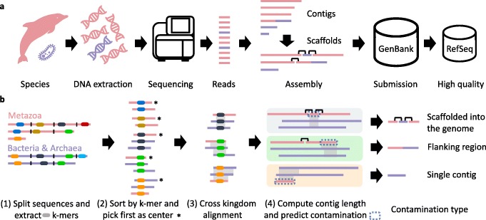
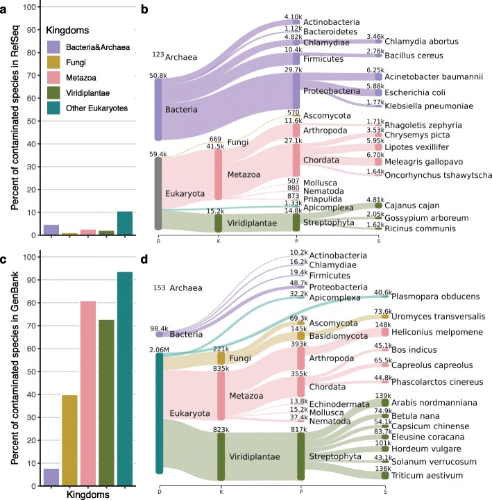
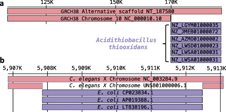
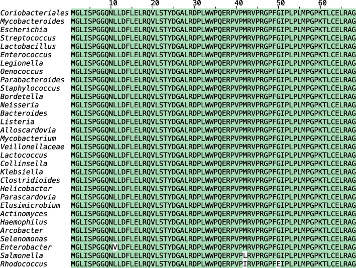
Verification
Claims the paper makes
The claims and statistics of this paper haven’t been checked yet.
Run the check — free, no account needed. It reads the open-access text and checks the sources it cites.
Claims other papers make about this one
Each is checked against this paper’s own text: does it say what the citing paper says it does?
In the citing paper
“The bacterial protein (UniProt: A0A1C5UEQ5), encoded on a contig of length 138,559 (GenBank: FMFM01000010), is unlikely to be a contaminant due to its length29.”
From Clustering predicted structures at the scale of the known protein universe (2023) · cites this paper as [29]
Self-check
Percentages, sums and reported differences re-computed from the paper’s own numbers.
The self-check needs the paper’s full text.
Citations
Cites
Fast gapped-read alignment with Bowtie 2
Langmead, B., & Salzberg, S. L. (2012) · Nature Methods · doi:10.1038/nmeth.1923 · Cited by 56,400
Self-citationBLAST+: architecture and applications
Camacho, C., Coulouris, G., Avagyan, V., Ma, N., Papadopoulos, J., Bealer, K., & Madden, T. L. (2009) · BMC Bioinformatics · doi:10.1186/1471-2105-10-421 · Cited by 22,074
Minimap2: Pairwise alignment for nucleotide sequences
Li, H. (2018) · Bioinformatics · doi:10.1093/bioinformatics/bty191 · Cited by 14,807
Jalview Version 2—a multiple sequence alignment editor and analysis workbench
Waterhouse, A. M., Procter, J. B., Martin, D. M. A., Clamp, M. E., & Barton, G. J. (2009) · Bioinformatics · doi:10.1093/bioinformatics/btp033 · Cited by 10,349
UniProt: a worldwide hub of protein knowledge
The UniProt Consortium (2019) · Nucleic Acids Research · doi:10.1093/nar/gky1049 · Cited by 7,698
O'Leary, N. A., Wright, M. W., Brister, J. R., Ciufo, S., Haddad, D., McVeigh, R., Rajput, B., Robbertse, B., Smith-White, B., Ako-Adjei, D., et al. (2016) · Nucleic Acids Research · doi:10.1093/nar/gkv1189 · Cited by 6,357
MMseqs2 enables sensitive protein sequence searching for the analysis of massive data sets
Steinegger, M., & Söding, J. (2017) · Nature Biotechnology · doi:10.1038/nbt.3988 · Cited by 4,840
Self-citationMash: fast genome and metagenome distance estimation using MinHash
Ondov, B. D., Treangen, T. J., Melsted, P., Mallonee, A. B., Bergman, N. H., Koren, S., & Phillippy, A. M. (2016) · Genome biology · doi:10.1186/s13059-016-0997-x · Cited by 3,373
Federhen, S. (2012) · Nucleic Acids Research · doi:10.1093/nar/gkr1178 · Cited by 1,436
Clustering huge protein sequence sets in linear time
Steinegger, M., & Söding, J. (2018) · Nature Communications · doi:10.1038/s41467-018-04964-5 · Cited by 875
Self-citation
Cited by
Sorted by citations
Items 101–200 of 270
Proteotranscriptomics – A facilitator in omics research
Levin, M., & Butter, F. (2022) · Computational and Structural Biotechnology Journal · doi:10.1016/j.csbj.2022.07.007 · Cited by 9
Horizontal Transfer of Bacterial Operons into Eukaryote Genomes
Kogay, R., Wolf, Y. I., & Koonin, E. V. (2025) · Genome Biology and Evolution · doi:10.1093/gbe/evaf055 · Cited by 8
Bioinformatic approaches to blood and tissue microbiome analyses: challenges and perspectives
Sirasani, J. P., Gardner, C., Jung, G., Lee, H., & Ahn, T.-H. (2025) · Briefings in Bioinformatics · doi:10.1093/bib/bbaf176 · Cited by 10
Quality Control in Metagenomics Data
Gihawi, A., Cardenas, R. P., Hurst, R., & Brewer, D. S. (2023) · Methods in molecular biology · doi:10.1007/978-1-0716-3072-3_2 · Cited by 8
Vancaester, E., & Blaxter, M. L. (2024) · Wellcome Open Research · doi:10.12688/wellcomeopenres.20730.1 · Cited by 8
The Azurin Coding Gene: Origin and Phylogenetic Distribution
Gammuto, L., Chiellini, C., Iozzo, M., Fani, R., & Petroni, G. (2021) · Microorganisms · doi:10.3390/microorganisms10010009 · Cited by 8
RAPiD: a rapid and accurate plant pathogen identification pipeline for on-site nanopore sequencing
Knobloch, S., Salimi, F., Buaya, A., Ploch, S., & Thines, M. (2024) · PeerJ · doi:10.7717/peerj.17893 · Cited by 8
Alvarez-Costes, S., Baker, C. S., Constantine, R., Carroll, E. L., Guhlin, J. G., Dutoit, L., Ferreira, S., Heimeier, D., Gemmell, N. J., Gillum, J. E., et al. (2025) · Molecular Ecology Resources · doi:10.1111/1755-0998.14109 · Cited by 8
In Vouchers We (Hope to) Trust: Unveiling Hidden Errors in GenBank 's Tetrapod Taxonomic Foundations
Carné, A., Vieites, D. R., & van den Burg, M. P. (2025) · Molecular Ecology · doi:10.1111/mec.17812 · Cited by 7
Chin, P.-J., Bhavsar, J., Bosma, T. J., MacDonald, M. L., Polson, S. W., & Khan, A. S. (2025) · mSphere · doi:10.1128/msphere.00286-25 · Cited by 8
Bamdam: a post-mapping authentication toolkit for ancient metagenomics
Sanctis, B. D., Mirchandani, C., Dong, H., Macleod, R., Corbett-Detig, R. B., & Wang, Y. (2025) · Genome biology · doi:10.1186/s13059-025-03879-x · Cited by 8
Filtering out the noise: metagenomic classifiers optimize ancient DNA mapping
Ravishankar, S., Pérez, V., Davidson, R., Roca‐Rada, X., Lan, D. M., Souilmi, Y., & Llamas, B. (2024) · Briefings in Bioinformatics · doi:10.1093/bib/bbae646 · Cited by 8
Metagenome Proteins and Database Contamination
Arkhipova, I. R. (2020) · mSphere · doi:10.1128/msphere.00854-20 · Cited by 7
A Large-Scale Study into Protist-Animal Interactions Based on Public Genomic Data Using DNA Barcodes
Xie, J., Tan, B., & Zhang, Y. (2023) · Animals · doi:10.3390/ani13142243 · Cited by 7
Efficient k-mer based curation of raw sequence data: application in Drosophila suzukii
Gautier, M. (2023) · Peer Community Journal · doi:10.24072/pcjournal.309 · Cited by 7
Contamination in sequence databases
Tang, L. (2020) · Nature Methods · doi:10.1038/s41592-020-0895-8 · Cited by 7
Smith, C. A. (2021) · Scientific Reports · doi:10.1038/s41598-021-89146-y · Cited by 7
Metagenomic profiling pipelines improve taxonomic classification for 16S amplicon sequencing dataPreprint
Faits, T., Odom-Mabey, A. R., Castro‐Nallar, E., Crandall, K. A., & Johnson, W. E. (2022) · bioRxiv (Cold Spring Harbor Laboratory) · doi:10.1101/2022.07.27.501757 · Cited by 7
SeqDPI : A 1D ‐ CNN approach for predicting binding affinity of kinase inhibitors
Mishra, V. P., Singh, Y. N., Khan, F., & Dutta, M. K. (2024) · Journal of Computational Chemistry · doi:10.1002/jcc.27518 · Cited by 6
Purcell, M., Ackland, J., Staples, K. J., Freeman, A. T., & Wilkinson, T. M. A. (2025) · European Respiratory Review · doi:10.1183/16000617.0284-2024 · Cited by 10
Genomic evidence for the widespread presence of GH45 cellulases among soil invertebrates
Muelbaier, H., Arthen, F., Collins, G. E., Hickler, T., Hohberg, K., Lehmitz, R., Pauchet, Y., Pfenninger, M., Potapov, A., Romahn, J., et al. (2024) · Molecular Ecology · doi:10.1111/mec.17351 · Cited by 6
Zhuang, Y., Li, S., Bao, D., Furlan, J. P. R., & Ruan, Z. (2024) · Journal of Infection · doi:10.1016/j.jinf.2024.106356 · Cited by 6
Briand, M., Jacques, M., & Dittmer, J. (2025) · Applied and Environmental Microbiology · doi:10.1128/aem.00913-25 · Cited by 6
Sharma, M., & Singh, P. (2021) · Springer eBooks · doi:10.1007/978-981-16-0811-7_9 · Cited by 6
Advances in identifying GM plants: toward the routine detection of ‘hidden’ and ‘new’ GMOs
Bertheau, Y. (2021) · Burleigh Dodds series in agricultural science · doi:10.19103/as.2021.0097.22 · Cited by 6
GTax: improving de novo transcriptome assembly by removing foreign RNA contamination
Alvarez, R. V., & Landsman, D. (2024) · Genome biology · doi:10.1186/s13059-023-03141-2 · Cited by 6
WGA-LP: a pipeline for whole genome assembly of contaminated reads
Rossi, N., Colautti, A., Iacumin, L., & Piazza, C. (2021) · Bioinformatics · doi:10.1093/bioinformatics/btab719 · Cited by 6
Aguirre-Carvajal, K., Cárdenas, S., Munteanu, C. R., & Armijos‐Jaramillo, V. (2025) · International Journal of Molecular Sciences · doi:10.3390/ijms26051795 · Cited by 6
Genome Reference Assembly for Bottlenecked Southern Australian Koalas
Blanchard, A. M., Emes, R. D., Greenwood, A. D., Holmes, N., Loose, M., McEwen, G. K., Meers, J., Speight, N., & Tarlinton, R. E. (2022) · Genome Biology and Evolution · doi:10.1093/gbe/evac176 · Cited by 5
GenomeFLTR: filtering reads made easy
Dotan, E., Alburquerque, M., Wygoda, E., Huchon, D., & Pupko, T. (2023) · Nucleic Acids Research · doi:10.1093/nar/gkad410 · Cited by 5
Rumore, J., Walker, M., Pagotto, F., Forbes, J. D., Peterson, C.-L., Tyler, A. D., Graham, M., Domselaar, G. V., Nadon, C. A., Reimer, A. R., et al. (2023) · BMC Genomics · doi:10.1186/s12864-023-09338-w · Cited by 5
Xie, J., & Zhang, Y. (2023) · Animals · doi:10.3390/ani13203172 · Cited by 5
Mittelberger, C., Moser, M., Hause, B., & Janik, K. (2024) · BMC Plant Biology · doi:10.1186/s12870-024-05087-4 · Cited by 5
Aylward, J., Wingfield, M. J., Roets, F., & Wingfield, B. D. (2021) · BioTechniques · doi:10.2144/btn-2021-0097 · Cited by 5
Detection of Parasites in Microbiomes using MetagenomicsPreprint
Kirstahler, P., Aarestrup, F. M., & Pamp, S. J. (2022) · bioRxiv (Cold Spring Harbor Laboratory) · doi:10.1101/2022.03.27.485979 · Cited by 5
Oskolkov, N., Jin, C., Clinton, S. L., Guinet, B., Wijnands, F., Johnson, E., Kutschera, V. E., Kinsella, C. M., Heintzman, P. D., & van der Valk, T. (2025) · bioRxiv (Cold Spring Harbor Laboratory) · doi:10.1101/2025.03.19.644176 · Cited by 5
Disentangling cobionts and contamination in long-read genomic data using sequence composition
Weber, C. C. (2024) · G3 Genes Genomes Genetics · doi:10.1093/g3journal/jkae187 · Cited by 4
Tran, V., Langschied, F., Muelbaier, H., Dosch, J., Arthen, F., Bálint, M., & Ebersberger, I. (2025) · Molecular Biology and Evolution · doi:10.1093/molbev/msaf120 · Cited by 7
A practical guide on environmental genomics for prokaryotic systematics
Ernster, N. M., & Rodriguez‐R, L. M. (2025) · Systematic and Applied Microbiology · doi:10.1016/j.syapm.2025.126643 · Cited by 4
Expanding the taxonomic range in the fecal metagenome
Allnutt, T. R., Roth‐Schulze, A. J., & Harrison, L. C. (2021) · BMC Bioinformatics · doi:10.1186/s12859-021-04212-6 · Cited by 4
Xu, M., Guo, L., Qi, Y., Shi, C., Liu, X., Chen, J., Han, J., Deng, L., Liu, X., & Fan, G. (2023) · Frontiers in Marine Science · doi:10.3389/fmars.2023.1087447 · Cited by 4
Carballo, J., Bellido, A. M., Selva, J. P., Zappacosta, D., Gallo, C. A., Albertini, E., Cáccamo, M., & Echenique, V. C. (2023) · Frontiers in Plant Science · doi:10.3389/fpls.2023.1133986 · Cited by 4
Decontamination of DNA sequences from a Streptomyces genome for optimal genome mining
de Oliveira, R. V. F., Garrido, L. M., & Padilla, G. (2025) · Brazilian Journal of Microbiology · doi:10.1007/s42770-024-01598-2 · Cited by 4
Accurate annotation of protein coding sequences with IDTAXA
Cooley, N. P., & Wright, E. S. (2021) · NAR Genomics and Bioinformatics · doi:10.1093/nargab/lqab080 · Cited by 4
Best BLAST hit alone cannot be used as evidence of fraud
Díaz‐Arce, N., & Rodríguez‐Ezpeleta, N. (2023) · Scientific Reports · doi:10.1038/s41598-022-26720-y · Cited by 4
Velsko, I. M., Fagernäs, Z., Tromp, M., Bedford, S., Buckley, H. R., Clark, G., Dudgeon, J., Flexner, J. L., Tamarii, A., Lewis, C. M., et al. (2023) · bioRxiv (Cold Spring Harbor Laboratory) · doi:10.1101/2023.10.18.563027 · Cited by 4
Wang, H., Meng, Y., Chen, X., Cui, X., Zuo, Q., Han, N., Liang, X., Shen, X., Li, C., Li, D., et al. (2025) · Advanced Science · doi:10.1002/advs.202511920 · Cited by 3
Chromosome-Level Genome Assembly of the Australian Rainforest Tree Rhodamnia argentea (Malletwood)
Chen, S. H., Jones, A., Lu‐Irving, P., Yap, J. S., van der Merwe, M. M., Bragg, J. G., & Edwards, R. J. (2024) · Genome Biology and Evolution · doi:10.1093/gbe/evae238 · Cited by 3
Teknonaturalist: A Snakemake Pipeline for Assessing Fungal Diversity From Plant Genome Bycatch
Bard, N. W., Davies, T. J., & Cronk, Q. (2024) · Molecular Ecology Resources · doi:10.1111/1755-0998.14056 · Cited by 3
High-Performance Genome Annotation for a Safer and Faster-Developing Phage Therapy
Culot, A., Abriat, G., & Furlong, K. P. (2025) · Viruses · doi:10.3390/v17030314 · Cited by 4
ContFree-NGS: Removing Reads from Contaminating Organisms in Next Generation Sequencing Data
Peres, F. V., & Riaño‐Pachón, D. M. (2021) · Lecture notes in computer science · doi:10.1007/978-3-030-91814-9_6 · Cited by 3
A chromosome-level genome assembly of Zasmidium syzygii isolated from banana leaves
van Westerhoven, A. C., Mehrabi, R., Talebı, R., Steentjes, M. B. F., Corcolon, B. M., Chong, P., Kema, G. H. J., & Seidl, M. F. (2023) · G3 Genes Genomes Genetics · doi:10.1093/g3journal/jkad262 · Cited by 3
Gauvin, P., Eme, D., Domaizon, I., & Rimet, F. (2025) · Metabarcoding and Metagenomics · doi:10.3897/mbmg.9.137772 · Cited by 4
NetSyn: prokaryotic genomic context exploration of protein families
Stam, M. R., Langlois, J., Chevalier, C., Mainguy, J., Reboul, G., Bastard, K., Médigue, C., & Vallenet, D. (2026) · Peer Community Journal · doi:10.24072/pcjournal.805 · Cited by 3
Biderre‐Petit, C., Charvy, J., Bronner, G., Chauvet, M., Debroas, D., Gardon, H., Hennequin, C., Jouan‐Dufournel, I., Moné, A., Monjot, A., et al. (2022) · Molecular Ecology Resources · doi:10.1111/1755-0998.13692 · Cited by 4
Tracing foreign sequences in plant transcriptomes and genomes using OCT4, a POU domain protein
Saffar, A., & Matin, M. M. (2021) · Molecular Genetics and Genomics · doi:10.1007/s00438-021-01768-z · Cited by 3
Chen, J., Yan, X., Sun, Y., Ren, Z., Yan, G., Wang, G., Liu, Y., Zhao, Z., Liu, Y., Tu, C., et al. (2022) · bioRxiv (Cold Spring Harbor Laboratory) · doi:10.1101/2022.03.03.482774 · Cited by 3
Accurate identification of de novo genes in plant genomes using machine learning algorithmsPreprint
Casola, C., Owoyemi, A. O., Pepper, A. E., & Ioerger, T. R. (2022) · bioRxiv (Cold Spring Harbor Laboratory) · doi:10.1101/2022.11.01.514720 · Cited by 3
Decode-gLM: Tools to Interpret, Audit, and Steer Genomic Language ModelsPreprint
Maiwald, A., Jedryszek, P., Draye, F., Schölkopf, B., Morris, G. M., & Crook, O. M. (2025) · bioRxiv (Cold Spring Harbor Laboratory) · doi:10.1101/2025.10.31.685860 · Cited by 6
Prediction of Interleukin-binding Sites Combining Multi-Source Features with Integrated Algorithm
Niu, X., & Feng, Y. (2025) · Current Bioinformatics · doi:10.2174/0115748936372515250801173324 · Cited by 2
Oskolkov, N., Jin, C., Clinton, S. L., Guinet, B., Wijnands, F., Johnson, E., Kutschera, V. E., Kinsella, C. M., Heintzman, P. D., & van der Valk, T. (2025) · GigaScience · doi:10.1093/gigascience/giaf108 · Cited by 2
Composite Genome Quality Index for Pathogenic Bacterial Genomes
Farooq, A., & Rafique, A. (2025) · Applied Microbiology · doi:10.3390/applmicrobiol5040144 · Cited by 2
González-Ramírez, I. S., Song, M. J., Mehlferber, E. C., & Mishler, B. D. (2026) · Applications in Plant Sciences · doi:10.1002/aps3.70064 · Cited by 2
Fu, Y., Zhuang, Y., Luo, S., & Xu, X. J. (2023) · Genes · doi:10.3390/genes14091678 · Cited by 2
Contaminated bacterial genome data in the public domains: Evidence and solution
Tang, B., Hu, X., & Yue, M. (2024) · Journal of Infection · doi:10.1016/j.jinf.2024.106369 · Cited by 2
Contamination Survey of Insect Genomic and Transcriptomic Data
Zhou, J., Zhang, X., Wang, Y., Liang, H., Yang, Y., Huang, X., & Deng, J. (2024) · Animals · doi:10.3390/ani14233432 · Cited by 2
Furlan, J. P. R., Schenkman, S., Sellera, F. P., Li, S., Zhuang, Y., & Ruan, Z. (2025) · The Lancet Microbe · doi:10.1016/j.lanmic.2024.101067 · Cited by 2
gymnotoa-db : a database and application to optimize functional annotation in gymnosperms
Mora‐Márquez, F., Hurtado, M., & de Heredia, U. L. (2025) · Database · doi:10.1093/database/baaf019 · Cited by 2
Apicortin, a Putative Apicomplexan-Specific Protein, Is Present in Deep-Branching Opisthokonts
Orosz, F. (2025) · Biology · doi:10.3390/biology14060620 · Cited by 2
Identification of bacteriophage DNA in human umbilical cord blood
Sequoia, J. A., Haddock, N. L., Gay, P. M., Barkal, L. J., Narasimhan, P., Martinez, N., Winn, V. D., & Bollyky, P. L. (2025) · JCI Insight · doi:10.1172/jci.insight.183123 · Cited by 3
Digesting the data: Proper validation in ancient metagenomic studies is essentialPreprint
Pach, A. L., Lanigan, L. T., Niemann, J., Pedersen, M. W., & Schroeder, H. (2024) · bioRxiv (Cold Spring Harbor Laboratory) · doi:10.1101/2024.02.27.581519 · Cited by 2
Use of the CHM13-T2T genome improves metagenomic analysis by minimizing host DNA contamination
Liu, D., Hu, J., Zhang, D., Ren, S., Zhao, L., Gao, H., Hu, S., Xu, S., & Liang, G. (2025) · mSystems · doi:10.1128/msystems.00840-25 · Cited by 1
Granados-Casas, A. O., Fernández-Bravo, A., Stchigel, A. M., & Cano, J. (2025) · BMC Genomics · doi:10.1186/s12864-025-12223-3 · Cited by 1
Nichols, S., Estandía, A., Young, C., Knowles, L. S., Palinauskas, V., Okamura, B., & Clegg, S. M. (2026) · International Journal for Parasitology · doi:10.1016/j.ijpara.2025.104768 · Cited by 1
Ruiz‐Ruiz, S., Piquer-Esteban, S., Pérez-Rocher, B., Pérez‐Brocal, V., Arnau, V., Artacho, A., Diaz, W., Jiménez‐Hernández, N., Pons, J., Castro, J. A., et al. (2026) · Scientific Reports · doi:10.1038/s41598-026-35033-3 · Cited by 1
Guccione, C., Sfiligoi, I., Gonzalez, A., Shaffer, J. P., Kazachkova, M., Weng, Y., McDonald, D., Shah, S. C., Minot, S. S., Paulson, T. G., et al. (2026) · BMC Genomics · doi:10.1186/s12864-026-12545-w · Cited by 1
MitoNGS: an online platform to analyze fish metabarcoding data in high resolution
Zhu, T., Sato, Y., Fukunaga, T., Miya, M. U., Iwasaki, W., & Yoshizawa, S. (2026) · Molecular Biology and Evolution · doi:10.1093/molbev/msag046 · Cited by 1
Horizontal gene transfer in Saccharomyces cerevisiae and other Saccharomycotina yeasts: a review
Grassi, A., Rogo, U., Fambrini, M., Pugliesi, C., & Agnolucci, M. (2026) · World Journal of Microbiology and Biotechnology · doi:10.1007/s11274-026-05118-2 · Cited by 1
Isotopes, Genetic Markers and Microbiome: Tools for seafood traceability
Patarnello, T., Russo, A., Pavlovič, B., Nerini, M., Marvasi, M., D’Alessandro, A., Cardazzo, B., Martino, M. E., Cavalieri, D., & Ogrinc, N. (2026) · Reviews in Fisheries Science & Aquaculture · doi:10.1080/23308249.2026.2713632 · Cited by 1
Edwards, R. J., Chen, S. H., Halliday, B., & Bragg, J. G. (2025) · Genome Biology and Evolution · doi:10.1093/gbe/evaf023 · Cited by 1
Xie, J., Zhang, Y., Wang, L., & Deng, Y. (2025) · Diversity · doi:10.3390/d17030186 · Cited by 1
Majernik, S. N., Beaver, L. P., & Bradley, P. H. (2025) · mSphere · doi:10.1128/msphere.00857-24 · Cited by 1
NGATHA carpel development genes evolved in the common ancestor of seed plants
Cota, I., Moschin, S., Offer, E., Martínez‐Fernández, I., Magnanimi, F., Ambrose, B. A., Nigris, S., Baldan, B., Ferrándiz, C., & Pelaz, S. (2025) · The Plant Journal · doi:10.1111/tpj.70488 · Cited by 1
Development of Metagenomic Methods for Health Monitoring of Endangered Species Using Fecal Samples
Sapino, R., Fernández‐González, Á., & Castresana, J. (2026) · Evolutionary Applications · doi:10.1111/eva.70199 · Cited by 1
Zhu, Q., & Mirarab, S. (2022) · Methods in molecular biology · doi:10.1007/978-1-0716-2691-7_7 · Cited by 1
Orosz, F. (2023) · Life · doi:10.3390/life13081664 · Cited by 1
Decontaminating reads, not contigs
Galtier, N. (2023) · Peer Community In Genomics · doi:10.24072/pci.genomics.100244 · Cited by 1
Liu, Y., Elworth, R. A. L., Jochum, M. D., Aagaard, K. M., & Treangen, T. J. (2021) · bioRxiv (Cold Spring Harbor Laboratory) · doi:10.1101/2021.05.06.442815 · Cited by 1
De novo transcriptome assembly and the effect of foreign RNA contaminationPreprint
Alvarez, R. V., & Landsman, D. (2022) · bioRxiv (Cold Spring Harbor Laboratory) · doi:10.1101/2022.11.07.515468 · Cited by 1
ecology, A. A., Polling, M., Onderz. Form., D., de Groot, G. A., Laros, I., & van den Heuvel-Greve, M. J. (2023) · doi:10.18174/629836 · Cited by 1
Ruoshi, Z. (2024) · eDiss (Georg-August-Universität Göttingen) · doi:10.53846/goediss-10594 · Cited by 1
Gut Fungi Possess a Conserved Toxin Immunity Gene of Bacterial OriginPreprint
Rizvi, S. M., Zhang, C., Freddolino, P. L., & Zhang, Y. (2020) · bioRxiv (Cold Spring Harbor Laboratory) · doi:10.1101/2020.10.15.341461
Measuring quality of DNA sequence data via degradation
Karr, A. F., Hauzel, J., Porter, A., & Schaefer, M. (2022) · PLoS ONE · doi:10.1371/journal.pone.0271970
Bolaji, A., & Duggan, A. T. (2023) · bioRxiv (Cold Spring Harbor Laboratory) · doi:10.1101/2023.09.26.559465
Hess, M. K., Hodgkinson, H. E., Hess, A. S., Zetouni, L., Budel, J. C. D. C., Henry, H., Donaldson, A., Bilton, T. P., van Stijn, T. C., Kirk, M. R., et al. (2023) · Figshare · doi:10.6084/m9.figshare.24159508
Hess, M. K., Hodgkinson, H. E., Hess, A. S., Zetouni, L., Budel, J. C. D. C., Henry, H., Donaldson, A., Bilton, T. P., van Stijn, T. C., Kirk, M. R., et al. (2023) · Figshare · doi:10.6084/m9.figshare.24159517
Hess, M. K., Zetouni, L., Hess, A. S., Budel, J. C. D. C., Dodds, K. G., Henry, H., Bräuning, R., McCulloch, A., Hickey, S. M., Johnson, P. L., et al. (2023) · Figshare · doi:10.6084/m9.figshare.23751869
Metadata
- Authors
- Steinegger, M., & Salzberg, S. L.
- Affiliations
- Seoul National University (KR)
- Johns Hopkins University (US)
- Corresponding author
- Steinegger, Martin
- Venue
- Genome Biology
- Published
- 2020-05-12
- Volume
- 21
- Issue
- 1
- Pages
- 115-115
- ISSN
- 1465-6906 (ISSN-L)
- 1474-760X
- PMC
- PMC7218494
- OpenAlex
- W3028050938
- Cited by
- 246
- References
- 34
- Citations by year
- 2026: 27 · 2025: 52 · 2024: 38 · 2023: 37 · 2022: 34 · 2021: 45 · 2020: 13
- FWCI
- 11.19
- Type
- journal article
- Language
- en
- Open access
- gold
- doi.org
- Locations
- Genome biology (publishedVersion, cc-by)
- bioRxiv (Cold Spring Harbor Laboratory) (acceptedVersion, cc-by-nc-nd)
- PubMed Central (publishedVersion, cc-by)
- PubMed (publishedVersion)
- DOAJ (DOAJ: Directory of Open Access Journals) (submittedVersion, cc-by-sa)
- Seoul National University Open Repository (Seoul National University) (submittedVersion)
- Funders
- National Science Foundation (1744309, R01-HG006677)
- National Institutes of Health (HG006677)
- National Human Genome Research Institute (R01HG006677)
- National Institute of General Medical Sciences (R35GM130151)
- Topics
- Life Sciences › Biochemistry, Genetics and Molecular Biology › Molecular Biology › Genomics and Phylogenetic Studies
- Life Sciences › Biochemistry, Genetics and Molecular Biology › Molecular Biology › Fractal and DNA sequence analysis
- Life Sciences › Biochemistry, Genetics and Molecular Biology › Genetics › Chromosomal and Genetic Variations
- Keywords
- GenBank, reference sequences, contamination detection
- MeSH terms
- Animals, Humans, Genome, Databases, Nucleic Acid, Mice, DNA Contamination
About these checks
Registry, retraction, Cites and statistics results are computed, not judged, with one exception: a printed reference that no index lists is read once by a model before it counts as a citation error. Claims are extracted by a model and kept only when their quote appears verbatim in the text; each cited source is then checked, with a model’s help, for whether it says what the claim relies on. Those verdicts can be wrong — read the claim’s quote and the cited source before relying on one.
Statistics and arithmetic are recomputed, not judged.
Confidence bands are shown only for fields calibrated on our annotated test set. None is calibrated yet, so no band is shown.
What counts as a claim ›How we measure confidence ›How we check this ›
Are you an author, or do you see a problem with this page? Request a review
