

The DOI matches its Crossref record; 36 of the 36 references it cites were found in a registry or an index.
Tissue source determines the differentiation potentials of mesenchymal stem cells: a comparative study of human mesenchymal stem cells from bone marrow and adipose tissue
Open accessAbstract
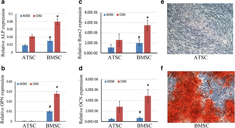
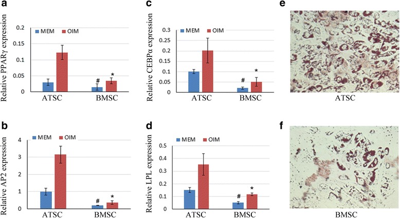
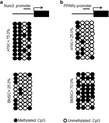
BACKGROUND: Mesenchymal stem cells (MSCs) possess intrinsic regeneration capacity as part of the repair process in response to injury, such as fracture or other tissue injury. Bone marrow and adipose tissue are the major sources of MSCs. However, which cell type is more effective and suitable for cell therapy remains to be answered. The intrinsic molecular mechanism supporting the assertion has also been lacking. METHODS: Human bone marrow-derived MSCs (BMSCs) and adipose tissue-derived MSCs (ATSCs) were isolated from bone marrow and adipose tissue obtained after total hip arthroplasty. ATSCs and BMSCs were incubated in standard growth medium. Trilineage differentiation including osteogenesis, adipogenesis, and chondrogenesis was performed by addition of relevant induction mediums. The expression levels of trilineage differentiation marker genes were evaluated by quantitative RT-PCR. The methylation status of CpG sites of Runx2, PPARγ, and Sox9 promoters were checked by bisulfite sequencing. In addition, ectopic bone formation and calvarial bone critical defect models were used to evaluate the bone regeneration ability of ATSCs and BMSCs in vivo. RESULTS: The results showed that BMSCs possessed stronger osteogenic and lower adipogenic differentiation potentials compared to ATSCs. There was no significant difference in the chondrogenic differentiation potential. The CpG sites of Runx2 promoter in BMSCs were hypomethylated, while in ATSCs they were hypermethylated. The CpG sites of PPARγ promoter in ATSCs were hypomethylated, while in BMSCs they were hypermethylated. The methylation status of Sox9 promoter in BMSCs was only slightly lower than that in ATSCs. CONCLUSIONS: The epigenetic memory obtained from either bone marrow or adipose tissue favored MSC differentiation along an osteoblastic or adipocytic lineage. The methylation status of the main transcription factors controlling MSC fate contributes to the differential differentiation capacities of different source-derived MSCs.
Overview
The claims and statistics of this paper haven’t been checked yet.
Run the check — free, no account needed. It reads the open-access text and checks the sources it cites.
Integrity signals
Registry
Cites
Statistics
Figures
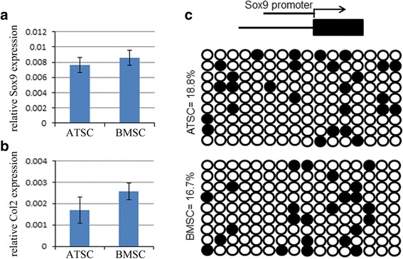
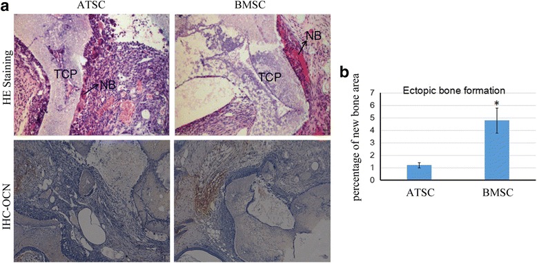
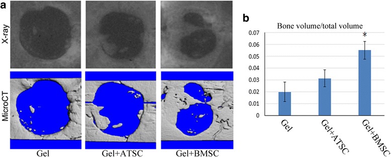
Verification
Claims the paper makes
The claims and statistics of this paper haven’t been checked yet.
Run the check — free, no account needed. It reads the open-access text and checks the sources it cites.
Claims other papers make about this one
Each is checked against this paper’s own text: does it say what the citing paper says it does?
The source backs part of the claim, not all of it — or backs it for a different population, condition or comparison.
In the citing paper
“Regarding bone tissue engineering, studies comparing the osteogenic potential of human BM-MSCs and ASCs have yielded mixed results.”
From Mesenchymal Stem/Stromal Cells: Immunomodulatory and Bone Regeneration Potential after Tumor Excision in Osteosarcoma Patients (2023) · cites this paper as [61]
Experimental — accuracy not yet measuredThe cited source says what the claim attributes to it.
In the citing paper
“Some suggest that BM-MSCs exhibit greater osteogenic capacity [58,59,60,61,62], while others propose that ASCs have equal or superior potential [58,63,64,65,66].”
From Mesenchymal Stem/Stromal Cells: Immunomodulatory and Bone Regeneration Potential after Tumor Excision in Osteosarcoma Patients (2023) · cites this paper as [61]
Self-check
Percentages, sums and reported differences re-computed from the paper’s own numbers.
The self-check needs the paper’s full text.
Citations
Cites
Multilineage Cells from Human Adipose Tissue: Implications for Cell-Based Therapies
Zuk, P. A., Zhu, M., Mizuno, H., Huang, J., Futrell, J. W., Katz, A. J., Benhaim, P., Lorenz, H. P., & Hedrick, M. H. (2001) · Tissue Engineering · doi:10.1089/107632701300062859 · Cited by 7,956
Stöckl, S., Göttl, C., Grifka, J. K., & Grässel, S. (2013) · Cellular Physiology and Biochemistry · doi:10.1159/000350089 · Cited by 4,670
Liao, J., Wei, Q., Zou, Y., Fan, J., Song, D., Cui, J., Zhang, W., Zhu, Y., Ma, C., Hu, X. Y., et al. (2017) · Cellular Physiology and Biochemistry · doi:10.1159/000471945 · Cited by 2,742
Self-citationChen, J., Sanberg, P. R., Li, Y., Wang, L., Lü, M., Willing, A. E., Sanchez‐Ramos, J., & Chopp, M. (2001) · Stroke · doi:10.1161/hs1101.098367 · Cited by 1,347
In Search of the In Vivo Identity of Mesenchymal Stem Cells
da Silva Meirelles, L., Caplan, A. I., & Nardi, N. B. (2008) · Stem Cells · doi:10.1634/stemcells.2007-1122 · Cited by 1,077
Puissant‐Lubrano, B., Barreau, C., Bourin, P., Clavel, C., Corre, J., Bousquet, C., Taureau, C., Cousin, B., Abbal, M., Laharrague, P. F., et al. (2005) · British Journal of Haematology · doi:10.1111/j.1365-2141.2005.05409.x · Cited by 957
Lee, R. H., Kim, B., Choi, I., Kim, H., Choi, H. S., Suh, K. T., Bae, Y. C., & Jung, J. S. (2004) · Cellular Physiology and Biochemistry · doi:10.1159/000080341 · Cited by 933
Human marrow stromal cell therapy for stroke in rat
Li, Y., Chen, J., Chen, X. G., Wang, L., Gautam, S. C., Xu, Y. X., Katakowski, M., Zhang, L. J., Lü, M., Janakiraman, N., et al. (2002) · Neurology · doi:10.1212/wnl.59.4.514 · Cited by 867
Mesenchymal Stem Cells as Therapeutics
Parekkadan, B., & Milwid, J. M. (2010) · Annual Review of Biomedical Engineering · doi:10.1146/annurev-bioeng-070909-105309 · Cited by 771
Human body epigenome maps reveal noncanonical DNA methylation variation
Schultz, M. D., He, Y., Whitaker, J. W., Hariharan, M., Mukamel, E. A., Leung, D., Rajagopal, N., Nery, J. R., Urich, M. A., Chen, H., et al. (2015) · Nature · doi:10.1038/nature14465 · Cited by 690
Haynesworth, S. E., Barer, M., & Caplan, A. I. (1992) · Bone · doi:10.1016/8756-3282(92)90363-2 · Cited by 645
Wagner, J. R., Busche, S., Ge, B., Kwan, T., Pastinen, T., & Blanchette, M. (2014) · Genome biology · doi:10.1186/gb-2014-15-2-r37 · Cited by 446
Hsiao, S. T.-F., Asgari, A., Lokmic, Z., Sinclair, R., Dusting, G. J., Lim, S. Y., & Dilley, R. J. (2012) · Stem Cells and Development · doi:10.1089/scd.2011.0674 · Cited by 410 · cited 1×
“In addition, significant differences in the cytokine secretome and chemokine receptor expression between ATSCs and BMSCs have also been reported [14, 15].”
Huang, J. I., Kazmi, N., Durbhakula, M. M., Hering, T. M., Yoo, J. U., & Johnstone, B. (2005) · Journal of Orthopaedic Research® · doi:10.1016/j.orthres.2005.03.008.1100230621 · Cited by 225
Mesenchymal stem cells homing to improve bone healing
Lin, W., Xu, L., Zwingenberger, S., Gibon, E., Goodman, S. B., & Li, G. (2017) · Journal of Orthopaedic Translation · doi:10.1016/j.jot.2017.03.002 · Cited by 222
Self-citationHicks, A. U., Lappalainen, R. S., Narkilahti, S., Suuronen, R., Corbett, D., Sivenius, J., Hovatta, O., & Jolkkonen, J. (2009) · European Journal of Neuroscience · doi:10.1111/j.1460-9568.2008.06599.x · Cited by 218
Zinn, R. L., Pruitt, K., Eguchi, S., Baylin, S. B., & Herman, J. G. (2007) · Cancer Research · doi:10.1158/0008-5472.can-06-3396 · Cited by 210
Stem cell transplantation in multiple sclerosis: current status and future prospects
the Stem Cells in Multiple Sclerosis (STEMS) Consensus Group, Martino, G., Franklin, R. J. M., Evercooren, A. B., & Kerr, D. A. (2010) · Nature Reviews Neurology · doi:10.1038/nrneurol.2010.35 · Cited by 181
Poloni, A., Maurizi, G., Leoni, P., Serrani, F., Mancini, S., Frontini, A., Zingaretti, M. C., Siquini, W., Sarzani, R., & Cinti, S. (2012) · Stem Cells · doi:10.1002/stem.1067 · Cited by 138
Stem cell therapy for cardiac repair: benefits and barriers
Joggerst, S. J., & Hatzopoulos, A. K. (2009) · Expert Reviews in Molecular Medicine · doi:10.1017/s1462399409001124 · Cited by 135
Elman, J. S., Li, M., Wang, F., Gimble, J. M., & Parekkadan, B. (2014) · Journal of Inflammation · doi:10.1186/1476-9255-11-1 · Cited by 126
Kia, N. A., Bahrami, A. R., Ebrahimi, M., Matin, M. M., Neshati, Z., Almohaddesin, M. R., Aghdami, N., & Bidkhori, H. R. (2010) · Journal of Molecular Neuroscience · doi:10.1007/s12031-010-9446-6 · Cited by 101
Rasmussen, J. G., Fröbert, O., Holst‐Hansen, C., Kastrup, J., Baandrup, U. T., Zachar, V., Fink, T., & Simonsen, U. (2013) · Cell Transplantation · doi:10.3727/096368912x659871 · Cited by 96
Epigenetic programming of mesenchymal stem cells from human adipose tissue
Boquest, A. C., Noer, A., & Collas, P. (2006) · Stem Cell Reviews and Reports · doi:10.1007/bf02698059 · Cited by 90
Huang, S., Xu, L., Zhang, Y., Sun, Y., & Li, G. (2015) · Cell Transplantation · doi:10.3727/096368915x687219 · Cited by 83
Self-citationAnalysis of Allogeneic and Syngeneic Bone Marrow Stromal Cell Graft Survival in the Spinal Cord
Swanger, S. A., Neuhuber, B., Himes, B. T., Bakshi, A., & Fischer, I. (2005) · Cell Transplantation · doi:10.3727/000000005783982594 · Cited by 71
Lineage-Specific Promoter DNA Methylation Patterns Segregate Adult Progenitor Cell Types
Sørensen, A. L., Barrand, S., West, F. D., Vekterud, K., Boquest, A. C., Ährlund‐Richter, L., Stice, S. L., & Collas, P. (2009) · Stem Cells and Development · doi:10.1089/scd.2009.0309 · Cited by 62
Cho, Y. D., Yoon, W. J., Kim, W. J., Woo, K. M., Baek, J. H., Lee, G., Ku, Y., van Wijnen, A. J., & Ryoo, H. (2014) · Journal of Biological Chemistry · doi:10.1074/jbc.m114.558064 · Cited by 62
Rui, Y., Lui, P. P. Y., Lee, W., & Chan, K. M. (2011) · International Orthopaedics · doi:10.1007/s00264-011-1417-1 · Cited by 56
Chung, C., Fujita, N., Kawahara, N., Yui, S., Nam, E., & Nishimura, R. (2013) · Journal of Veterinary Medical Science · doi:10.1292/jvms.12-0470 · Cited by 56
Danišovič, Ľ., Varga, I., Polák, Š., Ulicna, M., Böhmer, D., & Vojtaššák, J. (2009) · General Physiology and Biophysics · doi:10.4149/gpb_2009_01_56 · Cited by 55
DNA methylation of the Trip10 promoter accelerates mesenchymal stem cell lineage determination
Hsiao, S., Lee, K., Hsu, C., Tseng, M.-J., Jin, V. X., Sun, W.-S., Hung, Y.-C., Yeh, K.-T., Yan, P. S., Lai, Y.-Y., et al. (2010) · Biochemical and Biophysical Research Communications · doi:10.1016/j.bbrc.2010.08.048 · Cited by 46
Rui, Y., Xu, L., Chen, R., Zhang, T., Lin, S., Hou, Y., Liu, Y., Meng, F., Liu, Z., Ni, M., et al. (2015) · Scientific Reports · doi:10.1038/srep11056 · Cited by 45
Self-citationHoogduijn, M. J., & Dor, F. J. M. F. (2013) · Frontiers in Immunology · doi:10.3389/fimmu.2013.00144 · Cited by 25
Epigenetic Reprogramming of Mesenchymal Stem Cells
Leu, Y., Huang, T. H. ‐., & Hsiao, S. (2012) · Advances in experimental medicine and biology · doi:10.1007/978-1-4419-9967-2_10 · Cited by 21
Cooper, G. M., Durham, E. L., Cray, J., Bykowski, M. R., DeCesare, G. E., Smalley, M. A., Mooney, M. P., Campbell, P. G., & Losee, J. E. (2010) · Plastic & Reconstructive Surgery · doi:10.1097/prs.0b013e3181fad311 · Cited by 13
Cited by
Sorted by citations
Stem cell-based therapy for human diseases
Hoang, D. M., Pham, P. T., Bach, T. Q., Ngo, A. T. L., Nguyen, Q. T., Phan, T. T. K., Nguyen, G. H., Le, P. T. T., Hoang, V. T., Forsyth, N. R., et al. (2022) · Signal Transduction and Targeted Therapy · doi:10.1038/s41392-022-01134-4 · Cited by 1,074
Adipose-derived and bone marrow mesenchymal stem cells: A donor-matched comparison
Mohamed-Ahmed, S., Fristad, I., Lie, S. A., Suliman, S., Mustafa, K., Vindenes, H., & Idris, S. B. (2018) · Stem Cell Research & Therapy · doi:10.1186/s13287-018-0914-1 · Cited by 515
Challenges in Clinical Development of Mesenchymal Stromal/Stem Cells: Concise Review
Mastrolia, I., Foppiani, E. M., Murgia, A., Candini, O., Samarelli, A. V., Grisendi, G., Veronesi, E., Horwitz, E. M., & Dominici, M. (2019) · Stem Cells Translational Medicine · doi:10.1002/sctm.19-0044 · Cited by 353
Lee, Y., Chan, Y.-H., Hsieh, S., Lew, W.-Z., & Feng, S. (2019) · International Journal of Molecular Sciences · doi:10.3390/ijms20205015 · Cited by 246
Allogeneic vs. autologous mesenchymal stem/stromal cells in their medication practice
Li, C., Zhao, H., Cheng, L., & Wang, B. (2021) · Cell & Bioscience · doi:10.1186/s13578-021-00698-y · Cited by 200
Self-citation
Metadata
- Authors
- Xu, L., Liu, Y., Sun, Y., Wang, B., Xiong, Y., Lin, W., Wei, Q., Wang, H., He, W., Wang, B., & Li, G.
- Affiliations
- Guangzhou University of Chinese Medicine (CN)
- Chinese University of Hong Kong (HK)
- Corresponding author
- He, Wei
- Li, Gang
- Venue
- Stem Cell Research & Therapy
- Published
- 2017-12
- Volume
- 8
- Issue
- 1
- Pages
- 275-275
- ISSN
- 1757-6512 (ISSN-L)
- PMC
- PMC5718061
- OpenAlex
- W2773440515
- Cited by
- 284
- References
- 36
- Citations by year
- 2026: 10 · 2025: 26 · 2024: 29 · 2023: 50 · 2022: 39 · 2021: 52 · 2020: 34 · 2019: 30 · 2018: 14
- FWCI
- 12.25
- Type
- journal article
- Language
- en
- Open access
- gold
- doi.org
- Licence
- cc-by
- Locations
- Stem Cell Research & Therapy (publishedVersion, cc-by)
- Europe PMC (PubMed Central) (publishedVersion, cc-by)
- PubMed Central (publishedVersion, cc-by)
- PubMed (publishedVersion)
- DOAJ (DOAJ: Directory of Open Access Journals) (submittedVersion, cc-by-sa)
- Funders
- City University of Hong Kong
- Lui Che Woo Institute of Innovative Medicine
- National Natural Science Foundation of China (14119115, 81772322, 81430049, 81371946)
- Chinese University of Hong Kong
- Hong Kong Government
- Topics
- Life Sciences › Biochemistry, Genetics and Molecular Biology › Cell Biology › Mesenchymal stem cell research
- Physical Sciences › Engineering › Biomedical Engineering › Bone Tissue Engineering Materials
- Health Sciences › Dentistry › Periodontics › Periodontal Regeneration and Treatments
- Keywords
- mesenchymal stem cells, bone marrow-derived stem cells, adipose-derived stem cells, trilineage differentiation, osteogenic differentiation, adipogenesis, chondrogenic differentiation, DNA methylation, Runx2, PPARγ, SOX9, epigenetic memory, bone regeneration, cell therapy
- MeSH terms
- Adipose Tissue, Bone Marrow Cells, Bone Regeneration, Cell Differentiation, Humans, Organ Specificity, Osteogenesis, Promoter Regions, Genetic, Skull, Biomarkers, Gene Expression, CpG Islands, DNA Methylation, Arthroplasty, Replacement, Hip, Chondrogenesis, Mesenchymal Stem Cell Transplantation, PPAR gamma, Adipogenesis, Core Binding Factor Alpha 1 Subunit, SOX9 Transcription Factor, Mesenchymal Stem Cells, Primary Cell Culture
About these checks
Registry, retraction, Cites and statistics results are computed, not judged, with one exception: a printed reference that no index lists is read once by a model before it counts as a citation error. Claims are extracted by a model and kept only when their quote appears verbatim in the text; each cited source is then checked, with a model’s help, for whether it says what the claim relies on. Those verdicts can be wrong — read the claim’s quote and the cited source before relying on one.
Statistics and arithmetic are recomputed, not judged.
Confidence bands are shown only for fields calibrated on our annotated test set. None is calibrated yet, so no band is shown.
What counts as a claim ›How we measure confidence ›How we check this ›
Are you an author, or do you see a problem with this page? Request a review
