

The DOI matches its Crossref record; 4 of the 4 references it cites were found in a registry or an index.
Physiologic expansion of human heart-derived cells enhances therapeutic repair of injured myocardium
Abstract
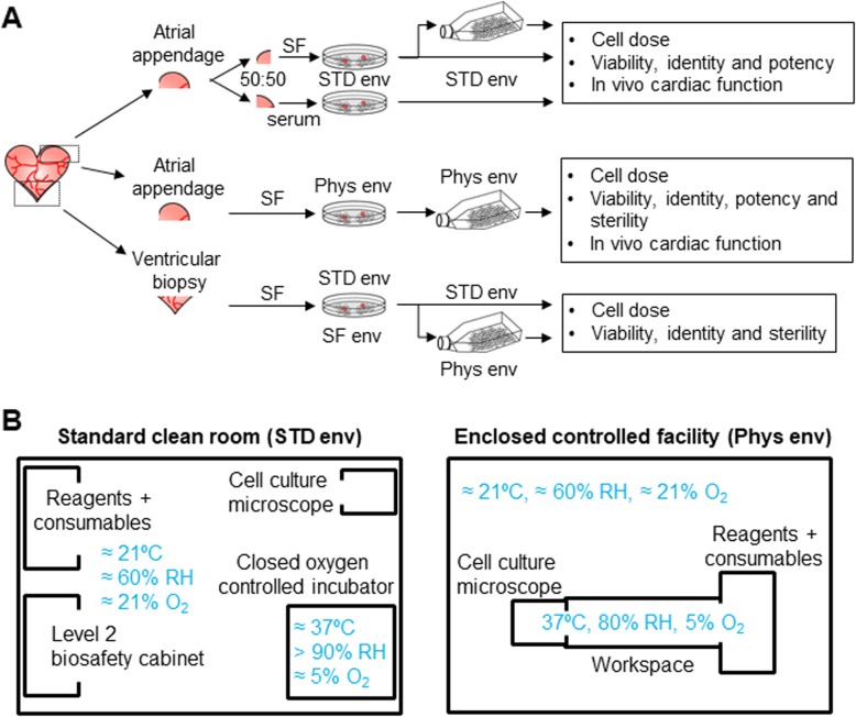
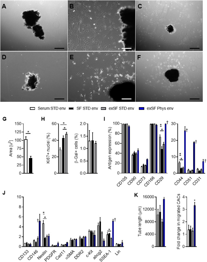
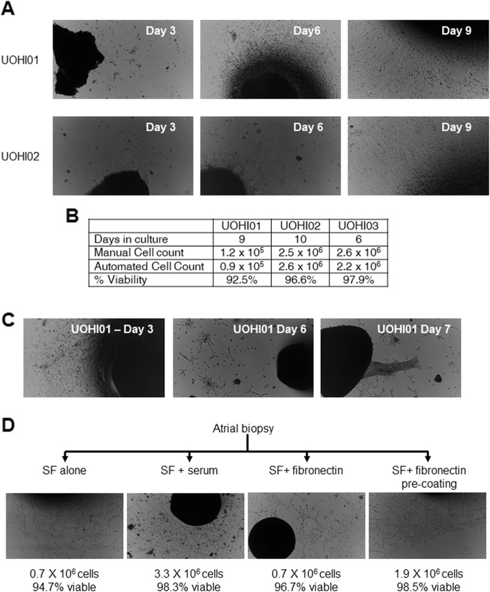
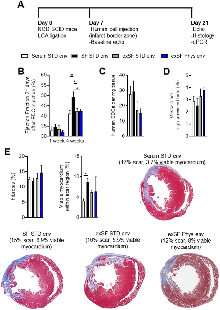
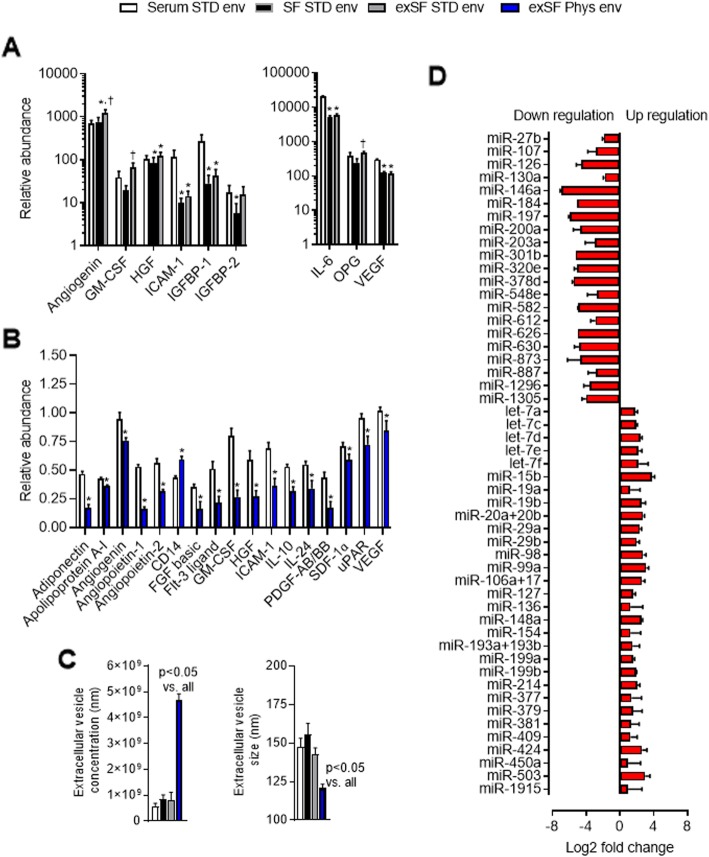
Background: Serum-free xenogen-free defined media and continuous controlled physiological cell culture conditions have been developed for stem cell therapeutics, but the effect of these conditions on the relative potency of the cell product is unknown. As such, we conducted a head-to-head comparison of cell culture conditions on human heart explant-derived cells using established in vitro measures of cell potency and in vivo functional repair. Methods: Heart explant-derived cells cultured from human atrial or ventricular biopsies within a serum-free xenogen-free media and a continuous physiological culture environment were compared to cells cultured under traditional (high serum) cell culture conditions in a standard clean room facility. Results: Transitioning from traditional high serum cell culture conditions to serum-free xenogen-free conditions had no effect on cell culture yields but provided a smaller, more homogenous, cell product with only minor antigenic changes. Culture within continuous physiologic conditions markedly boosted cell proliferation while increasing the expression of stem cell-related antigens and ability of cells to stimulate angiogenesis. Intramyocardial injection of physiologic cultured cells into immunodeficient mice 1 week after coronary ligation translated into improved cardiac function and reduced scar burden which was attributable to increased production of pro-healing cytokines, extracellular vesicles, and microRNAs. Conclusions: Continuous physiological cell culture increased cell growth, paracrine output, and treatment outcomes to provide the greatest functional benefit after experimental myocardial infarction.
Overview
The claims and statistics of this paper haven’t been checked yet.
Run the check — free, no account needed. It reads the open-access text and checks the sources it cites.
Integrity signals
Registry
Cites
Statistics
Figures
Verification
Claims the paper makes
The claims and statistics of this paper haven’t been checked yet.
Run the check — free, no account needed. It reads the open-access text and checks the sources it cites.
Claims other papers make about this one
Each is checked against this paper’s own text: does it say what the citing paper says it does?
In the citing paper
“The use of closed cell handling chambers to provide constant physiologically relevant oxygen can improve cell function (Mount et al., 2019; Wong et al., 2020).”
From Guidance document on Good cell and tissue culture practice 2.0 (GCCP 2.0) (2021) · cites this paper as Mount et al., 2019
Self-check
Percentages, sums and reported differences re-computed from the paper’s own numbers.
The self-check needs the paper’s full text.
Citations
Cites
International trial of the Edmonton protocol for islet transplantation
Shapiro, A. M. J., Ricordi, C., Hering, B. J., Auchincloss, H., Lindblad, R., Robertson, R. P., Secchi, A., Brendel, M. D., Berney, T., Brennan, D. C., et al. (2006) · New England Journal of Medicine · doi:10.1056/nejmoa061267 · Cited by 1,942
Functional screening identifies miRNAs inducing cardiac regeneration
Eulalio, A., Mano, M., Ferro, M. D., Zentilin, L., Sinagra, G., Zacchigna, S., & Giacca, M. (2012) · Nature · doi:10.1038/nature11739 · Cited by 1,029
Konstam, M. A., Kronenberg, M. W., Rousseau, M. F., Udelson, J. E., Melin, J., Stewart, D., Dolan, N., Edens, T. R., Ahn, S., & Kinan, D. (1993) · Circulation · doi:10.1161/01.cir.88.5.2277 · Cited by 316
Self-citationValidation of the Cardiosphere Method to Culture Cardiac Progenitor Cells from Myocardial Tissue
Davis, D. R., Zhang, Y., Smith, R. R., Cheng, K., Terrovitis, J., Malliaras, K., Li, T.-S., White, A., Makkar, R., & Marbán, E. (2009) · PLoS ONE · doi:10.1371/journal.pone.0007195 · Cited by 300
Self-citation
Research works printed but not linked: 55
- Alter DA, Ko DT, Tu JV, Stukel TA, Lee DS, Laupacis A, et al. The average lifespan of patients discharged from hospital with heart failure. J Gen Intern Med. 2012;27(9):1171–9. · DOI
- Lee DS, Schull MJ, Alter DA, Austin PC, Laupacis A, Chong A, et al. Early deaths in patients with heart failure discharged from the emergency department: a population-based analysis. Circ Heart Fail. 2010;3(2):228–35. · DOI
- Malliaras K, Makkar RR, Smith RR, Cheng K, Wu E, Bonow RO, et al. Intracoronary cardiosphere-derived cells after myocardial infarction: evidence of therapeutic regeneration in the final 1-year results of the CADUCEUS trial (CArdiosphere-Derived aUtologous stem CElls to reverse ventricUlar dySfunction). J Am Coll Cardiol. 2014;63(2):110–22. · DOI
- Makkar RR, Smith RR, Cheng K, Malliaras K, Thomson LE, Berman D, et al. Intracoronary cardiosphere-derived cells for heart regeneration after myocardial infarction (CADUCEUS): a prospective, randomised phase 1 trial. Lancet. 2012;379(9819):895–904. · DOI
- Ishigami S, Ohtsuki S, Eitoku T, Ousaka D, Kondo M, Kurita Y, et al. Intracoronary cardiac progenitor cells in single ventricle physiology: the PERSEUS (cardiac progenitor cell infusion to treat univentricular heart disease) randomized phase 2 trial. Circ Res. 2017;120(7):1162–73. · DOI
- Ishigami S, Ohtsuki S, Tarui S, Ousaka D, Eitoku T, Kondo M, et al. Intracoronary autologous cardiac progenitor cell transfer in patients with hypoplastic left heart syndrome: the TICAP prospective phase 1 controlled trial. Circ Res. 2015;116(4):653–64. · DOI
- Tarui S, Ishigami S, Ousaka D, Kasahara S, Ohtsuki S, Sano S, et al. Transcoronary infusion of cardiac progenitor cells in hypoplastic left heart syndrome: three-year follow-up of the Transcoronary Infusion of Cardiac Progenitor Cells in Patients With Single-Ventricle Physiology (TICAP) trial. J Thorac Cardiovasc Surg. 2015;150(5):1198–207 208 e1–2. · DOI
- Davis DR. Heart stem cells in the post-Anversa era. Eur Heart J. 2019;40(13):1039–41. · DOI
- Davis DR, Kizana E, Terrovitis J, Barth AS, Zhang Y, Smith RR, et al. Isolation and expansion of functionally-competent cardiac progenitor cells directly from heart biopsies. JMCC. 2010;49(2):312–21.
- Chimenti I, Gaetani R, Forte E, Angelini F, De FE, Zoccai GB, et al. Serum and supplement optimization for EU GMP-compliance in cardiospheres cell culture. J Cell Mol Med. 2014;18(4):624–34. · DOI
- Mendicino M, Bailey AM, Wonnacott K, Puri RK, Bauer SR. MSC-based product characterization for clinical trials: an FDA perspective. Cell Stem Cell. 2014;14(2):141–5. · DOI
- Shenje LT, Field LJ, Pritchard CA, Guerin CJ, Rubart M, Soonpaa MH, et al. Lineage tracing of cardiac explant derived cells. PLoS One. 2008;3(4):e1929. · DOI
- Andersen DC, Andersen P, Schneider M, Jensen HB, Sheikh SP. Murine “Cardiospheres” are not a source of stem cells with cardiomyogenic potential. Stem Cells. 2009;27(7):1571–81. · DOI
- Jochems CE, van der Valk JB, Stafleu FR, Baumans V. The use of fetal bovine serum: ethical or scientific problem? Altern Lab Anim. 2002;30(2):219–27. · DOI · Paper page
- Li TS, Cheng K, Malliaras K, Matsushita N, Sun B, Marban L, et al. Expansion of human cardiac stem cells in physiological oxygen improves cell production efficiency and potency for myocardial repair. Cardiovasc Res. 2011;89(1):157–65. · DOI
- Li TS, Cheng K, Malliaras K, Matsushita N, Sun B, Marban L, et al. Expansion of human cardiac stem cells in physiological oxygen improves cell production efficiency and potency for myocardial repair. Cardiovasc Res. 2011;89(1):157–65. · DOI
- Neutelings T, Lambert CA, Nusgens BV, Colige AC. Effects of mild cold shock (25 degrees C) followed by warming up at 37 degrees C on the cellular stress response. PLoS One. 2013;8(7):e69687. · DOI
- Galluzzi L, Yamazaki T, Kroemer G. Linking cellular stress responses to systemic homeostasis. Nat Rev Mol Cell Biol. 2018;19(11):731–45. · DOI
- Tilokee EL, Latham N, Jackson R, Mayfield AE, Ye B, Mount S, et al. Paracrine engineering of human explant-derived cardiac stem cells to over-express stromal-cell derived factor 1alpha enhances myocardial repair. Stem Cells. 2016;34(7):1826–35. · DOI
- Mayfield AE, Fitzpatrick ME, Latham N, Tilokee EL, Villanueva M, Mount S, et al. The impact of patient co-morbidities on the regenerative capacity of cardiac explant-derived stem cells. Stem Cell Res Ther. 2016;7(1):60. · DOI
- Jackson R, Tilokee EL, Latham N, Mount S, Rafatian G, Strydhorst J, et al. Paracrine engineering of human cardiac stem cells with insulin-like growth factor 1 enhances myocardial repair. J Am Heart Assoc. 2015;4(9):e002104. · DOI
- Mayfield AE, Tilokee EL, Latham N, McNeill B, Lam BK, Ruel M, et al. The effect of encapsulation of cardiac stem cells within matrix-enriched hydrogel capsules on cell survival, post-ischemic cell retention and cardiac function. Biomaterials. 2014;35(1):133–42. · DOI
- Molgat AS, Tilokee EL, Rafatian G, Vulesevic B, Ruel M, Milne R, et al. Hyperglycemia inhibits cardiac stem cell-mediated cardiac repair and angiogenic capacity. Circulation. 2014;130(11 Suppl 1):S70–S6. · DOI
- Latham N, Ye B, Jackson R, Lam B, Kuraitis D, Ruel M, et al. Human blood and cardiac stem cells synergize to enhance cardiac repair when cotransplanted into ischemic myocardium. Circulation. 2013;128(suppl1):S1–8.
- Ott HC, Matthiesen TS, Brechtken J, Grindle S, Goh SK, Nelson W, et al. The adult human heart as a source for stem cells: repair strategies with embryonic-like progenitor cells. Nat Clin Pract Cardiovasc Med. 2007;4(S1):S27–39. · DOI
- Kanda P, Alarcon EI, Yeuchyk T, Parent S, de Kemp RA, Variola F, et al. Deterministic encapsulation of human cardiac stem cells in variable composition nanoporous gel cocoons to enhance therapeutic repair of injured myocardium. ACS Nano. 2018;12(5):4338–50. · DOI
- Jackson R, Mount S, Ye B, Mayfield AE, Chan V, Boodhwani M, et al. Isolation of human explant derived cardiac stem cells from cryopreserved heart tissue. PLoS One. 2017;12(4):e0176000. · DOI
- Mayfield AE, Kanda P, Nantsios A, Parent S, Mount S, Dixit S, et al. Interleukin-6 mediates post-infarct repair by cardiac explant-derived stem cells. Theranostics. 2017;7(19):4850–61. · DOI
- Ibrahim AG, Cheng K, Marban E. Exosomes as critical agents of cardiac regeneration triggered by cell therapy. Stem Cell Reports. 2014;2(5):606–19. · DOI
- van Niel G, D'Angelo G, Raposo G. Shedding light on the cell biology of extracellular vesicles. Nat Rev Mol Cell Biol. 2018;19(4):213–28. · DOI · Paper page
- Lesizza P, Prosdocimo G, Martinelli V, Sinagra G, Zacchigna S, Giacca M. Single-dose Intracardiac injection of pro-regenerative micrornas improves cardiac function after myocardial infarction. Circ Res. 2017;120(8):1298–304. · DOI
- Xu J, Tang Y, Bei Y, Ding S, Che L, Yao J, et al. MiR-19b attenuates H2O2-induced apoptosis in rat H9C2 cardiomyocytes via targeting PTEN. Oncotarget. 2016;7(10):10870–8. · DOI
- Gao F, Kataoka M, Liu N, Liang T, Huang Z-P, Gu F, et al. Therapeutic role of miR-19a/19b in cardiac regeneration and protection from myocardial infarction. Nat Commun. 2019;10(1):1802. · DOI
- Raso A, Dirkx E, Philippen LE, Fernandez-Celis A, De Majo F, Sampaio-Pinto V, et al. Therapeutic delivery of miR-148a suppresses ventricular dilation in heart failure. Mol Ther. 2019;27(3):584–99. · DOI
- van Rooij E, Sutherland LB, Thatcher JE, DiMaio JM, Naseem RH, Marshall WS, et al. Dysregulation of micrornas after myocardial infarction reveals a role of Mir-29 in cardiac fibrosis. Proc Natl Acad Sci U S A. 2008;105(35):13027–32. · DOI
- Li Q, Xie J, Wang B, Li R, Bai J, Ding L, et al. Overexpression of microRNA-99a attenuates cardiac hypertrophy. PLoS One. 2016;11(2):e0148480. · DOI
- Bearzi C, Rota M, Hosoda T, Tillmanns J, Nascimbene A, De Angelis A, et al. Human cardiac stem cells. Proc Natl Acad Sci U S A. 2007;104(35):14068–73. · DOI
- Smith RR, Barile L, Cho HC, Leppo MK, Hare JM, Messina E, et al. Regenerative potential of cardiosphere-derived cells expanded from percutaneous endomyocardial biopsy specimens. Circulation. 2007;115:896–908. · DOI
- Sebastiao MJ, Menta R, Serra M, Palacios I, Alves PM, Sanchez B, et al. Human cardiac stem cells inhibit lymphocyte proliferation through paracrine mechanisms that correlate with indoleamine 2,3-dioxygenase induction and activity. Stem Cell Res Ther. 2018;9(1):290. · DOI
- Goodlin SJ. Palliative care in congestive heart failure. J Am Coll Cardiol. 2009;54(5):386–96. · DOI
- Capricor Therapeutics Provides Update on ALLSTAR Trial [press release]. May 12, 2017. 2017. https://www.prnewswire.com/news-releases/capricor-therapeutics-provides-update-on-allstar-trial-300456715.html .
- Rafatian G, Davis DR. Heart-derived cell therapy 2.0: paracrine strategies to increase therapeutic repair of injured myocardium. Stem Cells. 2018;36(12):1794–803. · DOI
- Mount S, Davis DR. Electrical effects of stem cell transplantation for ischaemic cardiomyopathy: friend or foe? J Physiol. 2016;594(9):2511–24. · DOI
- Bristow MR, Gilbert EM, Abraham WT, Adams KF, Fowler MB, Hershberger RE, et al. Carvedilol produces dose-related improvements in left ventricular function and survival in subjects with chronic heart failure. MOCHA Investigators. Circulation. 1996;94(11):2807–16. · DOI · Paper page
- Packer M, Bristow MR, Cohn JN, Colucci WS, Fowler MB, Gilbert EM, et al. The effect of carvedilol on morbidity and mortality in patients with chronic heart failure. U.S. Carvedilol Heart Failure Study Group. N Engl J Med. 1996;334(21):1349–55. · DOI · Paper page
- A randomized trial of beta-blockade in heart failure. The Cardiac Insufficiency Bisoprolol Study (CIBIS). CIBIS Investigators and Committees. Circulation. 1994;90(4):1765–73. https://www.ahajournals.org/doi/abs/10.1161/01.CIR.90.4.1765 . · DOI
- Konstam MA, Rousseau MF, Kronenberg MW, Udelson JE, Melin J, Stewart D, et al. Effects of the angiotensin converting enzyme inhibitor enalapril on the long-term progression of left ventricular dysfunction in patients with heart failure. SOLVD Investigators. Circulation. 1992;86(2):431–8. · DOI
- Greenberg B, Quinones MA, Koilpillai C, Limacher M, Shindler D, Benedict C, et al. Effects of long-term enalapril therapy on cardiac structure and function in patients with left ventricular dysfunction. Results of the SOLVD echocardiography substudy. Circulation. 1995;91(10):2573–81. · DOI
- Pfeffer MA, Lamas GA, Vaughan DE, Parisi AF, Braunwald E. Effect of captopril on progressive ventricular dilatation after anterior myocardial infarction. N Engl J Med. 1988;319(2):80–6. · DOI
- Wong M, Staszewsky L, Latini R, Barlera S, Volpi A, Chiang YT, et al. Valsartan benefits left ventricular structure and function in heart failure: Val-HeFT echocardiographic study. J Am Coll Cardiol. 2002;40(5):970–5. · DOI
- Sutton MG, Plappert T, Hilpisch KE, Abraham WT, Hayes DL, Chinchoy E. Sustained reverse left ventricular structural remodeling with cardiac resynchronization at one year is a function of etiology: quantitative Doppler echocardiographic evidence from the Multicenter InSync Randomized Clinical Evaluation (MIRACLE). Circulation. 2006;113(2):266–72. · DOI
- St John Sutton M, Ghio S, Plappert T, Tavazzi L, Scelsi L, Daubert C, et al. Cardiac resynchronization induces major structural and functional reverse remodeling in patients with New York Heart Association class I/II heart failure. Circulation. 2009;120(19):1858–65. · DOI
- Wikstrom G, Blomstrom-Lundqvist C, Andren B, Lonnerholm S, Blomstrom P, Freemantle N, et al. The effects of aetiology on outcome in patients treated with cardiac resynchronization therapy in the CARE-HF trial. Eur Heart J. 2009;30(7):782–8. · DOI
- Barsheshet A, Goldenberg I, Moss AJ, Eldar M, Huang DT, McNitt S, et al. Response to preventive cardiac resynchronization therapy in patients with ischaemic and nonischaemic cardiomyopathy in MADIT-CRT. Eur Heart J. 2011;32(13):1622–30. · DOI
- Vigneault P, Naud P, Qi X, Xiao J, Villeneuve L, Davis DR, et al. Calcium-dependent potassium channels control proliferation of cardiac progenitor cells and bone marrow-derived mesenchymal stem cells. J Physiol. 2018;596(12):2359–79. · DOI
Cited by
Sorted by citations
Guidance document on Good cell and tissue culture practice 2.0 (GCCP 2.0)
Pamies, D. (2021) · ALTEX · doi:10.14573/altex.2111011 · Cited by 100
Metadata
- Authors
- Mount, S., Kanda, P., Parent, S., Khan, S., Michie, C., Davila, L., Chan, V., Davies, R. A., Haddad, H., Courtman, D., Stewart, D. J., & Davis, D. R.
- ORCID
- Davis, Darryl R.
- Venue
- Stem Cell Research & Therapy
- Published
- 2019-11-04
- Volume
- 10
- Issue
- 1
- ISSN
- 1757-6512
- PMC
- PMC6829847
- Cited by
- 12
- References
- 4
- Language
- en
- Funders
- Canadian Institutes of Health Research (PJT-162088, MC2-121291, Frederick Banting and Charles Best Canada Graduate Scholarship)
- Stem Cell Network (NA)
About these checks
Registry, retraction, Cites and statistics results are computed, not judged, with one exception: a printed reference that no index lists is read once by a model before it counts as a citation error. Claims are extracted by a model and kept only when their quote appears verbatim in the text; each cited source is then checked, with a model’s help, for whether it says what the claim relies on. Those verdicts can be wrong — read the claim’s quote and the cited source before relying on one.
Statistics and arithmetic are recomputed, not judged.
Confidence bands are shown only for fields calibrated on our annotated test set. None is calibrated yet, so no band is shown.
What counts as a claim ›How we measure confidence ›How we check this ›
Are you an author, or do you see a problem with this page? Request a review
