

The DOI matches its Crossref record; 52 of the 52 references it cites were found in a registry or an index.
Osteoinductive micro-nano guided bone regeneration membrane for in situ bone defect repair
Open accessAbstract
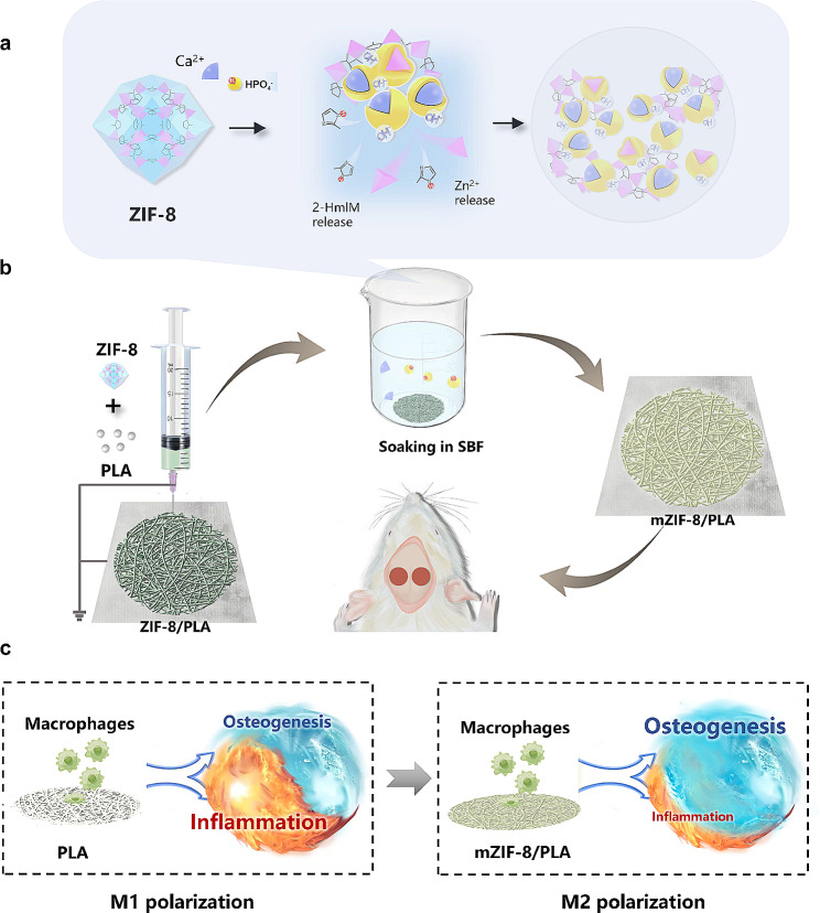
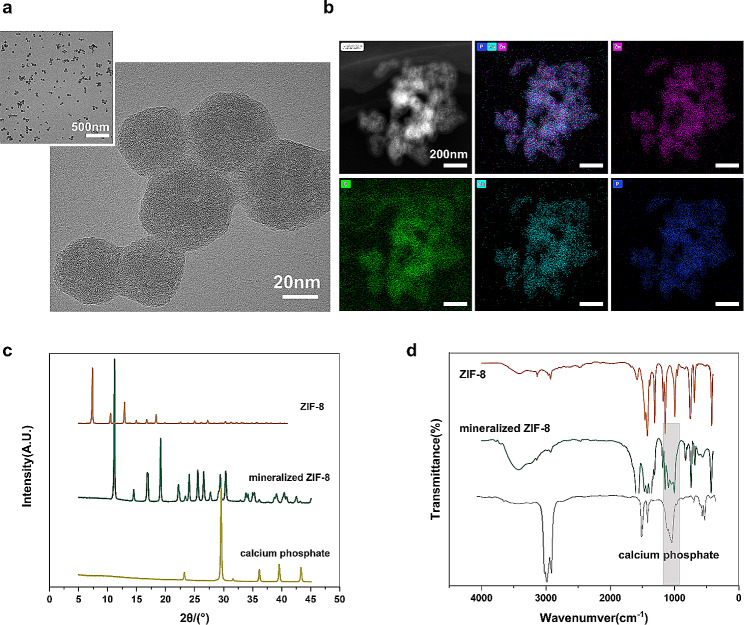
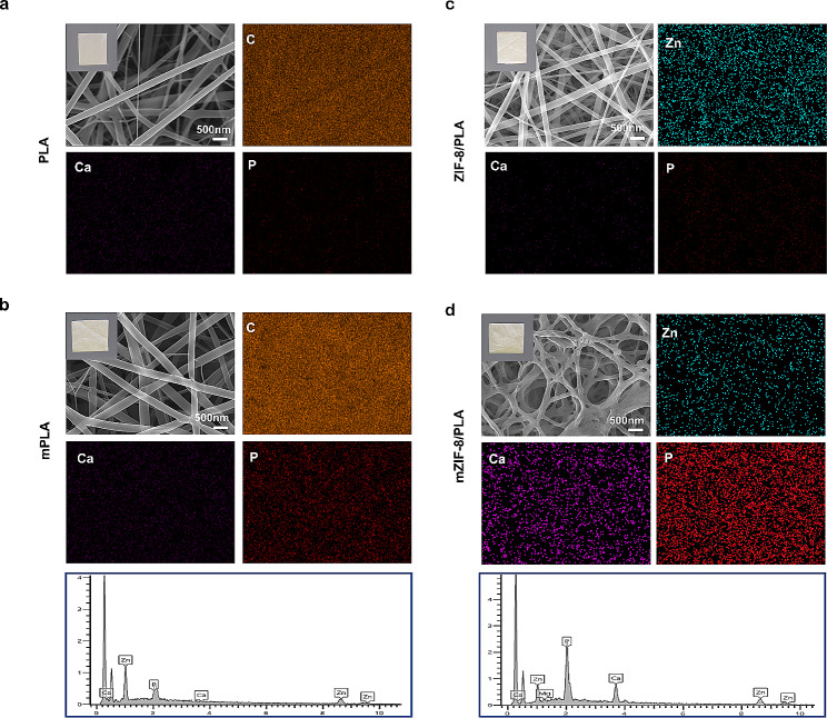
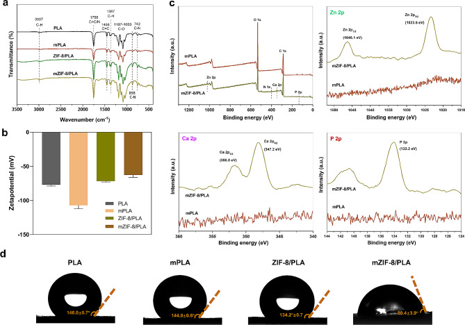
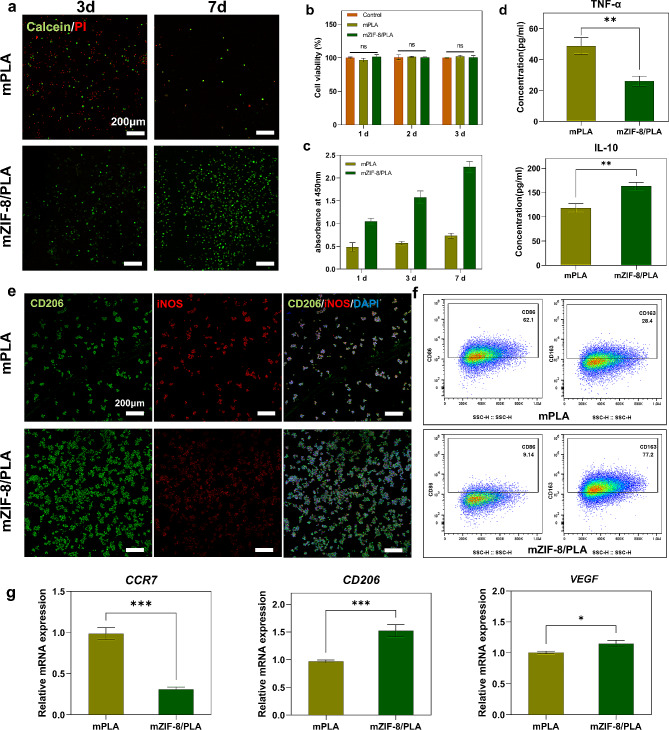
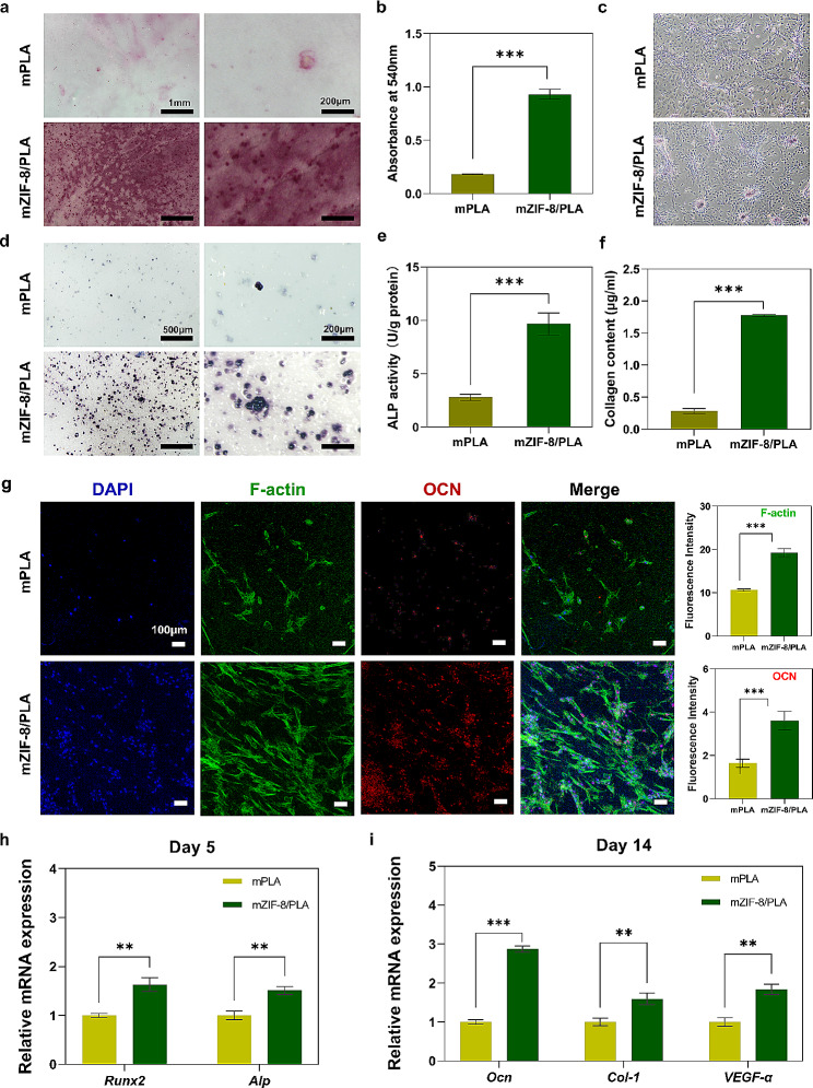
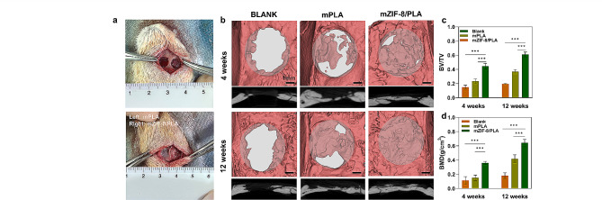
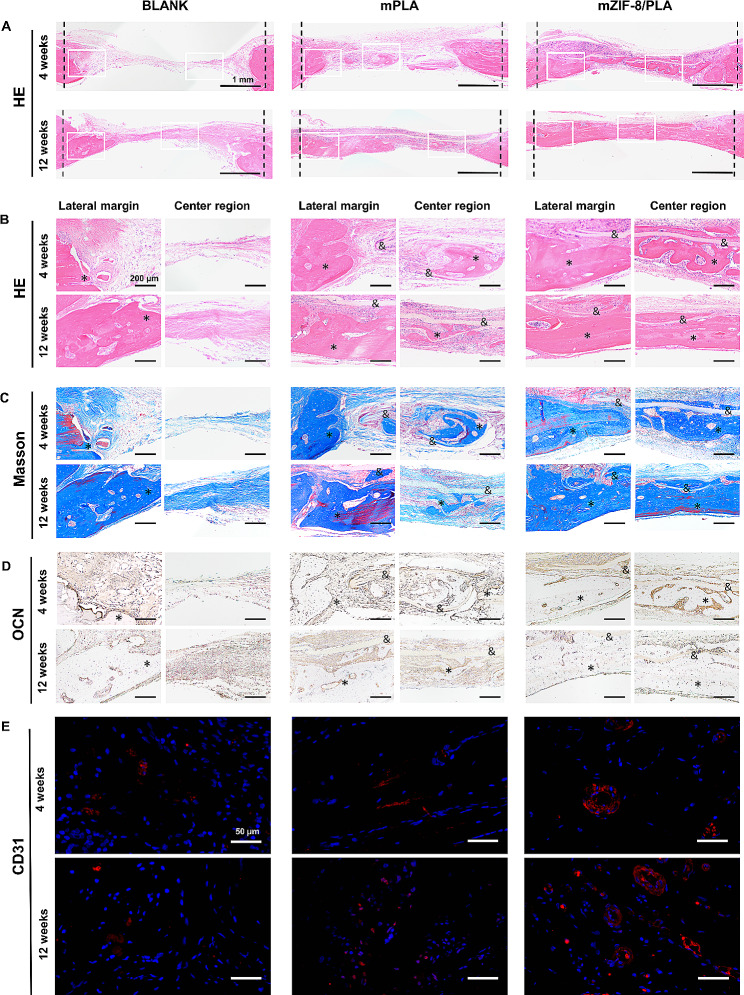
BACKGROUND: Biomaterials used in bone tissue engineering must fulfill the requirements of osteoconduction, osteoinduction, and osseointegration. However, biomaterials with good osteoconductive properties face several challenges, including inadequate vascularization, limited osteoinduction and barrier ability, as well as the potential to trigger immune and inflammatory responses. Therefore, there is an urgent need to develop guided bone regeneration membranes as a crucial component of tissue engineering strategies for repairing bone defects. METHODS: The mZIF-8/PLA membrane was prepared using electrospinning technology and simulated body fluid external mineralization method. Its ability to induce biomimetic mineralization was evaluated through TEM, EDS, XRD, FT-IR, zeta potential, and wettability techniques. The biocompatibility, osteoinduction properties, and osteo-immunomodulatory effects of the mZIF-8/PLA membrane were comprehensively evaluated by examining cell behaviors of surface-seeded BMSCs and macrophages, as well as the regulation of cellular genes and protein levels using PCR and WB. In vivo, the mZIF-8/PLA membrane's potential to promote bone regeneration and angiogenesis was assessed through Micro-CT and immunohistochemical staining. RESULTS: The mineralized deposition enhances hydrophilicity and cell compatibility of mZIF-8/PLA membrane. mZIF-8/PLA membrane promotes up-regulation of osteogenesis and angiogenesis related factors in BMSCs. Moreover, it induces the polarization of macrophages towards the M2 phenotype and modulates the local immune microenvironment. After 4-weeks of implantation, the mZIF-8/PLA membrane successfully bridges critical bone defects and almost completely repairs the defect area after 12-weeks, while significantly improving the strength and vascularization of new bone. CONCLUSIONS: The mZIF-8/PLA membrane with dual osteoconductive and immunomodulatory abilities could pave new research paths for bone tissue engineering.
Overview
The claims and statistics of this paper haven’t been checked yet.
Run the check — free, no account needed. It reads the open-access text and checks the sources it cites.
Integrity signals
Registry
Cites
Statistics
Figures
Verification
Claims the paper makes
The claims and statistics of this paper haven’t been checked yet.
Run the check — free, no account needed. It reads the open-access text and checks the sources it cites.
Claims other papers make about this one
Each is checked against this paper’s own text: does it say what the citing paper says it does?
The source backs part of the claim, not all of it — or backs it for a different population, condition or comparison.
In the citing paper
“In recent years, MOFs have provided an innovative platform for collaborative osteogenesis and immunomodulation, owing to their unique structural designability, high loading capacity, and controllable release properties [[38], [39], [40]].”
From Hydrogel scaffold encapsulating MSC-Exos and ZIF-8 promotes bone regeneration via coordinating osteogenesis and immunomodulation (2025) · cites this paper as [39]
Experimental — accuracy not yet measured
Self-check
Percentages, sums and reported differences re-computed from the paper’s own numbers.
The self-check needs the paper’s full text.
Citations
Cites
Cravillon, J., Münzer, S., Lohmeier, S., Feldhoff, A., Huber, K. P., & Wiebcke, M. (2009) · Chemistry of Materials · doi:10.1021/cm900166h · Cited by 1,408
Repair of Large Bone Defects with the Use of Autologous Bone Marrow Stromal Cells
Quarto, R., Mastrogiacomo, M., Cancedda, R., Кутепов, С. М., Mukhachev, V., Lavroukov, A., Kon, E., & Marcacci, M. (2001) · New England Journal of Medicine · doi:10.1056/nejm200102013440516 · Cited by 1,382
Brown, B. N., Valentin, J. E., Stewart-Akers, A. M., McCabe, G. P., & Badylak, S. F. (2009) · Biomaterials · doi:10.1016/j.biomaterials.2008.11.040 · Cited by 896 · cited 1×
“On the other hand, M2 macrophages release anti-inflammatory factors such as interleukin 10 (IL10) and interleukin 4 (IL4), promoting tissue regeneration [22].”
Backgroundda Silva, D., Kaduri, M., Poley, M., Adir, O., Krinsky, N., Shainsky‐Roitman, J., & Schroeder, A. (2018) · Chemical Engineering Journal · doi:10.1016/j.cej.2018.01.010 · Cited by 872
Biomaterial based modulation of macrophage polarization: a review and suggested design principles
Sridharan, R., Cameron, A. R., Kelly, D. J., Kearney, C. J., & O’Brien, F. J. (2015) · Materials Today · doi:10.1016/j.mattod.2015.01.019 · Cited by 826
Metal-organic frameworks for stimuli-responsive drug delivery
Wang, Y., Yan, J., Wen, N., Xiong, H., Cai, S., He, Q., Hu, Y., Peng, D., Liu, Z., & Liu, Y. (2019) · Biomaterials · doi:10.1016/j.biomaterials.2019.119619 · Cited by 676
Guihard, P. J., Danger, Y., Royer, B. B.-L., David, E., Brion, R., Delécrin, J., Richards, C. D., Chevalier, S., Rédiní, F., Heymann, D., et al. (2012) · Stem Cells · doi:10.1002/stem.1040 · Cited by 477
Miron, R. J., & Zhang, Y. F. (2012) · Journal of Dental Research · doi:10.1177/0022034511435260 · Cited by 346
Self-citationQiu, P., Li, M., Chen, K., Fang, B., Chen, P., Tang, Z., Lin, X., & Fan, S. (2019) · Biomaterials · doi:10.1016/j.biomaterials.2019.119552 · Cited by 340
OsteoMacs: Key players around bone biomaterials
Miron, R. J., & Bosshardt, D. D. (2015) · Biomaterials · doi:10.1016/j.biomaterials.2015.12.017 · Cited by 328
Cited by
Sorted by citations
Si, Y., Li, X., Dong, S., Gao, X., Zhu, Y., Mo, L., Wang, Z., Wang, S., & Zhang, C. (2025) · Bioactive Materials · doi:10.1016/j.bioactmat.2025.08.026 · Cited by 17
“Wang et al. suggest that the immunomodulatory mechanism of ZIF-8 may involve ions released during its biodegradation as well as its induced biomineralization processes [39].”
Self-citation
Metadata
- Authors
- Wang, B., Xie, X., Jiang, W., Zhan, Y., Zhang, Y., Guo, Y., Wang, Z., Guo, N., Guo, K., & Sun, J.
- Affiliations
- Huazhong University of Science and Technology (CN)
- Wuhan Union Hospital (CN)
- Corresponding author
- Guo, Nengqiang
- Guo, Ke
- Sun, Jiaming
- Venue
- Stem Cell Research & Therapy
- Published
- 2024-05-07
- Volume
- 15
- Issue
- 1
- Pages
- 135-135
- ISSN
- 1757-6512 (ISSN-L)
- PMC
- PMC11077813
- OpenAlex
- W4396689525
- Cited by
- 27
- References
- 52
- Citations by year
- 2026: 7 · 2025: 16 · 2024: 4
- FWCI
- 2.96
- Type
- journal article
- Language
- en
- Open access
- gold
- doi.org
- Locations
- Stem Cell Research & Therapy (publishedVersion, cc-by)
- PubMed Central (publishedVersion, cc-by)
- PubMed (publishedVersion)
- DOAJ (DOAJ: Directory of Open Access Journals) (submittedVersion)
- Funders
- National Key R&D Program of China (2019YFA0110500)
- National Natural Science Foundation of China (82020108020, 82072198, 81873941, 81701922)
- Health Commission of Hubei Province scientific research project (WJ2021Z008)
- Jingshan Union Hospital program (2023-XHJS-004)
- Topics
- Physical Sciences › Engineering › Biomedical Engineering › Bone Tissue Engineering Materials
- Health Sciences › Dentistry › Oral Surgery › Dental Implant Techniques and Outcomes
- Health Sciences › Dentistry › Periodontics › Periodontal Regeneration and Treatments
- Keywords
- metal-organic framework derived materials, polylactic acid, biomimetic mineralization, osteoinduction, osteoimmunomodulation, macrophage polarization, M2 macrophages, bone marrow mesenchymal stem cells, angiogenesis, bone defect repair, critical-size bone defect, bone tissue engineering
- MeSH terms
- Animals, Biocompatible Materials, Bone Regeneration, Membranes, Artificial, Osteogenesis, Polyesters, Tissue Engineering, Guided Tissue Regeneration, Mice, Rats, Tissue Scaffolds, Mesenchymal Stem Cells
About these checks
Registry, retraction, Cites and statistics results are computed, not judged, with one exception: a printed reference that no index lists is read once by a model before it counts as a citation error. Claims are extracted by a model and kept only when their quote appears verbatim in the text; each cited source is then checked, with a model’s help, for whether it says what the claim relies on. Those verdicts can be wrong — read the claim’s quote and the cited source before relying on one.
Statistics and arithmetic are recomputed, not judged.
Confidence bands are shown only for fields calibrated on our annotated test set. None is calibrated yet, so no band is shown.
What counts as a claim ›How we measure confidence ›How we check this ›
Are you an author, or do you see a problem with this page? Request a review
