

The DOI matches its Crossref record; 3 of the 3 references it cites were found in a registry or an index.
Sex-specific response to A1BG loss results in female dilated cardiomyopathy
Open accessPublished version of a preprint
Abstract
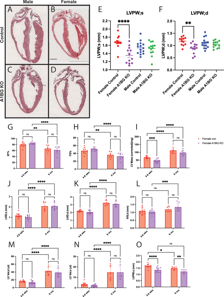
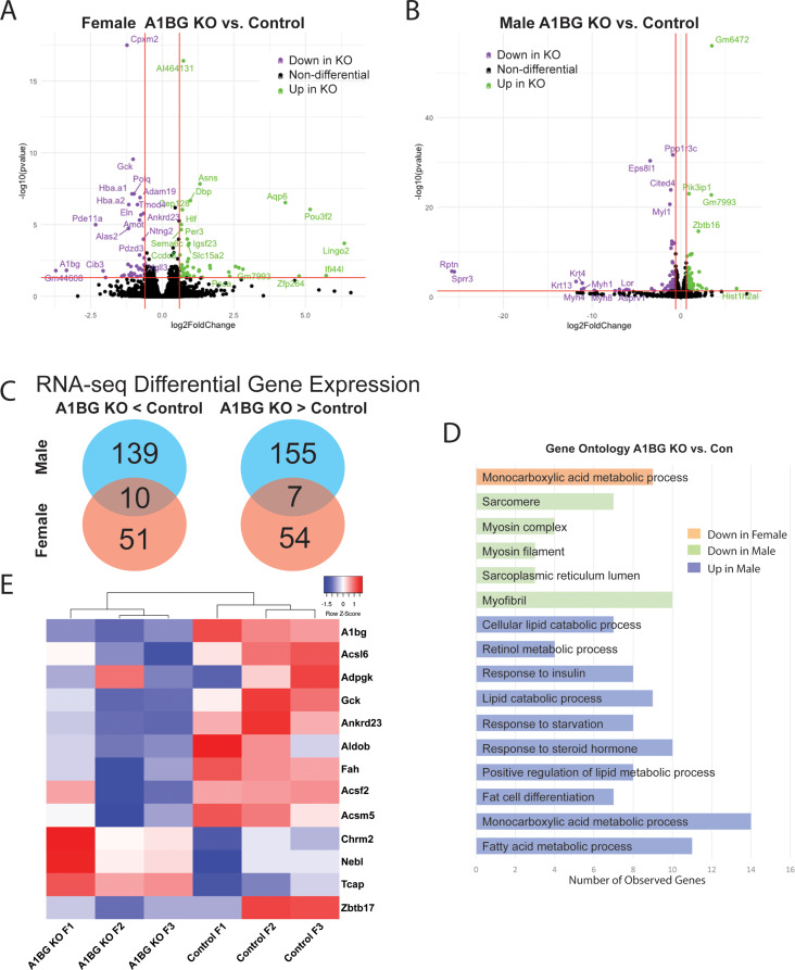
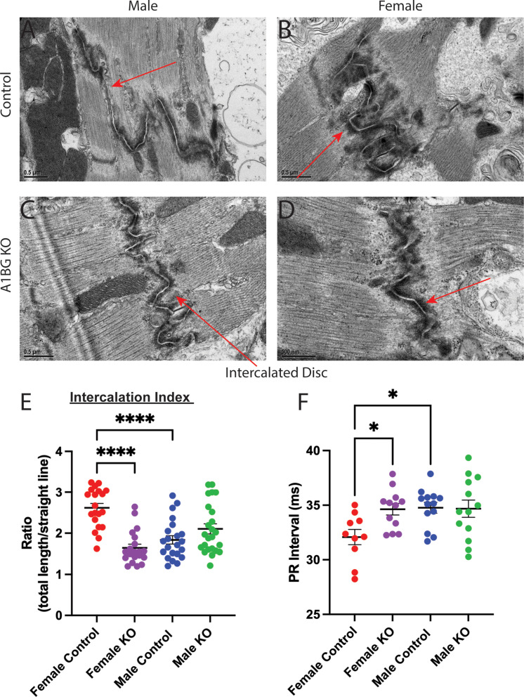
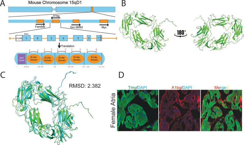
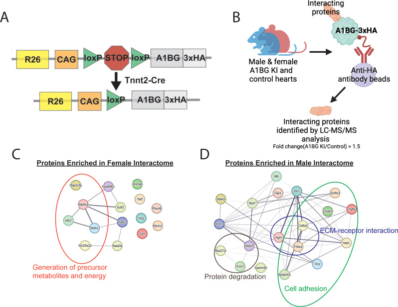
Background: Cardiac disease often manifests with sex-specific differences in frequency, pathology, and progression. However, the molecular mechanisms underlying these differences remain incompletely understood. The glycoprotein A1BG has emerged as a female-specific regulator of cardiac structure and integrity, yet its precise role in the female heart is not well characterized. Methods: To investigate the sex-specific role of A1BG in the heart, we generated both a conditional A1bg knockout allele and an A1bg Rosa26 knockin allele. We employed histological analysis, electrocardiography, RNA sequencing (RNA-seq), transmission electron microscopy (TEM), western blotting, mass spectrometry, and immunohistochemistry to assess structural, functional, and molecular phenotypes. Results: Loss of A1BG in cardiomyocytes leads to persistent structural remodeling in female, but not male, hearts. Despite preserved systolic function in female A1bg CM/CM mice left ventricular dilation and wall thinning are evident and sustained over time, consistent with early-stage dilated cardiomyopathy (DCM). Transcriptomic analyses reveal that A1BG regulates key metabolic pathways in females, including glucose-6-phosphate and acetyl-CoA metabolism. TEM imaging highlights sex-specific disruption of intercalated disc architecture in female cardiomyocytes. These findings suggest that the absence of A1BG initiates chronic pathological remodeling in female hearts, potentially predisposing them to DCM under stress or aging. Conclusion: A1BG is essential for maintaining ventricular structural integrity in female, but not male, hearts, leading to a chronic remodeling consistent with early-stage DCM.
Overview
The claims and statistics of this paper haven’t been checked yet.
Run the check — free, no account needed. It reads the open-access text and checks the sources it cites.
Integrity signals
Registry
Cites
Statistics
Figures
Verification
Claims the paper makes
The claims and statistics of this paper haven’t been checked yet.
Run the check — free, no account needed. It reads the open-access text and checks the sources it cites.
Self-check
Percentages, sums and reported differences re-computed from the paper’s own numbers.
The self-check needs the paper’s full text.
Citations
Cites
Highly accurate protein structure prediction with AlphaFold
Jumper, J., Evans, R., Pritzel, A., Green, T., Figurnov, M., Ronneberger, O., Tunyasuvunakool, K., Bates, R., Žídek, A., Potapenko, A., et al. (2021) · Nature · doi:10.1038/s41586-021-03819-2 · Cited by 43,764
Luczak, E. D., & Leinwand, L. A. (2009) · Annual Review of Physiology · doi:10.1146/annurev.physiol.010908.163156 · Cited by 143
Gelsolin Regulates Cardiac Remodeling After Myocardial Infarction Through DNase I–Mediated Apoptosis
Li, G. H., Shi, Y., Chen, Y., Sun, M., Sader, S., Maekawa, Y., Arab, S., Dawood, F., Chen, M., De Couto, G., et al. (2009) · Circulation Research · doi:10.1161/circresaha.108.172882 · Cited by 90
Research works printed but not linked: 93
- Rubin JB. The spectrum of sex differences in cancer. Trends Cancer. 2022;8(4):303–15. · DOI
- Kiyohara C, Ohno Y. Sex differences in lung cancer susceptibility: a review. Gend Med. 2010;7(5):381–401. · DOI
- Dunn SE, Perry WA, Klein SL. Mechanisms and consequences of sex differences in immune responses. Nat Rev Nephrol. 2024;20(1):37–55. · DOI
- Lala A, et al. Sex differences in heart failure. J Card Fail. 2022;28(3):477–98. · DOI
- Wilkinson NM, et al. Sex differences in immunity. Annu Rev Immunol. 2022;40:75–94. · DOI
- Regitz-Zagrosek V, Kararigas G. Mechanistic pathways of sex differences in cardiovascular disease. Physiol Rev. 2017;97(1):1–37. · DOI
- Conlon FL, Arnold AP. Sex chromosome mechanisms in cardiac development and disease. Nat Cardiovasc Res. 2023;2(4):340–50. · DOI
- Shufelt CL, et al. Sex-Specific physiology and cardiovascular disease. Adv Exp Med Biol. 2018;1065:433–54. · DOI
- Ji H, et al. Sex differences in myocardial and vascular aging. Circ Res. 2022;130(4):566–77. · DOI
- Ventura-Clapier R, et al. Sex in basic research: concepts in the cardiovascular field. Cardiovasc Res. 2017;113(7):711–24. · DOI
- Siokatas G et al. Sex-Related effects on cardiac development and disease. J Cardiovasc Dev Dis, 2022. 9(3). · DOI
- Walker CJ, et al. Matters of the heart: cellular sex differences. J Mol Cell Cardiol. 2021;160:42–55. · DOI · Paper page
- Leinwand LA. Sex is a potent modifier of the cardiovascular system. J Clin Invest. 2003;112(3):302–7. · DOI
- Dasinger JH, Alexander BT. Gender differences in developmental programming of cardiovascular diseases. Clin Sci (Lond). 2016;130(5):337–48. · DOI
- Lindsey ML, et al. Sex as a biological variable for cardiovascular physiology. Am J Physiol Heart Circ Physiol. 2024;326(3):H459–69. · DOI
- Usselman CW, et al. Guidelines on the use of sex and gender in cardiovascular research. Am J Physiol Heart Circ Physiol. 2024;326(1):H238–55. · DOI
- Beale AL, et al. Sex differences in cardiovascular pathophysiology: why women are overrepresented in heart failure with preserved ejection fraction. Circulation. 2018;138(2):198–205. · DOI
- Ravens U. Sex differences in cardiac electrophysiology. Can J Physiol Pharmacol. 2018;96(10):985–90. · DOI
- Tadros R, et al. Sex differences in cardiac electrophysiology and clinical arrhythmias: epidemiology, therapeutics, and mechanisms. Can J Cardiol. 2014;30(7):783–92. · DOI
- Schultheiss HP, et al. Dilated cardiomyopathy. Nat Rev Dis Primers. 2019;5(1):32. · DOI · Paper page
- Ware SM, et al. The genetic architecture of pediatric cardiomyopathy. Am J Hum Genet. 2022;109(2):282–98. · DOI
- Jefferies JL, Towbin JA. Dilated Cardiomyopathy Lancet. 2010;375(9716):752–62. · DOI
- Boucek MM, et al. The registry of the international society for heart and lung transplantation: fourth official pediatric Report–2000. J Heart Lung Transpl. 2001;20(1):39–52. · DOI
- Hunt SA, et al. ACC/AHA guidelines for the evaluation and management of chronic heart failure in the adult: executive summary. J Heart Lung Transpl. 2002;21(2):189–203. · DOI
- Mozaffarian D, et al. Heart disease and stroke statistics–2015 update: a report from the American heart association. Circulation. 2015;131(4):e29–322.
- Greenberg MJ, Tardiff JC. Complexity in genetic cardiomyopathies and new approaches for mechanism-based precision medicine. J Gen Physiol, 2021. 153(3). · DOI
- Xiang D, et al. Protective effects of Estrogen on cardiovascular disease mediated by oxidative stress. Oxid Med Cell Longev. 2021;2021:p5523516. · DOI
- Knowlton AA, Lee AR. Estrogen and the cardiovascular system. Pharmacol Ther. 2012;135(1):54–70. · DOI
- Iorga A, et al. The protective role of Estrogen and Estrogen receptors in cardiovascular disease and the controversial use of Estrogen therapy. Biol Sex Differ. 2017;8(1):33. · DOI · Paper page
- Rodgers JL et al. Cardiovascular risks associated with gender and aging. J Cardiovasc Dev Dis, 2019. 6(2). · DOI
- Argiro A, et al. Sex-Related differences in genetic cardiomyopathies. J Am Heart Assoc. 2022;11(9):e024947. · DOI
- Shi W, et al. Cardiac proteomics reveals sex chromosome-dependent differences between males and females that arise prior to gonad formation. Dev Cell. 2021;56(21):3019–e30347. · DOI
- Jiao K, et al. An essential role of Bmp4 in the atrioventricular septation of the mouse heart. Genes Dev. 2003;17(19):2362–7. · DOI · Paper page
- Dorr KM, et al. Casz1 is required for cardiomyocyte G1-to-S phase progression during mammalian cardiac development. Development. 2015;142(11):2037–47. · DOI
- Palay SL, et al. Fixation of neural tissues for electron microscopy by perfusion with solutions of osmium tetroxide. J Cell Biol. 1962;12(2):385–410. · DOI
- Reynolds ES. The use of lead citrate at high pH as an electron-opaque stain in electron microscopy. J Cell Biol. 1963;17(1):208–12. · DOI
- Emerson JI et al. Sex differences in mouse cardiac electrophysiology revealed by simultaneous imaging of Excitation-Contraction coupling. J Cardiovasc Dev Dis, 2023. 10(12). · DOI
- Shi W, et al. Cardiac proteomics reveals sex chromosome-dependent differences between males and females that arise prior to gonad formation. Dev Cell; 2021. · DOI
- Waldron L, et al. The cardiac TBX5 interactome reveals a chromatin remodeling network essential for cardiac septation. Dev Cell. 2016;36(3):262–75. · DOI
- Miner GE, et al. PLIN5 interacts with FATP4 at membrane contact sites to promote lipid droplet-to-mitochondria fatty acid transport. Dev Cell. 2023;58(14):1250–e12656. · DOI
- Zhu X, et al. Effect of Metformin on cardiac metabolism and longevity in aged female mice. Front Cell Dev Biol. 2020;8:626011. · DOI
- Zhang TY, et al. Effect of aging and sex on cardiovascular structure and function in wildtype mice assessed with echocardiography. Sci Rep. 2021;11(1):22800. · DOI
- Abo Alrob O, Lopaschuk GD. Role of coa and acetyl-CoA in regulating cardiac fatty acid and glucose oxidation. Biochem Soc Trans. 2014;42(4):1043–51. · DOI
- Shao D, et al. Increasing fatty acid oxidation prevents High-Fat Diet-Induced cardiomyopathy through regulating Parkin-Mediated mitophagy. Circulation. 2020;142(10):983–97. · DOI
- Peterson LR, et al. Sex differences in myocardial oxygen and glucose metabolism. J Nucl Cardiol. 2007;14(4):573–81. · DOI
- de las Fuentes L, et al. Myocardial fatty acid metabolism: independent predictor of left ventricular mass in hypertensive heart disease. Hypertension. 2003;41(1):83–7. · DOI
- Davila-Roman VG, et al. Altered myocardial fatty acid and glucose metabolism in idiopathic dilated cardiomyopathy. J Am Coll Cardiol. 2002;40(2):271–7. · DOI
- Flam E, et al. Integrated landscape of cardiac metabolism in end-stage human nonischemic dilated cardiomyopathy. Nat Cardiovasc Res. 2022;1(9):817–29.
- Carvajal K, Moreno-Sanchez R. Heart metabolic disturbances in cardiovascular diseases. Arch Med Res. 2003;34(2):89–99. · DOI
- Trico D, et al. Abnormal glucose tolerance is associated with a reduced myocardial metabolic flexibility in patients with dilated cardiomyopathy. J Diabetes Res. 2016;2016:3906425. · DOI
- Estigoy CB, et al. Intercalated discs: multiple proteins perform multiple functions in non-failing and failing human hearts. Biophys Rev. 2009;1(1):43. · DOI
- Vermij SH, Abriel H, van Veen TA. Refining the molecular organization of the cardiac intercalated disc. Cardiovasc Res. 2017;113(3):259–75. · DOI
- Vite A, Radice GL. N-cadherin/catenin complex as a master regulator of intercalated disc function. Cell Commun Adhes. 2014;21(3):169–79. · DOI
- Delmar M, Makita N. Cardiac connexins, mutations and arrhythmias. Curr Opin Cardiol. 2012;27(3):236–41. · DOI
- Deranek AE, Klass MM, Tardiff JC. Moving beyond simple answers to complex disorders in sarcomeric cardiomyopathies: the role of integrated systems. Pflugers Arch. 2019;471(5):661–71. · DOI
- Ito Y, et al. Disorganization of intercalated discs in dilated cardiomyopathy. Sci Rep. 2021;11(1):11852. · DOI
- Rockman HA, Koch WJ, Lefkowitz RJ. Seven-transmembrane-spanning receptors and heart function. Nature. 2002;415(6868):206–12. · DOI
- Lakdawala NK, Givertz MM. Dilated cardiomyopathy with conduction disease and arrhythmia. Circulation. 2010;122(5):527–34. · DOI
- Asimaki A, Saffitz JE. Remodeling of cell-cell junctions in arrhythmogenic cardiomyopathy. Cell Commun Adhes. 2014;21(1):13–23. · DOI
- Moise N et al. Intercalated disk nanoscale structure regulates cardiac conduction. J Gen Physiol, 2021. 153(8). · DOI
- Corrado D, Basso C, Thiene G. Arrhythmogenic right ventricular cardiomyopathy: an update. Heart. 2009;95(9):766–73. · DOI
- Gutstein DE, et al. Conduction slowing and sudden arrhythmic death in mice with cardiac-restricted inactivation of connexin43. Circ Res. 2001;88(3):333–9. · DOI
- Siegersma KR, et al. Deep neural networks reveal novel sex-specific electrocardiographic features relevant for mortality risk. Eur Heart J Digit Health. 2022;3(2):245–54. · DOI
- Liu S, et al. Gender differences in the electrophysiological characteristics of atrioventricular conduction system and their clinical implications. Scand Cardiovasc J. 2001;35(5):313–7. · DOI
- Jeevaratnam K, et al. Differences in sino-atrial and atrio-ventricular function with age and sex attributable to the Scn5a+/- mutation in a murine cardiac model. Acta Physiol (Oxf). 2010;200(1):23–33. · DOI
- Rodgers JL, et al. Sex differences in murine cardiac pathophysiology with hyperoxia exposure. J Cell Physiol. 2019;234(2):1491–501. · DOI
- Shi W et al. CHD4 and SMYD1 repress common transcriptional programs in the developing heart. Development, 2024. 151(8). · DOI
- Dorr KM, Conlon FL. Proteomic-based approaches to cardiac development and disease. Curr Opin Chem Biol. 2019;48:150–7. · DOI
- Kennedy L, et al. Formation of a TBX20-CASZ1 protein complex is protective against dilated cardiomyopathy and critical for cardiac homeostasis. PLoS Genet. 2017;13(9):e1007011. · DOI
- Blenck CL, et al. The importance of biological sex and Estrogen in rodent models of cardiovascular health and disease. Circ Res. 2016;118(8):1294–312. · DOI
- Ojeda NB, et al. Estrogen protects against increased blood pressure in postpubertal female growth restricted offspring. Hypertension. 2007;50(4):679–85. · DOI
- Spoladore R et al. Metabolic approaches for the treatment of dilated cardiomyopathy. J Cardiovasc Dev Dis, 2023. 10(7). · DOI
- Varma E, et al. Translational control of Ybx1 expression regulates cardiac function in response to pressure overload in vivo. Basic Res Cardiol. 2023;118(1):25. · DOI
- Wu LF, et al. Global profiling of protein lysine malonylation in mouse cardiac hypertrophy. J Proteom. 2022;266:104667. · DOI
- Ku HJ, et al. IDH2 deficiency promotes mitochondrial dysfunction and cardiac hypertrophy in mice. Free Radic Biol Med. 2015;80:84–92. · DOI
- Alsina KM, et al. Loss of protein phosphatase 1 regulatory subunit PPP1R3A promotes atrial fibrillation. Circulation. 2019;140(8):681–93. · DOI
- Cordero P, et al. Pathologic gene network rewiring implicates PPP1R3A as a central regulator in pressure overload heart failure. Nat Commun. 2019;10(1):2760. · DOI
- Schrickel JW, et al. Lack of Gelsolin promotes perpetuation of atrial fibrillation in the mouse heart. J Interv Card Electrophysiol. 2009;26(1):3–10. · DOI
- Frazier AE, et al. Fatal perinatal mitochondrial cardiac failure caused by recurrent de Novo duplications in the ATAD3 locus. Med. 2021;2(1):49–73. · DOI
- Xu M, et al. Berberine protects Kawasaki disease-induced human coronary artery endothelial cells dysfunction by inhibiting of oxidative and Endoplasmic reticulum stress. Vascul Pharmacol. 2020;127:106660. · DOI
- Pan Y, et al. Characterization of differentially expressed plasma proteins in patients with acute myocardial infarction. J Proteom. 2020;227:103923. · DOI
- Shankar TS, et al. Cardiac-specific deletion of voltage dependent anion channel 2 leads to dilated cardiomyopathy by altering calcium homeostasis. Nat Commun. 2021;12(1):4583. · DOI
- Palatinus JA, et al. GJA1-20k rescues Cx43 localization and arrhythmias in arrhythmogenic cardiomyopathy. Circ Res. 2023;132(6):744–6. · DOI · Paper page
- Davies GE, et al. Genetic variation in the COL6A1 region is associated with congenital heart defects in trisomy 21 (Down’s syndrome). Ann Hum Genet. 1995;59(3):253–69. · DOI
- Verdonschot JAJ, et al. A mutation update for the FLNC gene in myopathies and cardiomyopathies. Hum Mutat. 2020;41(6):1091–111. · DOI
- Teekakirikul P, et al. Genetic resiliency associated with dominant lethal TPM1 mutation causing atrial septal defect with high heritability. Cell Rep Med. 2022;3(2):100501. · DOI
- Xu R, et al. Samm50 promotes hypertrophy by regulating Pink1-Dependent mitophagy signaling in neonatal cardiomyocytes. Front Cardiovasc Med. 2021;8:748156. · DOI
- Jaouadi H, et al. Identification of two variants in AGRN and RPL3L genes in a patient with catecholaminergic polymorphic ventricular tachycardia suggesting new candidate disease genes and digenic inheritance. Clin Case Rep. 2022;10(2):e05339. · DOI
- Olive M, et al. Myoglobinopathy is an adult-onset autosomal dominant myopathy with characteristic sarcoplasmic inclusions. Nat Commun. 2019;10(1):1396. · DOI
- Munjal C, et al. TGF-beta mediates early angiogenesis and latent fibrosis in an Emilin1-deficient mouse model of aortic valve disease. Dis Model Mech. 2014;7(8):987–96. · DOI
- Guan YZ et al. Identification of the Potential Molecular Mechanism of TGFBI Gene in Persistent Atrial Fibrillation. Comput Math Methods Med, 2022. 2022: p. 1643674. · DOI
- Wu G, et al. Truncating variants in OBSCN gene associated with Disease-Onset and outcomes of hypertrophic cardiomyopathy. Circ Genom Precis Med. 2021;14(5):e003401. · DOI
- Chen P, et al. Intracellular calcium current disorder and disease phenotype in OBSCN mutant iPSC-based cardiomyocytes in arrhythmogenic right ventricular cardiomyopathy. Theranostics. 2020;10(24):11215–29. · DOI
Metadata
- Authors
- Emerson, J. I., Shi, W., & Conlon, F. L.
- Affiliations
- University of North Carolina at Chapel Hill (US)
- Corresponding author
- Conlon, Frank L.
- Venue
- Biology of Sex Differences
- Published
- 2025-04-23
- Volume
- 16
- Issue
- 1
- Pages
- 27-27
- ISSN
- 2042-6410 (ISSN-L)
- PMC
- PMC12016195
- OpenAlex
- W4400772688
- Cited by
- 1
- References
- 3
- Citations by year
- 2026: 1
- FWCI
- 0.70
- Type
- journal article
- Language
- en
- Open access
- gold
- doi.org
- Locations
- Biology of Sex Differences (publishedVersion, cc-by)
- Research Square (acceptedVersion, cc-by)
- PubMed Central (publishedVersion, cc-by)
- PubMed (publishedVersion)
- PubMed Central (submittedVersion, cc-by)
- Carolina Digital Repository (University of North Carolina at Chapel Hill) (submittedVersion, cc-by)
- DOAJ (DOAJ: Directory of Open Access Journals) (submittedVersion, cc-by-sa)
- Funders
- National Heart, Lung, and Blood Institute (R01HL126509)
- Topics
- Health Sciences › Medicine › Cardiology and Cardiovascular Medicine › Cardiomyopathy and Myosin Studies
- Health Sciences › Medicine › Cardiology and Cardiovascular Medicine › Cardiac electrophysiology and arrhythmias
- Health Sciences › Medicine › Cardiology and Cardiovascular Medicine › Cardiac Fibrosis and Remodeling
- Keywords
- dilated cardiomyopathy, sex differences, cardiomyocyte function, intercalated discs, conditional knockout mice, left ventricle
About these checks
Registry, retraction, Cites and statistics results are computed, not judged, with one exception: a printed reference that no index lists is read once by a model before it counts as a citation error. Claims are extracted by a model and kept only when their quote appears verbatim in the text; each cited source is then checked, with a model’s help, for whether it says what the claim relies on. Those verdicts can be wrong — read the claim’s quote and the cited source before relying on one.
Statistics and arithmetic are recomputed, not judged.
Confidence bands are shown only for fields calibrated on our annotated test set. None is calibrated yet, so no band is shown.
What counts as a claim ›How we measure confidence ›How we check this ›
Are you an author, or do you see a problem with this page? Request a review
