

The DOI matches its Crossref record; 135 of the 135 references it cites were found in a registry or an index.
Lipid metabolism and storage in neuroglia: role in brain development and neurodegenerative diseases
Open accessAbstract
The importance of neuroglia in maintaining normal brain function under physiological and pathological conditions has been supported by growing evidence in recent years. The most important issues regarding glial metabolism and function include the cooperation between glial populations and neurons, morphological and functional changes in pathological states, and the role in the onset and progression of neurodegenerative diseases. Although lipid accumulation and further lipid droplet production in neurodegenerative disease brain models have been observed for a long time, the dynamic development of brain lipid droplet research in recent years suggests its role in the development and progression of neurodegenerative diseases was previously underestimated. First recognized as organelles of lipid storage, lipid droplets (LDs) have emerged as an important organelle in metabolic diseases, inflammation, and host defense. Dynamic changes in lipid metabolism within neurons and glial cells resulting in lipid accumulation and lipid droplet formation are present in brain models of various neurodegenerative diseases, yet their role in the brain remains largely unexplored. This paper first reviews the metabolism and accumulation of several major lipids in the brain and discusses the regulation of lipid accumulation in different types of brain cells. We explore the potential role of intracellular lipid accumulation in the pathogenesis of neurodegeneration, starting from lipid metabolism and LDs biogenesis in glial cells, and discuss several pathological factors that promote lipid droplet formation, mainly focusing on oxidative stress, energy metabolism and glial cell-neuron coupling, which are closely related to the etiology and progression of neurodegenerative diseases. Finally, the directions and challenges of intracellular lipid metabolism in glial cells in neurodegeneration are discussed.
Overview
The claims and statistics of this paper haven’t been checked yet.
Run the check — free, no account needed. It reads the open-access text and checks the sources it cites.
Integrity signals
Registry
Cites
Statistics
Figures
Verification
Claims the paper makes
The claims and statistics of this paper haven’t been checked yet.
Run the check — free, no account needed. It reads the open-access text and checks the sources it cites.
Claims other papers make about this one
Each is checked against this paper’s own text: does it say what the citing paper says it does?
The cited source says what the claim attributes to it.
In the citing paper
“Importantly, cholesterol dysregulation6–9 and lipid dyshomeostasis8,10–15 have been identified in several neurodegenerative diseases.”
From GRAMD1B is a regulator of lipid homeostasis, autophagic flux and phosphorylated tau (2025) · cites this paper as [11]
The check wasn’t confident either way, so it gives no verdict — never disputed.
In the citing paper
“In our studies, we have found that iNeurons are more resistant to the proteostatic stress induced by heavy metal toxicity.164”
From The lysosome and proteostatic stress at the intersection of pediatric neurological disorders and adult neurodegenerative diseases (2025) · cites this paper as [164]
Experimental — accuracy not yet measured
Self-check
Percentages, sums and reported differences re-computed from the paper’s own numbers.
The self-check needs the paper’s full text.
Citations
Cites
1 retracted
Puglielli, L., Ellis, B. C., Saunders, A. J., & Kovacs, D. M. (2003) · Journal of Biological Chemistry · doi:10.1074/jbc.m300466200 · Cited by 265
RetractedA Unique Microglia Type Associated with Restricting Development of Alzheimer’s Disease
Keren-Shaul, H., Spinrad, A., Weiner, A., Matcovitch-Natan, O., Dvir-Szternfeld, R., Ulland, T. K., David, E., Baruch, K., Lara-Astaiso, D., Toth, B., et al. (2017) · Cell · doi:10.1016/j.cell.2017.05.018 · Cited by 5,493
Apolipoprotein E: Cholesterol Transport Protein with Expanding Role in Cell Biology
Mahley, R. W. (1988) · Science · doi:10.1126/science.3283935 · Cited by 4,028
Fission and selective fusion govern mitochondrial segregation and elimination by autophagy
Twig, G., Elorza, A., Molina, A. J. A., Mohamed, H., Wikstrom, J. D., Walzer, G., Stiles, L., Haigh, S. E., Katz, S., Las, G., et al. (2008) · The EMBO Journal · doi:10.1038/sj.emboj.7601963 · Cited by 3,082
Pellerin, L., & Magistretti, P. J. (1994) · Proceedings of the National Academy of Sciences · doi:10.1073/pnas.91.22.10625 · Cited by 2,927 · cited 1×
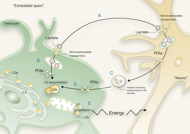
“The Astrocyte Neuron Lactate Shuttle (ANLS) Hypothesis [78] proposes that lactate is transported from glial to neurons via monocarboxylate transporters (MCTs) in Drosophila [79] and mice [80–82].”
BackgroundDynamics and functions of lipid droplets
Olzmann, J. A., & Carvalho, P. (2019) · Nature Reviews Molecular Cell Biology · doi:10.1038/s41580-018-0085-z · Cited by 2,740
Apolipoprotein E: Far More Than a Lipid Transport Protein
Mahley, R. W., & Rall, S. C. (2000) · Annual Review of Genomics and Human Genetics · doi:10.1146/annurev.genom.1.1.507 · Cited by 1,836
CNS synaptogenesis promoted by glia-derived cholesterol
Mauch, D. H., Nägler, K., Schumacher, S., Göritz, C., Müller, E.-C., Otto, A., & Pfrieger, F. W. (2001) · Science · doi:10.1126/science.294.5545.1354 · Cited by 1,711
Lipid Droplets and Cellular Lipid Metabolism
Walther, T. C., & Farese, R. V. (2012) · Annual Review of Biochemistry · doi:10.1146/annurev-biochem-061009-102430 · Cited by 1,701
Oligodendroglia metabolically support axons and contribute to neurodegeneration
Lee, Y., Morrison, B. M., Li, Y., Lengacher, S., Farah, M. H., Hoffman, P. N., Liu, Y., Tsingalia, A., Jin, L., Zhang, P.-W., et al. (2012) · Nature · doi:10.1038/nature11314 · Cited by 1,664
Cited by
Sorted by citations
Lipids and lipid metabolism in cellular senescence: Emerging targets for age-related diseases
Zeng, Q., Gong, Y., Zhu, N., Shi, Y., Zhang, C., & Li, Q. (2024) · Ageing Research Reviews · doi:10.1016/j.arr.2024.102294 · Cited by 189
Brain energy metabolism: A roadmap for future research
Rae, C. D., Baur, J. A., Borges, K., Dienel, G. A., Díaz‐García, C. M., Douglass, S. R., Drew, K. L., Duarte, J. M. N., Durán, J., Kann, O., et al. (2024) · Journal of Neurochemistry · doi:10.1111/jnc.16032 · Cited by 113
Apolipoprotein E in Alzheimer’s disease: molecular insights and therapeutic opportunities
Belaidi, A. A., Bush, A. I., & Ayton, S. (2025) · Molecular Neurodegeneration · doi:10.1186/s13024-025-00843-y · Cited by 91
Zhang, Y., Zou, M., Wu, H., Zhu, J., & Jin, T. (2024) · Neurobiology of Disease · doi:10.1016/j.nbd.2024.106710 · Cited by 75
Molecular imaging: design mechanism and bioapplications
Chen, L., Lyu, Y., Zhang, X., Zheng, L., Li, Q., Ding, D., Chen, F., Li, W., Li, W., Zhang, Y., et al. (2023) · Science China Chemistry · doi:10.1007/s11426-022-1461-3 · Cited by 69
Metadata
- Authors
- Yang, D., Wang, X., Zhang, L., Fang, Y., Zheng, Q., Liu, X., Yu, W., Chen, S., Ying, J., & Hua, F.
- Affiliations
- Second Affiliated Hospital of Nanchang University (CN)
- First Affiliated Hospital of Nanchang University (CN)
- Corresponding author
- Ying, Jun
- Hua, Fuzhou
- Venue
- Cell & Bioscience
- Published
- 2022-07-12
- Volume
- 12
- Issue
- 1
- Pages
- 106-106
- ISSN
- 2045-3701 (ISSN-L)
- PMC
- PMC9277953
- OpenAlex
- W4285093609
- Cited by
- 179
- References
- 135
- Citations by year
- 2026: 46 · 2025: 64 · 2024: 40 · 2023: 28 · 2022: 1
- FWCI
- 3.60
- Type
- journal article
- Language
- en
- Open access
- gold
- doi.org
- Locations
- Cell & Bioscience (publishedVersion, cc-by)
- PubMed Central (publishedVersion, cc-by)
- PubMed (publishedVersion)
- DOAJ (DOAJ: Directory of Open Access Journals) (submittedVersion, cc-by-sa)
- Funders
- National Natural Science Foundation of China (82060219, 81860259)
- Natural Science Foundation of Jiangxi Province (20212ACB216009, 20202BAB206033, 20212BAB216048)
- Nanchang University (20192BAB215042)
- Topics
- Life Sciences › Biochemistry, Genetics and Molecular Biology › Biochemistry › Lipid metabolism and biosynthesis
- Life Sciences › Neuroscience › Neurology › Neuroinflammation and Neurodegeneration Mechanisms
- Life Sciences › Neuroscience › Neurology › Neurological diseases and metabolism
- Keywords
- glial cells, lipid metabolism, lipid droplets, neurodegenerative diseases, brain development, lipid accumulation, oxidative stress, energy metabolism, lipid droplet formation, intracellular lipid metabolism
About these checks
Registry, retraction, Cites and statistics results are computed, not judged, with one exception: a printed reference that no index lists is read once by a model before it counts as a citation error. Claims are extracted by a model and kept only when their quote appears verbatim in the text; each cited source is then checked, with a model’s help, for whether it says what the claim relies on. Those verdicts can be wrong — read the claim’s quote and the cited source before relying on one.
Statistics and arithmetic are recomputed, not judged.
Confidence bands are shown only for fields calibrated on our annotated test set. None is calibrated yet, so no band is shown.
What counts as a claim ›How we measure confidence ›How we check this ›
Are you an author, or do you see a problem with this page? Request a review
