

The DOI matches its Crossref record; 66 of the 66 references it cites were found in a registry or an index.
Complement 3(+)-astrocytes are highly abundant in prion diseases, but their abolishment led to an accelerated disease course and early dysregulation of microglia
Open accessAbstract
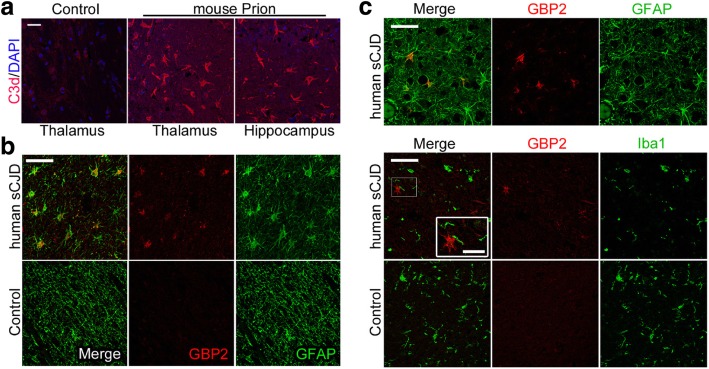
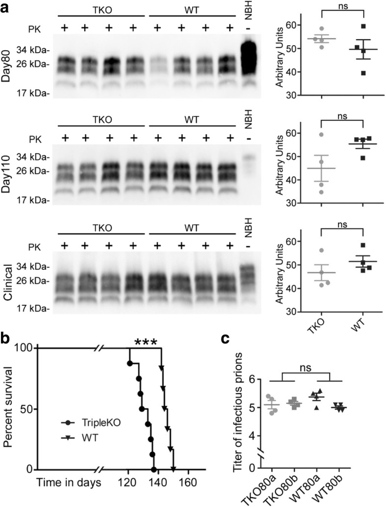
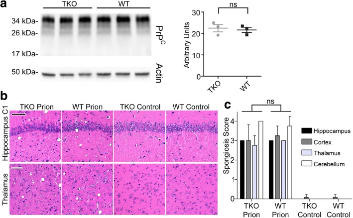
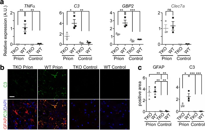
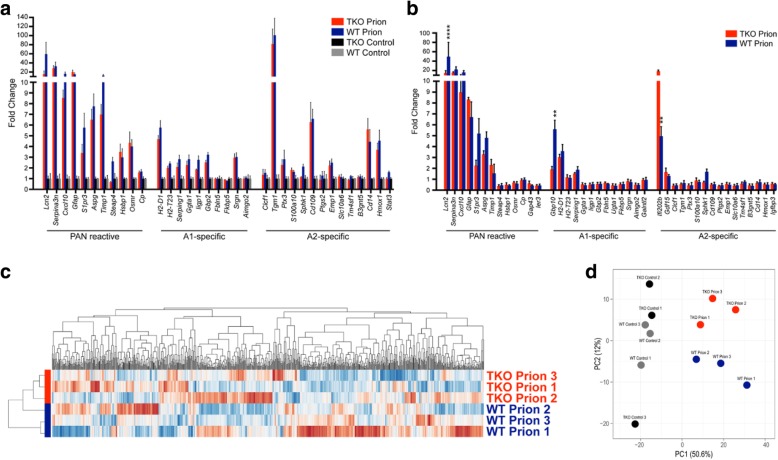
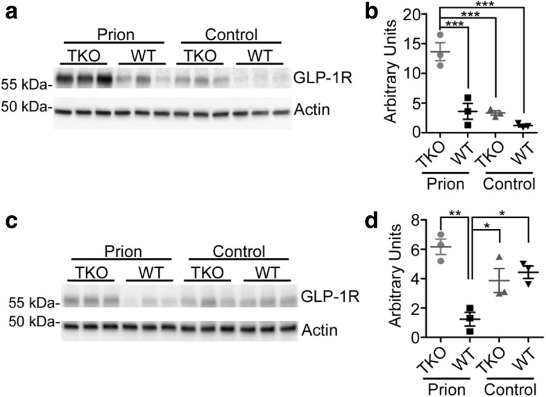
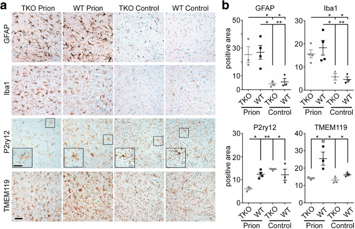
Astrogliosis and activation of microglia are hallmarks of prion diseases in humans and animals. Both were viewed to be rather independent events in disease pathophysiology, with proinflammatory microglia considered to be the potential neurotoxic species at late disease stages. Recent investigations have provided substantial evidence that a proinflammatory microglial cytokine cocktail containing TNF-α, IL-1α and C1qa reprograms a subset of astrocytes to change their expression profile and phenotype, thus becoming neurotoxic (designated as A1-astrocytes). Knockout or antibody blockage of the three cytokines abolish formation of A1-astrocytes, therefore, this pathway is of high therapeutic interest in neurodegenerative diseases. Since astrocyte polarization profiles have never been investigated in prion diseases, we performed several analyses and could show that C3 + -PrP Sc -reactive-astrocytes, which may represent a subtype of A1-astrocytes, are highly abundant in prion disease mouse models and human prion diseases. To investigate their impact on prion disease pathophysiology and to evaluate their potential therapeutic targeting, we infected TNF-α, IL-1α, and C1qa Triple-KO mice (TKO-mice), which do not transit astrocytes into A1, with prions. Although formation of C3 + -astrocytes was significantly reduced in prion infected Triple-KO-mice, this did not affect the amount of PrP Sc deposition or titers of infectious prions. Detailed characterization of the astrocyte activation signature in thalamus tissue showed that astrocytes in prion diseases are highly activated, showing a mixed phenotype that is distinct from other neurodegenerative diseases and were therefore termed C3 + -PrP Sc -reactive-astrocytes. Unexpectedly, Triple-KO led to a significant acceleration of prion disease course. While pan-astrocyte and -microglia marker upregulation was unchanged compared to WT-brains, microglial homeostatic markers were lost early in disease in TKO-mice, pointing towards important functions of different glia cell types in prion diseases.
Overview
The claims and statistics of this paper haven’t been checked yet.
Run the check — free, no account needed. It reads the open-access text and checks the sources it cites.
Integrity signals
Registry
Cites
Statistics
Figures
Verification
Claims the paper makes
The claims and statistics of this paper haven’t been checked yet.
Run the check — free, no account needed. It reads the open-access text and checks the sources it cites.
Claims other papers make about this one
Each is checked against this paper’s own text: does it say what the citing paper says it does?
The source backs part of the claim, not all of it — or backs it for a different population, condition or comparison.
In the citing paper
“The loss of homeostatic microglial proteins is visible in immunofluorescence double-staining of IBA1 and TMEM119 in terminally sick mice and mock-infected healthy control brains, and shows the reciprocal regulation of both proteins in prion disease (Figure 5A) [55,73,88,89,90,91].”
From Loss of Homeostatic Microglia Signature in Prion Diseases (2022) · cites this paper as [73]
Experimental — accuracy not yet measuredThe cited source says what the claim attributes to it.
In the citing paper
“Several proinflammatory cytokines are upregulated in the brain during the prion disease course including TNF α, IL 1α, and C1qa [46,69,70,71,72,73].”
From Loss of Homeostatic Microglia Signature in Prion Diseases (2022) · cites this paper as [73]
In the citing paper
“Microglia and astrocytes keep close contacts in the healthy brain but also in terminal prion disease, although both cell types are considerably dysregulated (Figure 5B) [73].”
From Loss of Homeostatic Microglia Signature in Prion Diseases (2022) · cites this paper as [73]
In the citing paper
“We recently showed that astrocytes in murine and human prion diseases express the A1-signature proteins complement 3 and GBP2 [73].”
From Loss of Homeostatic Microglia Signature in Prion Diseases (2022) · cites this paper as [73]
In the citing paper
“As previously shown, TMEM119-positive processes are highly reduced at the terminal stage of prion disease in the hippocampus, and almost completely lost in the thalamus during terminal prion disease (Figure 5) [73].”
From Loss of Homeostatic Microglia Signature in Prion Diseases (2022) · cites this paper as [73]
In the citing paper
“While the homeostatic markers are still abundant in the hippocampus, this signature is almost completely lost in the thalamus, an effect which is especially severe for P2RY12 (Figure 4) [47,73,97].”
From Loss of Homeostatic Microglia Signature in Prion Diseases (2022) · cites this paper as [73]
In the citing paper
“In contrast, astrocytes in prion disease showed a mixed phenotype with an expression of both A1 and A2 proteins that is unique in prion disorders and differs from other neurodegenerative diseases [73].”
From Loss of Homeostatic Microglia Signature in Prion Diseases (2022) · cites this paper as [73]
In the citing paper
“Prion disease-associated astrogliosis is also prominent in human prion diseases as shown by the upregulation of GFAP and YKL-40 (or Chitinase-3-like protein 1; CHI3L1) (Figure 6B) [73,107].”
From Loss of Homeostatic Microglia Signature in Prion Diseases (2022) · cites this paper as [73]
In the citing paper
“GFAP is highly upregulated in astrocytes upon disease [73,75,107].”
From Loss of Homeostatic Microglia Signature in Prion Diseases (2022) · cites this paper as [73]
In the citing paper
“We recently showed that the abundance of homeostatic microglia proteins such as TMEM119 and P2RY12 is significantly reduced in terminal prion disease (Figure 4) [47,73,97].”
From Loss of Homeostatic Microglia Signature in Prion Diseases (2022) · cites this paper as [73]
In the citing paper
“Moreover, in our recent experiments, the extensive loss of the homeostatic signature in prion diseases stands out remarkably [47,73,97].”
From Loss of Homeostatic Microglia Signature in Prion Diseases (2022) · cites this paper as [73]
Self-check
Percentages, sums and reported differences re-computed from the paper’s own numbers.
The self-check needs the paper’s full text.
Citations
Cites
Neurotoxic reactive astrocytes are induced by activated microglia
Liddelow, S. A., Guttenplan, K. A., Clarke, L. E., Bennett, F. C., Bohlen, C. J., Schirmer, L., Bennett, M. L., Münch, A. E., Chung, W.-S., Peterson, T. C., et al. (2017) · Nature · doi:10.1038/nature21029 · Cited by 7,971
Self-citationNeuroinflammation in Alzheimer's disease
Heneka, M. T., Carson, M. J., Khoury, J. E., Landreth, G. E., Brosseron, F., Feinstein, D. L., Jacobs, A. H., Wyss-Coray, T., Vitorica, J., Ransohoff, R. M., et al. (2015) · The Lancet Neurology · doi:10.1016/s1474-4422(15)70016-5 · Cited by 6,226
ImageJ2: ImageJ for the next generation of scientific image data
Rueden, C., Schindelin, J., Hiner, M., DeZonia, B. E., Walter, A., Arena, E. T., & Eliceiri, K. W. (2017) · BMC Bioinformatics · doi:10.1186/s12859-017-1934-z · Cited by 6,087
Novel Proteinaceous Infectious Particles Cause Scrapie
Prusiner, S. B. (1982) · Science · doi:10.1126/science.6801762 · Cited by 5,221
Metsalu, T., & Vilo, J. (2015) · Nucleic Acids Research · doi:10.1093/nar/gkv468 · Cited by 3,770
Krasemann, S., Madore, C., Cialic, R., Baufeld, C., Calcagno, N., El Fatimy, R., Beckers, L., O’Loughlin, E., Xu, Y., Fanek, Z., et al. (2017) · Immunity · doi:10.1016/j.immuni.2017.08.008 · Cited by 2,979
Self-citationIdentification of a unique TGF-β–dependent molecular and functional signature in microglia
Butovsky, O., Jedrychowski, M. P., Moore, C. S., Cialic, R., Lanser, A. J., Gabriely, G., Koeglsperger, T., Dake, B., Wu, P. M., Doykan, C. E., et al. (2014) · Nature Neuroscience · doi:10.1038/nn.3599 · Cited by 2,543
Self-citationGenomic analysis of reactive astrogliosis
Zamanian, J. L., Xu, L., Foo, L. C., Nouri, N., Zhou, L., Giffard, R. G., & Barres, B. A. (2012) · The Journal of Neuroscience · doi:10.1523/jneurosci.6221-11.2012 · Cited by 2,404
Reactive Astrocytes: Production, Function, and Therapeutic Potential
Liddelow, S. A., & Barres, B. A. (2017) · Immunity · doi:10.1016/j.immuni.2017.06.006 · Cited by 2,336
Self-citationMice devoid of PrP are resistant to scrapie
Büeler, H., Aguzzi, A., Sailer, A. W., Greiner, R., Autenried, P., Aguet, M., & Weissmann, C. (1993) · Cell · doi:10.1016/0092-8674(93)90360-3 · Cited by 2,107
Cited by
Sorted by citations
Items 101–151 of 151
Makarava, N., Mychko, O., Molesworth, K., Chang, J. C., Henry, R. J., Tsymbalyuk, N., Gerzanich, V., Simard, J. M., Loane, D. J., & Baskakov, I. V. (2023) · bioRxiv (Cold Spring Harbor Laboratory) · doi:10.1101/2023.02.01.526708 · Cited by 2
Astrocitos: acción e interacción en el Sistema Nervioso
Padilla-Gil, D. N. (2024) · Revista Med · doi:10.18359/rmed.6870 · Cited by 1
Innate immune signaling and functions in astrocytes
Guo, A. X., Fisher, T. M., Comandante-Lou, N., Jager, P. L. D., & Liddelow, S. A. (2026) · Nature Immunology · doi:10.1038/s41590-026-02561-z · Cited by 5
Self-citationEnfermedades priónicas y enfermedades similares a priones
Juárez, J. C. C., Batres-Godoy, S. S., Calderón, M. J., Solís-Aguilar, R., Palencia, A. E., & Silva-Arévalo, G. (2022) · Neurología, Neurocirugía y Psiquiatría · doi:10.35366/108765 · Cited by 1
Hay, A. J. D., Popichak, K. A., Zabel, M. D., & Moreno, J. A. (2023) · Journal of Visualized Experiments · doi:10.3791/65565 · Cited by 1
Risen, S. J., Wahl, D., LaRocca, T. J., & Moreno, J. A. (2025) · Neuroglia · doi:10.3390/neuroglia6020022 · Cited by 1
A Model of Epileptogenesis in Rhinal Cortex-Hippocampus Organotypic Slice Cultures
Valente, C. A., Meda, F. J., Carvalho, M., & Sebastião, A. M. (2021) · Journal of Visualized Experiments · doi:10.3791/61330-v · Cited by 1
Chemical inhibition of pathological reactive astrocytes promotes neural protectionPreprint
Clayton, B. L. L., Kristell, J. D., Allan, K. C., Karl, M. T., Garrison, E. D., Maeno-Hikichi, Y., Sturno, A. M., Shick, H. E., Miller, R. H., & Tesar, P. J. (2021) · bioRxiv (Cold Spring Harbor Laboratory) · doi:10.1101/2021.11.03.467083 · Cited by 1
Loss of region-specific glial homeostatic signature in prion diseasesPreprint
Makarava, N., Chang, J. C., Molesworth, K., & Baskakov, I. V. (2019) · bioRxiv (Cold Spring Harbor Laboratory) · doi:10.21203/rs.2.17016/v1 · Cited by 1
Transgenic Mice Modelling in Prion Diseases
Bradford, B. M., Mabbott, N. A., & Diack, A. B. (2023) · Springer eBooks · doi:10.1007/978-3-031-20565-1_15
Walz, W. (2023) · Springer eBooks · doi:10.1007/978-3-031-48105-5_13
Regional Patterns of Neurodegeneration in a Mouse Model of ProteinopathyPreprint
Shepilov, D., Khan, I. A., Chen, H. C., dos Santos, Á., Rowley, C., Harding, E. C., Merkle, F. T., & Administrator, S. P. (2025) · SSRN Electronic Journal · doi:10.1101/2025.08.17.670564
Reactive Microglia and Astrocytes as Therapeutic Targets in Prion Diseases
Makarava, N., Kushwaha, R., & Baskakov, I. V. (2025) · Sub-cellular biochemistry/Subcellular biochemistry · doi:10.1007/978-3-031-97055-9_8 · Cited by 1
Carroll, J. A., Race, B., Williams, K., Striebel, J. F., & Chesebro, B. W. (2020) · Figshare · doi:10.6084/m9.figshare.12269870
Carroll, J. A., Race, B., Williams, K., Striebel, J. F., & Chesebro, B. W. (2020) · Figshare · doi:10.6084/m9.figshare.12269843
Martirosyan, A., Ansari, R. A., Pestana, F., Hebestreit, K., Gasparyan, H., Aleksanyan, R., Hnatova, S., Poovathingal, S. K., Marneffe, C., Thal, D. R., et al. (2024) · Figshare · doi:10.6084/m9.figshare.26670129
Martirosyan, A., Ansari, R. A., Pestana, F., Hebestreit, K., Gasparyan, H., Aleksanyan, R., Hnatova, S., Poovathingal, S. K., Marneffe, C., Thal, D. R., et al. (2024) · Figshare · doi:10.6084/m9.figshare.26670132
Martirosyan, A., Ansari, R. A., Pestana, F., Hebestreit, K., Gasparyan, H., Aleksanyan, R., Hnatova, S., Poovathingal, S. K., Marneffe, C., Thal, D. R., et al. (2024) · Figshare · doi:10.6084/m9.figshare.26670120
Martirosyan, A., Ansari, R. A., Pestana, F., Hebestreit, K., Gasparyan, H., Aleksanyan, R., Hnatova, S., Poovathingal, S. K., Marneffe, C., Thal, D. R., et al. (2024) · Figshare · doi:10.6084/m9.figshare.26670141
Martirosyan, A., Ansari, R. A., Pestana, F., Hebestreit, K., Gasparyan, H., Aleksanyan, R., Hnatova, S., Poovathingal, S. K., Marneffe, C., Thal, D. R., et al. (2024) · Figshare · doi:10.6084/m9.figshare.26670165
Martirosyan, A., Ansari, R. A., Pestana, F., Hebestreit, K., Gasparyan, H., Aleksanyan, R., Hnatova, S., Poovathingal, S. K., Marneffe, C., Thal, D. R., et al. (2024) · Figshare · doi:10.6084/m9.figshare.26670117
Vicente-Acosta, A., Giménez-Cassina, A., Díaz‐Nido, J., & Loría, F. (2022) · Figshare · doi:10.6084/m9.figshare.19588803
Martirosyan, A., Ansari, R. A., Pestana, F., Hebestreit, K., Gasparyan, H., Aleksanyan, R., Hnatova, S., Poovathingal, S. K., Marneffe, C., Thal, D. R., et al. (2024) · Figshare · doi:10.6084/m9.figshare.26670162
Martirosyan, A., Ansari, R. A., Pestana, F., Hebestreit, K., Gasparyan, H., Aleksanyan, R., Hnatova, S., Poovathingal, S. K., Marneffe, C., Thal, D. R., et al. (2024) · Figshare · doi:10.6084/m9.figshare.26670147
Carroll, J. A., Race, B., Williams, K., Striebel, J. F., & Chesebro, B. W. (2020) · Figshare · doi:10.6084/m9.figshare.12269852
Makarava, N., Mychko, O., Chang, J. C., Molesworth, K., & Baskakov, I. V. (2021) · Figshare · doi:10.6084/m9.figshare.14583799
Martirosyan, A., Ansari, R. A., Pestana, F., Hebestreit, K., Gasparyan, H., Aleksanyan, R., Hnatova, S., Poovathingal, S. K., Marneffe, C., Thal, D. R., et al. (2024) · Figshare · doi:10.6084/m9.figshare.26670126
Martirosyan, A., Ansari, R. A., Pestana, F., Hebestreit, K., Gasparyan, H., Aleksanyan, R., Hnatova, S., Poovathingal, S. K., Marneffe, C., Thal, D. R., et al. (2024) · Figshare · doi:10.6084/m9.figshare.26670108
Martirosyan, A., Ansari, R. A., Pestana, F., Hebestreit, K., Gasparyan, H., Aleksanyan, R., Hnatova, S., Poovathingal, S. K., Marneffe, C., Thal, D. R., et al. (2024) · Figshare · doi:10.6084/m9.figshare.26670138
Martirosyan, A., Ansari, R. A., Pestana, F., Hebestreit, K., Gasparyan, H., Aleksanyan, R., Hnatova, S., Poovathingal, S. K., Marneffe, C., Thal, D. R., et al. (2024) · Figshare · doi:10.6084/m9.figshare.26670150
Martirosyan, A., Ansari, R. A., Pestana, F., Hebestreit, K., Gasparyan, H., Aleksanyan, R., Hnatova, S., Poovathingal, S. K., Marneffe, C., Thal, D. R., et al. (2024) · Figshare · doi:10.6084/m9.figshare.26670153
Martirosyan, A., Ansari, R. A., Pestana, F., Hebestreit, K., Gasparyan, H., Aleksanyan, R., Hnatova, S., Poovathingal, S. K., Marneffe, C., Thal, D. R., et al. (2024) · Figshare · doi:10.6084/m9.figshare.26670156
Martirosyan, A., Ansari, R. A., Pestana, F., Hebestreit, K., Gasparyan, H., Aleksanyan, R., Hnatova, S., Poovathingal, S. K., Marneffe, C., Thal, D. R., et al. (2024) · Figshare · doi:10.6084/m9.figshare.26670123
Martirosyan, A., Ansari, R. A., Pestana, F., Hebestreit, K., Gasparyan, H., Aleksanyan, R., Hnatova, S., Poovathingal, S. K., Marneffe, C., Thal, D. R., et al. (2024) · Figshare · doi:10.6084/m9.figshare.26670111
Martirosyan, A., Ansari, R. A., Pestana, F., Hebestreit, K., Gasparyan, H., Aleksanyan, R., Hnatova, S., Poovathingal, S. K., Marneffe, C., Thal, D. R., et al. (2024) · Figshare · doi:10.6084/m9.figshare.26670168
Carroll, J. A., Race, B., Williams, K., Striebel, J. F., & Chesebro, B. W. (2020) · Figshare · doi:10.6084/m9.figshare.12269858
Martirosyan, A., Ansari, R. A., Pestana, F., Hebestreit, K., Gasparyan, H., Aleksanyan, R., Hnatova, S., Poovathingal, S. K., Marneffe, C., Thal, D. R., et al. (2024) · Figshare · doi:10.6084/m9.figshare.26670114
Martirosyan, A., Ansari, R. A., Pestana, F., Hebestreit, K., Gasparyan, H., Aleksanyan, R., Hnatova, S., Poovathingal, S. K., Marneffe, C., Thal, D. R., et al. (2024) · Figshare · doi:10.6084/m9.figshare.26670144
Martirosyan, A., Ansari, R. A., Pestana, F., Hebestreit, K., Gasparyan, H., Aleksanyan, R., Hnatova, S., Poovathingal, S. K., Marneffe, C., Thal, D. R., et al. (2024) · Figshare · doi:10.6084/m9.figshare.26670159
Martirosyan, A., Ansari, R. A., Pestana, F., Hebestreit, K., Gasparyan, H., Aleksanyan, R., Hnatova, S., Poovathingal, S. K., Marneffe, C., Thal, D. R., et al. (2024) · Figshare · doi:10.6084/m9.figshare.26670135
Makarava, N., Mychko, O., Chang, J. C., Molesworth, K., & Baskakov, I. V. (2021) · Figshare · doi:10.6084/m9.figshare.14583817
Carroll, J. A., Race, B., Williams, K., Striebel, J. F., & Chesebro, B. W. (2020) · Figshare · doi:10.6084/m9.figshare.12269864
APOE genotype differentially modulates prion pathology in a mouse model
Lizińczyk, A. M., Pankiewicz, J. E., Cullina, W. L., Franco, L. A., Sullivan, P. M., & Sadowski, M. J. (2026) · Acta Neuropathologica Communications · doi:10.1186/s40478-025-02207-5
Tang, X., Cui, H., & Cui, H. (2026) · Biofabrication · doi:10.1088/1758-5090/ae4895
Poulot-Becq-Giraudon, Y. (2024) · theses.fr (ABES) · doi:10.70675/0d7a8b92z8393z4d76z82a8z87efe0790346
A neural stem cell–derived 3D spheroid model that recapitulates prion infection and pathology
Park, H. J., Kim, H., Byun, S., & Ryou, C. (2026) · Materials Today Bio · doi:10.1016/j.mtbio.2026.103378
Luo, H., Jin, M., Xu, K., Zheng, H., Zhang, W., Gu, X., Deng, C., Chen, Z., Xiong, C., & Zhang, X. (2026) · Investigative Ophthalmology & Visual Science · doi:10.1167/iovs.67.6.49
Zhu, Y.-X., Bradford, B. M., & Mabbott, N. (2026) · Frontiers in Neuroscience · doi:10.3389/fnins.2026.1924347
Li, W., Liu, Y., Zhang, H., Zhang, H., Shao, W., Hu, T., Liao, Y., Zhang, W., & Kang, X. (2026) · Biology Direct · doi:10.1186/s13062-026-00974-2 · Cited by 1
Time-dependent transcriptomic response to scrapie exposure in ovine MSC-derived neuron-like cells
Hernaiz, A., Gil-Guerrero, J., Marín, B., Vázquez, F. J., Badiola, J. J., Zaragoza, P., Bolea, R., & Martín‐Burriel, I. (2026) · Frontiers in Cellular and Infection Microbiology · doi:10.3389/fcimb.2026.1938678
Metadata
- Authors
- Hartmann, K., Sepulveda-Falla, D., Rose, I. V. L., Madore, C., Muth, C., Matschke, J., Butovsky, O., Liddelow, S., Glatzel, M., & Krasemann, S.
- Affiliations
- University Medical Center Hamburg-Eppendorf (DE)
- NYU Langone Health (US)
- Brigham and Women's Hospital (US)
- The University of Melbourne (AU)
- Corresponding author
- Krasemann, Susanne
- ORCID
- Venue
- Acta Neuropathologica Communications
- Published
- 2019-05-22
- Volume
- 7
- Issue
- 1
- Pages
- 83-83
- ISSN
- 2051-5960 (ISSN-L)
- PMC
- PMC6530067
- OpenAlex
- W2947673562
- Cited by
- 141
- References
- 66
- Citations by year
- 2026: 9 · 2025: 17 · 2024: 25 · 2023: 20 · 2022: 18 · 2021: 25 · 2020: 24 · 2019: 3
- FWCI
- 5.79
- Type
- journal article
- Language
- en
- Open access
- gold
- doi.org
- Locations
- Acta Neuropathologica Communications (publishedVersion, cc-by)
- PubMed Central (publishedVersion, cc-by)
- PubMed (publishedVersion)
- HAL (submittedVersion)
- DOAJ (DOAJ: Directory of Open Access Journals) (submittedVersion, cc-by-sa)
- Minerva Access (University of Melbourne) (submittedVersion, cc-by)
- Funders
- Werner Otto Stiftung (9/91)
- Topics
- Life Sciences › Biochemistry, Genetics and Molecular Biology › Molecular Biology › Prion Diseases and Protein Misfolding
- Life Sciences › Neuroscience › Neurology › Neuroinflammation and Neurodegeneration Mechanisms
- Life Sciences › Neuroscience › Cellular and Molecular Neuroscience › Alzheimer's disease research and treatments
- Keywords
- prion disease, A1 astrocytes, astrocyte polarization, microglial activation, TNF-α, IL-1α, C1QA, triple knockout mice, prion pathogenesis, neuroinflammation, glial interactions, thalamus, neurodegeneration
- MeSH terms
- Aged, Animals, Astrocytes, Complement C3, Female, Humans, Creutzfeldt-Jakob Syndrome, Male, Mice, Inbred C57BL, Middle Aged, Cytokines, Prion Diseases, Microglia, Mice, Knockout, Disease Progression, PrPSc Proteins
About these checks
Registry, retraction, Cites and statistics results are computed, not judged, with one exception: a printed reference that no index lists is read once by a model before it counts as a citation error. Claims are extracted by a model and kept only when their quote appears verbatim in the text; each cited source is then checked, with a model’s help, for whether it says what the claim relies on. Those verdicts can be wrong — read the claim’s quote and the cited source before relying on one.
Statistics and arithmetic are recomputed, not judged.
Confidence bands are shown only for fields calibrated on our annotated test set. None is calibrated yet, so no band is shown.
What counts as a claim ›How we measure confidence ›How we check this ›
Are you an author, or do you see a problem with this page? Request a review
