

The DOI matches its Crossref record; 66 of the 66 references it cites were found in a registry or an index.
Complement 3(+)-astrocytes are highly abundant in prion diseases, but their abolishment led to an accelerated disease course and early dysregulation of microglia
Open accessAbstract
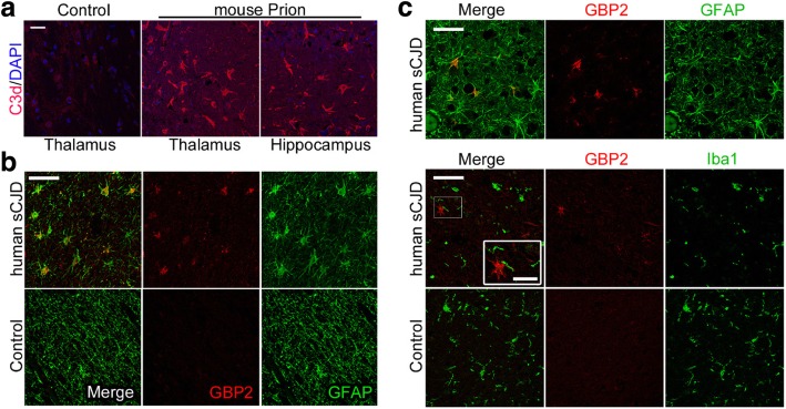
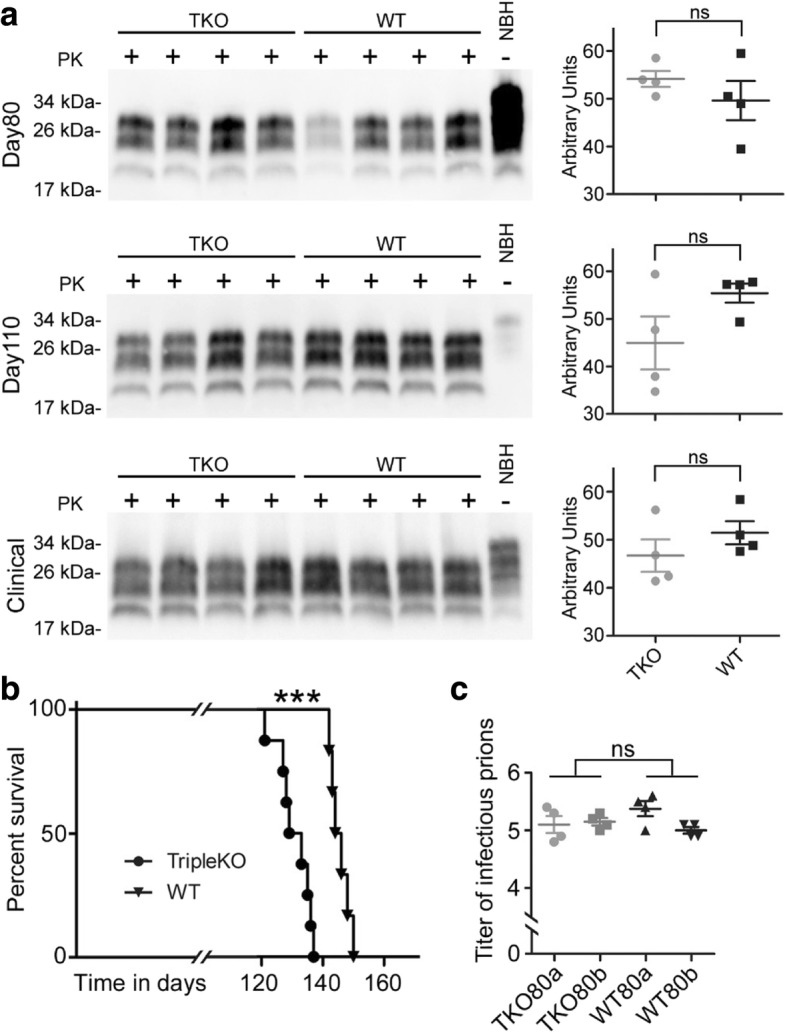
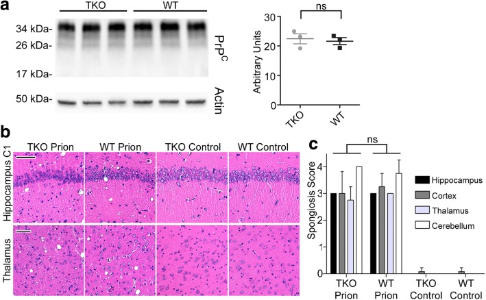
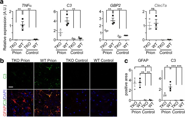
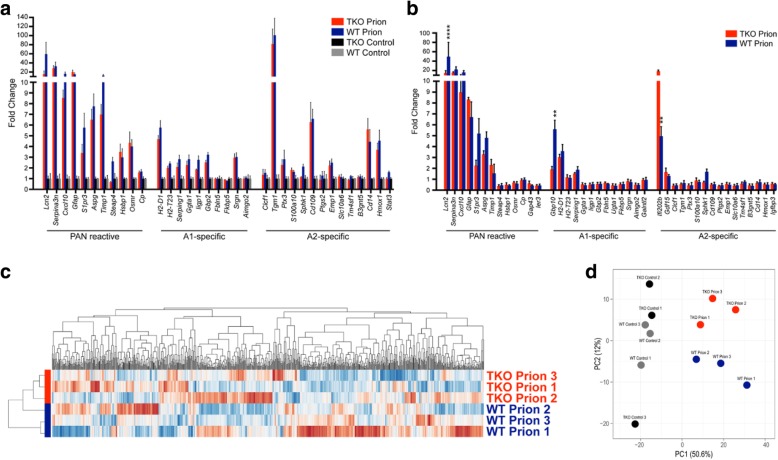
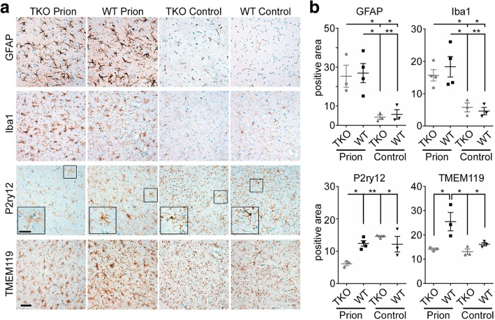
Astrogliosis and activation of microglia are hallmarks of prion diseases in humans and animals. Both were viewed to be rather independent events in disease pathophysiology, with proinflammatory microglia considered to be the potential neurotoxic species at late disease stages. Recent investigations have provided substantial evidence that a proinflammatory microglial cytokine cocktail containing TNF-α, IL-1α and C1qa reprograms a subset of astrocytes to change their expression profile and phenotype, thus becoming neurotoxic (designated as A1-astrocytes). Knockout or antibody blockage of the three cytokines abolish formation of A1-astrocytes, therefore, this pathway is of high therapeutic interest in neurodegenerative diseases. Since astrocyte polarization profiles have never been investigated in prion diseases, we performed several analyses and could show that C3 + -PrP Sc -reactive-astrocytes, which may represent a subtype of A1-astrocytes, are highly abundant in prion disease mouse models and human prion diseases. To investigate their impact on prion disease pathophysiology and to evaluate their potential therapeutic targeting, we infected TNF-α, IL-1α, and C1qa Triple-KO mice (TKO-mice), which do not transit astrocytes into A1, with prions. Although formation of C3 + -astrocytes was significantly reduced in prion infected Triple-KO-mice, this did not affect the amount of PrP Sc deposition or titers of infectious prions. Detailed characterization of the astrocyte activation signature in thalamus tissue showed that astrocytes in prion diseases are highly activated, showing a mixed phenotype that is distinct from other neurodegenerative diseases and were therefore termed C3 + -PrP Sc -reactive-astrocytes. Unexpectedly, Triple-KO led to a significant acceleration of prion disease course. While pan-astrocyte and -microglia marker upregulation was unchanged compared to WT-brains, microglial homeostatic markers were lost early in disease in TKO-mice, pointing towards important functions of different glia cell types in prion diseases.
Overview
The claims and statistics of this paper haven’t been checked yet.
Run the check — free, no account needed. It reads the open-access text and checks the sources it cites.
Integrity signals
Registry
Cites
Statistics
Figures
Verification
Claims the paper makes
The claims and statistics of this paper haven’t been checked yet.
Run the check — free, no account needed. It reads the open-access text and checks the sources it cites.
Claims other papers make about this one
Each is checked against this paper’s own text: does it say what the citing paper says it does?
The source backs part of the claim, not all of it — or backs it for a different population, condition or comparison.
In the citing paper
“The loss of homeostatic microglial proteins is visible in immunofluorescence double-staining of IBA1 and TMEM119 in terminally sick mice and mock-infected healthy control brains, and shows the reciprocal regulation of both proteins in prion disease (Figure 5A) [55,73,88,89,90,91].”
From Loss of Homeostatic Microglia Signature in Prion Diseases (2022) · cites this paper as [73]
Experimental — accuracy not yet measuredThe cited source says what the claim attributes to it.
In the citing paper
“Several proinflammatory cytokines are upregulated in the brain during the prion disease course including TNF α, IL 1α, and C1qa [46,69,70,71,72,73].”
From Loss of Homeostatic Microglia Signature in Prion Diseases (2022) · cites this paper as [73]
In the citing paper
“Microglia and astrocytes keep close contacts in the healthy brain but also in terminal prion disease, although both cell types are considerably dysregulated (Figure 5B) [73].”
From Loss of Homeostatic Microglia Signature in Prion Diseases (2022) · cites this paper as [73]
In the citing paper
“We recently showed that astrocytes in murine and human prion diseases express the A1-signature proteins complement 3 and GBP2 [73].”
From Loss of Homeostatic Microglia Signature in Prion Diseases (2022) · cites this paper as [73]
In the citing paper
“As previously shown, TMEM119-positive processes are highly reduced at the terminal stage of prion disease in the hippocampus, and almost completely lost in the thalamus during terminal prion disease (Figure 5) [73].”
From Loss of Homeostatic Microglia Signature in Prion Diseases (2022) · cites this paper as [73]
In the citing paper
“While the homeostatic markers are still abundant in the hippocampus, this signature is almost completely lost in the thalamus, an effect which is especially severe for P2RY12 (Figure 4) [47,73,97].”
From Loss of Homeostatic Microglia Signature in Prion Diseases (2022) · cites this paper as [73]
In the citing paper
“In contrast, astrocytes in prion disease showed a mixed phenotype with an expression of both A1 and A2 proteins that is unique in prion disorders and differs from other neurodegenerative diseases [73].”
From Loss of Homeostatic Microglia Signature in Prion Diseases (2022) · cites this paper as [73]
In the citing paper
“Prion disease-associated astrogliosis is also prominent in human prion diseases as shown by the upregulation of GFAP and YKL-40 (or Chitinase-3-like protein 1; CHI3L1) (Figure 6B) [73,107].”
From Loss of Homeostatic Microglia Signature in Prion Diseases (2022) · cites this paper as [73]
In the citing paper
“GFAP is highly upregulated in astrocytes upon disease [73,75,107].”
From Loss of Homeostatic Microglia Signature in Prion Diseases (2022) · cites this paper as [73]
In the citing paper
“We recently showed that the abundance of homeostatic microglia proteins such as TMEM119 and P2RY12 is significantly reduced in terminal prion disease (Figure 4) [47,73,97].”
From Loss of Homeostatic Microglia Signature in Prion Diseases (2022) · cites this paper as [73]
In the citing paper
“Moreover, in our recent experiments, the extensive loss of the homeostatic signature in prion diseases stands out remarkably [47,73,97].”
From Loss of Homeostatic Microglia Signature in Prion Diseases (2022) · cites this paper as [73]
Self-check
Percentages, sums and reported differences re-computed from the paper’s own numbers.
The self-check needs the paper’s full text.
Citations
Cites
Neurotoxic reactive astrocytes are induced by activated microglia
Liddelow, S. A., Guttenplan, K. A., Clarke, L. E., Bennett, F. C., Bohlen, C. J., Schirmer, L., Bennett, M. L., Münch, A. E., Chung, W.-S., Peterson, T. C., et al. (2017) · Nature · doi:10.1038/nature21029 · Cited by 7,971
Self-citationNeuroinflammation in Alzheimer's disease
Heneka, M. T., Carson, M. J., Khoury, J. E., Landreth, G. E., Brosseron, F., Feinstein, D. L., Jacobs, A. H., Wyss-Coray, T., Vitorica, J., Ransohoff, R. M., et al. (2015) · The Lancet Neurology · doi:10.1016/s1474-4422(15)70016-5 · Cited by 6,226
ImageJ2: ImageJ for the next generation of scientific image data
Rueden, C., Schindelin, J., Hiner, M., DeZonia, B. E., Walter, A., Arena, E. T., & Eliceiri, K. W. (2017) · BMC Bioinformatics · doi:10.1186/s12859-017-1934-z · Cited by 6,087
Novel Proteinaceous Infectious Particles Cause Scrapie
Prusiner, S. B. (1982) · Science · doi:10.1126/science.6801762 · Cited by 5,221
Metsalu, T., & Vilo, J. (2015) · Nucleic Acids Research · doi:10.1093/nar/gkv468 · Cited by 3,770
Krasemann, S., Madore, C., Cialic, R., Baufeld, C., Calcagno, N., El Fatimy, R., Beckers, L., O’Loughlin, E., Xu, Y., Fanek, Z., et al. (2017) · Immunity · doi:10.1016/j.immuni.2017.08.008 · Cited by 2,979
Self-citationIdentification of a unique TGF-β–dependent molecular and functional signature in microglia
Butovsky, O., Jedrychowski, M. P., Moore, C. S., Cialic, R., Lanser, A. J., Gabriely, G., Koeglsperger, T., Dake, B., Wu, P. M., Doykan, C. E., et al. (2014) · Nature Neuroscience · doi:10.1038/nn.3599 · Cited by 2,543
Self-citationGenomic analysis of reactive astrogliosis
Zamanian, J. L., Xu, L., Foo, L. C., Nouri, N., Zhou, L., Giffard, R. G., & Barres, B. A. (2012) · The Journal of Neuroscience · doi:10.1523/jneurosci.6221-11.2012 · Cited by 2,404
Reactive Astrocytes: Production, Function, and Therapeutic Potential
Liddelow, S. A., & Barres, B. A. (2017) · Immunity · doi:10.1016/j.immuni.2017.06.006 · Cited by 2,336
Self-citationMice devoid of PrP are resistant to scrapie
Büeler, H., Aguzzi, A., Sailer, A. W., Greiner, R., Autenried, P., Aguet, M., & Weissmann, C. (1993) · Cell · doi:10.1016/0092-8674(93)90360-3 · Cited by 2,107
Cited by
Sorted by citations
Astrocyte Reactivity: Subtypes, States, and Functions in CNS Innate Immunity
Sofroniew, M. V. (2020) · Trends in Immunology · doi:10.1016/j.it.2020.07.004 · Cited by 721
Functional roles of reactive astrocytes in neuroinflammation and neurodegeneration
Patani, R., Hardingham, G. E., & Liddelow, S. A. (2023) · Nature Reviews Neurology · doi:10.1038/s41582-023-00822-1 · Cited by 531
Self-citationRoles of neuropathology-associated reactive astrocytes: a systematic review
Lawrence, J. M., Schardien, K., Wigdahl, B., & Nonnemacher, M. R. (2023) · Acta Neuropathologica Communications · doi:10.1186/s40478-023-01526-9 · Cited by 433
Astrocytes in human central nervous system diseases: a frontier for new therapies
Verkhratsky, A., Butt, A., Li, B., Illes, P., Zorec, R., Semyanov, A., Tang, Y., & Sofroniew, M. V. (2023) · Signal Transduction and Targeted Therapy · doi:10.1038/s41392-023-01628-9 · Cited by 428
A1/A2 astrocytes in central nervous system injuries and diseases: Angels or devils?
Fan, Y., & Huo, J. (2021) · Neurochemistry International · doi:10.1016/j.neuint.2021.105080 · Cited by 336
Microglia and Astrocytes in Disease: Dynamic Duo or Partners in Crime?
Liddelow, S. A., Marsh, S. E., & Stevens, B. (2020) · Trends in Immunology · doi:10.1016/j.it.2020.07.006 · Cited by 226
Self-citationSmith, H., Freeman, O. J., Butcher, A. J., Holmqvist, S., Humoud, I., Schätzl, T., Hughes, D. T., Verity, N., Swinden, D., Hayes, J., et al. (2020) · Neuron · doi:10.1016/j.neuron.2019.12.014 · Cited by 223
Neurotoxic Reactive Astrocytes Drive Neuronal Death after Retinal Injury
Guttenplan, K. A., Stafford, B. K., El-Danaf, R. N., Adler, D. I., Münch, A. E., Weigel, M. K., Huberman, A. D., & Liddelow, S. A. (2020) · Cell Reports · doi:10.1016/j.celrep.2020.107776 · Cited by 221
Self-citationKim, H., Leng, K., Park, J., Sorets, A. G., Kim, S., Shostak, A., Embalabala, R. J., Mlouk, K., Katdare, K. A., Rose, I. V. L., et al. (2022) · Nature Communications · doi:10.1038/s41467-022-34412-4 · Cited by 201
Self-citationMicroglia and Astrocyte Function and Communication: What Do We Know in Humans?
Garland, E. F., Hartnell, I. J., & Boche, D. (2022) · Frontiers in Neuroscience · doi:10.3389/fnins.2022.824888 · Cited by 165
Molecular and metabolic heterogeneity of astrocytes and microglia
Hasel, P., Aisenberg, W. H., Bennett, F. C., & Liddelow, S. A. (2023) · Cell Metabolism · doi:10.1016/j.cmet.2023.03.006 · Cited by 135
Self-citationUnravelling cell type-specific responses to Parkinson’s Disease at single cell resolution
Martirosyan, A., Ansari, R. A., Pestana, F., Hebestreit, K., Gasparyan, H. A., Aleksanyan, R., Hnatova, S., Poovathingal, S. K., Marneffe, C., Thal, D. R., et al. (2024) · Molecular Neurodegeneration · doi:10.1186/s13024-023-00699-0 · Cited by 145
Microglia-Astrocyte Communication in Alzheimer’s Disease
Wu, Y., & Eisel, U. L. M. (2023) · Journal of Alzheimer s Disease · doi:10.3233/jad-230199 · Cited by 125
Bidirectional Communication Between Microglia and Astrocytes in Neuroinflammation
Bhusal, A., Afridi, R., Lee, W., & Suk, K. (2022) · Current Neuropharmacology · doi:10.2174/1570159x21666221129121715 · Cited by 113
Taylor, X., Cisternas, P., You, Y., You, Y., Xiang, S., Marambio, Y. G., Zhang, J., Vidal, R., & Lasagna‐Reeves, C. A. (2020) · Journal of Neuroinflammation · doi:10.1186/s12974-020-01900-7 · Cited by 93
Diversity of Reactive Astrogliosis in CNS Pathology: Heterogeneity or Plasticity?
Moulson, A. J., Squair, J. W., Franklin, R. J. M., Tetzlaff, W., & Assinck, P. (2021) · Frontiers in Cellular Neuroscience · doi:10.3389/fncel.2021.703810 · Cited by 97
King, A., Szekely, B., Calapkulu, E., Ali, H., Rios, F., Jones, S., & Troakes, C. (2020) · Brain Sciences · doi:10.3390/brainsci10080503 · Cited by 93
Astrocytes in myelination and remyelination
Molina-González, I., & Miron, V. E. (2019) · Neuroscience Letters · doi:10.1016/j.neulet.2019.134532 · Cited by 83
RNA-seq and network analysis reveal unique glial gene expression signatures during prion infection
Carroll, J. A., Race, B., Williams, K., Striebel, J., & Chesebro, B. (2020) · Molecular Brain · doi:10.1186/s13041-020-00610-8 · Cited by 66
Makarava, N., Chang, J. C.-Y., Molesworth, K., & Baskakov, I. V. (2020) · Neurobiology of Disease · doi:10.1016/j.nbd.2020.104783 · Cited by 64
Scheckel, C., Imeri, M., Schwarz, P., & Aguzzi, A. (2020) · eLife · doi:10.7554/elife.62911 · Cited by 57
Pons‐Espinal, M., Blasco-Agell, L., Fernández‐Carasa, I., Andrés‐Benito, P., di Domenico, A., Richaud‐Patín, Y., Baruffi, V., Marruecos, L., Espinosa, L., Garrido, A., et al. (2024) · JCI Insight · doi:10.1172/jci.insight.163359 · Cited by 69
She Doesn’t Even Go Here: The Role of Inflammatory Astrocytes in CNS Disorders
Reid, J. K., & Kuipers, H. F. (2021) · Frontiers in Cellular Neuroscience · doi:10.3389/fncel.2021.704884 · Cited by 66
Activated endothelial cells induce a distinct type of astrocytic reactivity
Taylor, X., Cisternas, P., Jury, N., Martínez, P., Huang, X., You, Y., Redding‐Ochoa, J., Vidal, R., Zhang, J., Troncoso, J. C., et al. (2022) · Communications Biology · doi:10.1038/s42003-022-03237-8 · Cited by 63
The spatial landscape of glial pathology and T cell response in Parkinson’s disease substantia nigra
Ma, M., Paryani, F., Jakubiak, K. A., Xia, S., Antoku, S., Kannan, A., Lee, J., Madden, N., Kumar, S. S., Li, J., et al. (2025) · Nature Communications · doi:10.1038/s41467-025-62478-3 · Cited by 61
Spatiotemporal transcriptomic maps of mouse intracerebral hemorrhage at single-cell resolution
Xiang, R., Wang, J., Chen, Z., Tao, J. Z., Peng, Q., Ding, R., Zhou, T., Tu, Z., Yang, T., Yang, T., et al. (2025) · Neuron · doi:10.1016/j.neuron.2025.04.026 · Cited by 51
Chen, M., Hu, Z., Ding, J., Du, R., & Hu, G. (2021) · Brain Behavior and Immunity · doi:10.1016/j.bbi.2021.04.003 · Cited by 45
Mechanisms of astrocyte aging in reactivity and disease
Gildea, H. K., & Liddelow, S. A. (2025) · Molecular Neurodegeneration · doi:10.1186/s13024-025-00810-7 · Cited by 45
Self-citationThe degree of astrocyte activation is predictive of the incubation time to prion disease
Makarava, N., Mychko, O., Chang, J. C., Molesworth, K., & Baskakov, I. V. (2021) · Acta Neuropathologica Communications · doi:10.1186/s40478-021-01192-9 · Cited by 43
Microglia deficiency accelerates prion disease but does not enhance prion accumulation in the brain
Bradford, B. M., McGuire, L. I., Hume, D. A., Pridans, C., & Mabbott, N. A. (2022) · Glia · doi:10.1002/glia.24244 · Cited by 44
A phenotypic screening platform for identifying chemical modulators of astrocyte reactivity
Clayton, B. L. L., Kristell, J. D., Allan, K. C., Cohn, E. F., Karl, M. T., Jerome, A. D., Garrison, E. D., Maeno-Hikichi, Y., Sturno, A. M., Kerr, A., et al. (2024) · Nature Neuroscience · doi:10.1038/s41593-024-01580-z · Cited by 43
Sinha, A., Kushwaha, R., Molesworth, K., Mychko, O., Makarava, N., & Baskakov, I. V. (2021) · Cells · doi:10.3390/cells10071728 · Cited by 42
Neuroinflammation in Prion Disease
Li, B., Chen, M., & Zhu, C. (2021) · International Journal of Molecular Sciences · doi:10.3390/ijms22042196 · Cited by 51
Labib, D., Wang, Z., Prakash, P. S., Zimmer, M., Smith, M. D., Frazel, P. W., Barbar, L., Sapar, M. L., Calabresi, P. A., Peng, J., et al. (2022) · Frontiers in Molecular Neuroscience · doi:10.3389/fnmol.2022.870085 · Cited by 45
Self-citationRosberg, R., Smoląg, K. I., Sjölund, J., Johansson, E., Bergelin, C., Wahldén, J., Pantazopoulou, V., Ceberg, C. P., Pietras, K., Blom, A. M., et al. (2024) · JCI Insight · doi:10.1172/jci.insight.179854 · Cited by 43
Posttranslational modifications define course of prion strain adaptation and disease phenotype
Makarava, N., Chang, J. C., Molesworth, K., & Baskakov, I. V. (2020) · Journal of Clinical Investigation · doi:10.1172/jci138677 · Cited by 39
Utilising Induced Pluripotent Stem Cells in Neurodegenerative Disease Research: Focus on Glia
Albert, K., Niskanen, J., Kälvälä, S., & Lehtonen, Š. (2021) · International Journal of Molecular Sciences · doi:10.3390/ijms22094334 · Cited by 40
Astrocytes: The Stars in Neurodegeneration?
Dittlau, K. S., & Freude, K. (2024) · Biomolecules · doi:10.3390/biom14030289 · Cited by 39
Chistyakov, D. V., Gavrish, G. E., Goriainov, S. V., Chistyakov, V. V., Astakhova, A. A., Azbukina, N. V., & Sergeeva, M. G. (2020) · International Journal of Molecular Sciences · doi:10.3390/ijms21051780 · Cited by 40
Non-cell autonomous astrocyte-mediated neuronal toxicity in prion diseases
Kushwaha, R., Sinha, A., Makarava, N., Molesworth, K., & Baskakov, I. V. (2021) · Acta Neuropathologica Communications · doi:10.1186/s40478-021-01123-8 · Cited by 41
Markers of A1 astrocytes stratify to molecular sub-types in sporadic Creutzfeldt–Jakob disease brain
Ugalde, C. L., Lewis, V., Stehmann, C., McLean, C., Lawson, V., Collins, S. J., & Hill, A. F. (2020) · Brain Communications · doi:10.1093/braincomms/fcaa029 · Cited by 36
Challenges and Opportunities of Targeting Astrocytes to Halt Neurodegenerative Disorders
Valori, C. F., Possenti, A., Brambilla, L., & Rossi, D. (2021) · Cells · doi:10.3390/cells10082019 · Cited by 39
The far-reaching HAND of cART: cART effects on astrocytes
Gonzalez, H., Podany, A. T., Al‐Harthi, L., & Wallace, J. (2020) · Journal of Neuroimmune Pharmacology · doi:10.1007/s11481-020-09907-w · Cited by 32
An Overview of Astrocyte Responses in Genetically Induced Alzheimer’s Disease Mouse Models
Spanos, F., & Liddelow, S. A. (2020) · Cells · doi:10.3390/cells9112415 · Cited by 32
Self-citationShinjyo, N., Kagaya, W., & Pekna, M. (2021) · Frontiers in Cellular Neuroscience · doi:10.3389/fncel.2021.710390 · Cited by 33
Zhang, S., Zhan, L., Li, X., Yang, Z., Luo, Y., & Zhao, H. (2021) · International Journal of Biological Sciences · doi:10.7150/ijbs.62001 · Cited by 31
Rupareliya, V. P., Singh, A. A., Butt, A. M., A, H., & Kumar, H. (2023) · European Journal of Pharmacology · doi:10.1016/j.ejphar.2023.176048 · Cited by 29
Heterogeneity of Astrocyte Reactivity
Clayton, B. L. L., & Liddelow, S. A. (2025) · Annual Review of Neuroscience · doi:10.1146/annurev-neuro-112723-031738 · Cited by 33
Self-citationReactive astrocytes associated with prion disease impair the blood brain barrier
Kushwaha, R., Li, Y., Makarava, N., Pandit, N. P., Molesworth, K., Birukov, K. G., & Baskakov, I. V. (2023) · Neurobiology of Disease · doi:10.1016/j.nbd.2023.106264 · Cited by 30
The Neurotropic Parasite Toxoplasma gondii Induces Astrocyte Polarization Through NFκB Pathway
Jin, Y., Yao, Y., El-Ashram, S. A., Tian, J., Shen, J., & Ji, Y. (2019) · Frontiers in Medicine · doi:10.3389/fmed.2019.00267 · Cited by 26
Dose-dependent long-term effects of a single radiation event on behaviour and glial cells
Ung, M., Garrett, L., Dalke, C., Leitner, V., Dragosa, D., Hladik, D., Neff, F., Wagner, F., Zitzelsberger, H. F., Miller, G., et al. (2020) · International Journal of Radiation Biology · doi:10.1080/09553002.2021.1857455 · Cited by 26
Mitochondrial spongiotic brain disease: astrocytic stress and harmful rapamycin and ketosis effect
Ignatenko, O., Nikkanen, J., Kononov, A., Zamboni, N., Ince-Dunn, G., & Suomalainen, A. (2020) · Life Science Alliance · doi:10.26508/lsa.202000797 · Cited by 22
Vicente-Acosta, A., Giménez-Cassina, A., Díaz‐Nido, J., & Loría, F. (2022) · Journal of Neuroinflammation · doi:10.1186/s12974-022-02442-w · Cited by 19
Choi, H.-R., Ha, J. S., Kim, E.-A. L., Cho, S., & Yang, S.-J. (2022) · BMB Reports · doi:10.5483/bmbrep.2022.55.9.061 · Cited by 27
Lange, J., Gillham, O., Flower, M. D., Ging, H., Eaton, S. D., Kapadia, S., Neueder, A., Duchen, M. R., Ferretti, P., & Tabrizi, S. J. (2023) · Progress in Neurobiology · doi:10.1016/j.pneurobio.2023.102448 · Cited by 23
Cieri, M. B., Villarreal, A., Cuautle, D. G., Mailing, I., & Ramos, A. J. (2023) · Journal of Neurochemistry · doi:10.1111/jnc.15941 · Cited by 23
Prion protein and prion disease at a glance
Zhu, C., & Aguzzi, A. (2021) · Journal of Cell Science · doi:10.1242/jcs.245605 · Cited by 27
Makarava, N., Mychko, O., Molesworth, K., Chang, J. C., Henry, R. J., Tsymbalyuk, N., Gerzanich, V., Simard, J. M., Loane, D. J., & Baskakov, I. V. (2023) · Cells · doi:10.3390/cells12172172 · Cited by 19
The Effects of Immune System Modulation on Prion Disease Susceptibility and Pathogenesis
Mabbott, N. A., Bradford, B. M., Pal, R., Young, R. A., & Donaldson, D. S. (2020) · International Journal of Molecular Sciences · doi:10.3390/ijms21197299 · Cited by 22
Ng, W., & Ng, S. (2022) · Translational Neurodegeneration · doi:10.1186/s40035-022-00332-y · Cited by 23
Zhang, Z., Niu, K., Huang, T., Guo, J., Xarbat, G., Gong, X., Gao, Y., Liu, F., Shan, C., Su, W., et al. (2025) · npj Parkinson s Disease · doi:10.1038/s41531-024-00846-4 · Cited by 21
Autoimmune inflammation triggers aberrant astrocytic calcium signaling to impair synaptic plasticity
Baraibar, A. M., Colomer, T., Moreno‐García, A., Bernal‐Chico, A., Sánchez-Martín, E., Utrilla, C., Serrat, R., Gómez, E. S., Rodríguez-Antigüedad, A., Araque, A., et al. (2024) · Brain Behavior and Immunity · doi:10.1016/j.bbi.2024.07.010 · Cited by 18
Astrocyte in prion disease: a double-edged sword
Schätzl, H. M., Tahir, W., & Thapa, S. (2021) · Neural Regeneration Research · doi:10.4103/1673-5374.332202 · Cited by 20
Intranasally delivered mesenchymal stromal cells decrease glial inflammation early in prion disease
Hay, A. J. D., Latham, A. S., Mumford, G., Hines, A. D., Risen, S. J., Gordon, E., Siebenaler, C., Gilberto, V. S., Zabel, M. D., & Moreno, J. A. (2023) · Frontiers in Neuroscience · doi:10.3389/fnins.2023.1158408 · Cited by 17
Tolédano, A., Rodríguez‐Casado, A., Álvarez, M., & Toledano‐Díaz, A. (2024) · Brain Sciences · doi:10.3390/brainsci14111101 · Cited by 19
Therapeutic Potential of Astrocyte Purinergic Signalling in Epilepsy and Multiple Sclerosis
Nobili, P., Shen, W., Milicevic, K. D., Pristov, J. B., Audinat, É., & Nikolic, L. M. (2022) · Frontiers in Pharmacology · doi:10.3389/fphar.2022.900337 · Cited by 17
On the reactive states of astrocytes in prion diseases
Baskakov, I. V. (2021) · Prion · doi:10.1080/19336896.2021.1930852 · Cited by 19
Gage, M., Gard, M., & Thippeswamy, T. (2022) · Frontiers in Cell and Developmental Biology · doi:10.3389/fcell.2022.867949 · Cited by 18
Pankiewicz, J. E., Lizińczyk, A. M., Franco, L. A., Diaz, J. R., Martá-Ariza, M., & Sadowski, M. J. (2021) · Acta Neuropathologica Communications · doi:10.1186/s40478-021-01261-z · Cited by 15
Tao, X., Chen, H., Zhu, Z., Ren, T., Zhen, H., Sun, X., Song, Y., Xu, X., Song, Z., & Liu, J. (2025) · International Journal of Biological Macromolecules · doi:10.1016/j.ijbiomac.2025.140289 · Cited by 14
Neutrophils-astrocyte interactions in central nervous system inflammation
Yuan, B., Zhang, X., Liu, L., Chai, Y., Zhang, J., & Chen, X. (2025) · Cell Death and Disease · doi:10.1038/s41419-025-07945-x · Cited by 15
Kwon, S. H., Parthiban, S., Tippani, M., Divecha, H. R., Eagles, N. J., Lobana, J. S., Williams, S. R., Mak, M., Bharadwaj, R. A., Kleinman, J. E., et al. (2023) · GEN Biotechnology · doi:10.1089/genbio.2023.0019 · Cited by 14
Reddaway, J., & Brydges, N. M. (2020) · Molecular and Cellular Neuroscience · doi:10.1016/j.mcn.2020.103567 · Cited by 17
Loss of Homeostatic Microglia Signature in Prion Diseases
Wang, Y., Hartmann, K., Thies, E., Mohammadi, B., Altmeppen, H., Sepulveda-Falla, D., Glatzel, M., & Krasemann, S. (2022) · Cells · doi:10.3390/cells11192948 · Cited by 12
“While the homeostatic markers are still abundant in the hippocampus, this signature is almost completely lost in the thalamus, an effect which is especially severe for P2RY12 (Figure 4) [47,73,97].”
BackgroundInfluentialSelf-citationYuan, T., Orock, A. M., & Meerveld, B. G. (2021) · American Journal of Physiology-Gastrointestinal and Liver Physiology · doi:10.1152/ajpgi.00307.2021 · Cited by 12
Tanaka, M., Yamasaki, T., Hasebe, R., Suzuki, A., & Horiuchi, M. (2020) · PLoS ONE · doi:10.1371/journal.pone.0234147 · Cited by 13
Hay, A. J. D., Popichak, K. A., Mumford, G., Shirley, P. M., Bian, J., Wolfrath, L., Lei, S., Eggers, M., Nicholson, E. M., Tjalkens, R. B., et al. (2025) · PLoS Pathogens · doi:10.1371/journal.ppat.1012582 · Cited by 12
Hong, Y., Wu, W., Wang, S., Hao, Q., Zheng, H., Li, S., Zhang, X., & Sun, R. (2021) · Neuropharmacology · doi:10.1016/j.neuropharm.2021.108704 · Cited by 10
Adipose-derived mesenchymal stromal cells decrease prion-induced glial inflammation in vitro
Hay, A. J. D., Murphy, T. J., Popichak, K. A., Zabel, M. D., & Moreno, J. A. (2022) · Scientific Reports · doi:10.1038/s41598-022-26628-7 · Cited by 12
Innate Immune Tolerance in Microglia Does Not Impact on Central Nervous System Prion Disease
Pal, R., Bradford, B. M., & Mabbott, N. A. (2022) · Frontiers in Cellular Neuroscience · doi:10.3389/fncel.2022.918883 · Cited by 7
Reactive astrocytes in prion diseases: Friend or foe?
Makarava, N., Kushwaha, R., & Baskakov, I. V. (2024) · PLoS Pathogens · doi:10.1371/journal.ppat.1012286 · Cited by 8
Intravitreal delivery of NMO-IgG causes primary retinal damage in the absence of optic nerve injury
Chen, B., Zhou, H., Sun, M., Yang, W., Li, Q., Yang, K., Song, H., Xu, Q., Xu, X., Li, Y., et al. (2025) · Journal of Neuroinflammation · doi:10.1186/s12974-025-03380-z · Cited by 5
Kushwaha, R., Molesworth, K., Makarava, N., & Baskakov, I. V. (2025) · Acta Neuropathologica Communications · doi:10.1186/s40478-025-02028-6 · Cited by 7
Interactions between Cytokines and the Pathogenesis of Prion Diseases: Insights and Implications
Lemos, G. G. M., Moura-do-Nascimento, R., Amaral-do-Nascimento, M., Miceli, A. C., & Vieira, T. C. R. G. (2024) · Brain Sciences · doi:10.3390/brainsci14050413 · Cited by 6
Supportive care or exhausted neglect: the role of microglia at the end stage of prion disease
Lawson, V. (2024) · Journal of Clinical Investigation · doi:10.1172/jci186940 · Cited by 4
A Model of Epileptogenesis in Rhinal Cortex-Hippocampus Organotypic Slice Cultures
Valente, C. A., Meda, F. J., Carvalho, M., & Sebastião, A. M. (2021) · Journal of Visualized Experiments · doi:10.3791/61330 · Cited by 5
The Role of Glial Cells in Neurobiology and Prion Neuropathology
Hay, A. J. D., Popichak, K. A., Moreno, J. A., & Zabel, M. D. (2024) · Cells · doi:10.3390/cells13100832 · Cited by 6
Ma, B., Ren, J., & Qian, X. (2025) · Bioengineering · doi:10.3390/bioengineering12020104 · Cited by 6
Complete Microglia Deficiency Accelerates Prion Disease Without Enhancing CNS Prion AccumulationPreprint
Bradford, B. M., McGuire, L. I., Hume, D. A., Pridans, C., & Mabbott, N. A. (2021) · SSRN Electronic Journal · doi:10.2139/ssrn.3761455 · Cited by 5
Kaur, N., & Singh, J. (2025) · International Journal of Molecular Sciences · doi:10.3390/ijms26041576 · Cited by 4
Latham, A. S., Geer, C. E., Ackart, D. F., Weninger, K. N., Gross, C. C., Podell, B. K., Basaraba, R. J., & Moreno, J. A. (2024) · PLoS ONE · doi:10.1371/journal.pone.0307577 · Cited by 5
Astrocytes réactifs et maladies cérébrales
Poulot-Becq-Giraudon, Y., Sauvage, M.-A. C.-D., & Escartin, C. (2022) · médecine/sciences · doi:10.1051/medsci/2022104 · Cited by 6
An Update on Parkinson’s Disease and its Neurodegenerative Counterparts
Adam, H., Gopinath, S. C. B., Arshad, M. K. M., Adam, T., Subramanıam, S., & Hashim, U. (2023) · Current Medicinal Chemistry · doi:10.2174/0929867330666230403085733 · Cited by 4
Kim, H., Leng, K., Park, J., Sorets, A. G., Kim, S., Shostak, A., Sturgeon, S. M., Neal, E. H., McMahon, D. G., Schrag, M., et al. (2022) · bioRxiv (Cold Spring Harbor Laboratory) · doi:10.1101/2022.02.21.481336 · Cited by 4
Astrocytic and microglial phenotypes in focal cortical dysplasia
Goyal, A., Rao, S. J., Philip, M., Arivazhagan, A., Saini, J. S., Vishwanathan, L. G., Asranna, A., Kenchaiah, R., Mundlamuri, R. C., Sadashiva, N., et al. (2025) · Epilepsy Research · doi:10.1016/j.eplepsyres.2025.107663 · Cited by 2
Role of Astrocytic and Microglial Phenotype in the Biology of Hippocampal Sclerosis
Goyal, A., Rao, S. J., Saha, A. K., Sugur, H. S., Poulose, R., Shirke, O., Mohanraj, N., Mehta, B., Philip, M., Arivazhagan, A., et al. (2025) · Neuropathology and Applied Neurobiology · doi:10.1111/nan.70044 · Cited by 2
Multi-target approaches to prion disease drug discovery: a status update
Nikolić, L., & Legname, G. (2026) · Expert Opinion on Drug Discovery · doi:10.1080/17460441.2026.2667912 · Cited by 2
Strain-Dependent Morphology of Reactive Astrocytes in Human- and Animal-Vole-Adapted Prions
Bruno, R., Riccardi, G., Iacobone, F., Chiarotti, F., Pirisinu, L., Vanni, I., Marcon, S., D’Agostino, C., Giovannelli, M., Parchi, P., et al. (2023) · Biomolecules · doi:10.3390/biom13050757 · Cited by 2
Innate Immune Status of Glia Modulates Prion Propagation in Early Stage of Infection
Kang, S., Kim, C., Aiken, J. M., & McKenzie, D. I. (2023) · Cells · doi:10.3390/cells12141878 · Cited by 2
Expanding cell-to-cell interactions
Silvin, A., & Ginhoux, F. (2021) · Science · doi:10.1126/science.abh2810 · Cited by 2
Metadata
- Authors
- Hartmann, K., Sepulveda-Falla, D., Rose, I. V. L., Madore, C., Muth, C., Matschke, J., Butovsky, O., Liddelow, S., Glatzel, M., & Krasemann, S.
- Affiliations
- University Medical Center Hamburg-Eppendorf (DE)
- NYU Langone Health (US)
- Brigham and Women's Hospital (US)
- The University of Melbourne (AU)
- Corresponding author
- Krasemann, Susanne
- ORCID
- Venue
- Acta Neuropathologica Communications
- Published
- 2019-05-22
- Volume
- 7
- Issue
- 1
- Pages
- 83-83
- ISSN
- 2051-5960 (ISSN-L)
- PMC
- PMC6530067
- OpenAlex
- W2947673562
- Cited by
- 141
- References
- 66
- Citations by year
- 2026: 9 · 2025: 17 · 2024: 25 · 2023: 20 · 2022: 18 · 2021: 25 · 2020: 24 · 2019: 3
- FWCI
- 5.79
- Type
- journal article
- Language
- en
- Open access
- gold
- doi.org
- Locations
- Acta Neuropathologica Communications (publishedVersion, cc-by)
- PubMed Central (publishedVersion, cc-by)
- PubMed (publishedVersion)
- HAL (submittedVersion)
- DOAJ (DOAJ: Directory of Open Access Journals) (submittedVersion, cc-by-sa)
- Minerva Access (University of Melbourne) (submittedVersion, cc-by)
- Funders
- Werner Otto Stiftung (9/91)
- Topics
- Life Sciences › Biochemistry, Genetics and Molecular Biology › Molecular Biology › Prion Diseases and Protein Misfolding
- Life Sciences › Neuroscience › Neurology › Neuroinflammation and Neurodegeneration Mechanisms
- Life Sciences › Neuroscience › Cellular and Molecular Neuroscience › Alzheimer's disease research and treatments
- Keywords
- prion disease, A1 astrocytes, astrocyte polarization, microglial activation, TNF-α, IL-1α, C1QA, triple knockout mice, prion pathogenesis, neuroinflammation, glial interactions, thalamus, neurodegeneration
- MeSH terms
- Aged, Animals, Astrocytes, Complement C3, Female, Humans, Creutzfeldt-Jakob Syndrome, Male, Mice, Inbred C57BL, Middle Aged, Cytokines, Prion Diseases, Microglia, Mice, Knockout, Disease Progression, PrPSc Proteins
About these checks
Registry, retraction, Cites and statistics results are computed, not judged, with one exception: a printed reference that no index lists is read once by a model before it counts as a citation error. Claims are extracted by a model and kept only when their quote appears verbatim in the text; each cited source is then checked, with a model’s help, for whether it says what the claim relies on. Those verdicts can be wrong — read the claim’s quote and the cited source before relying on one.
Statistics and arithmetic are recomputed, not judged.
Confidence bands are shown only for fields calibrated on our annotated test set. None is calibrated yet, so no band is shown.
What counts as a claim ›How we measure confidence ›How we check this ›
Are you an author, or do you see a problem with this page? Request a review
