

The DOI matches its Crossref record; 74 of the 75 references it cites were found in a registry or an index.
Nonprostatic diseases on PSMA PET imaging: a spectrum of benign and malignant findings
Open accessAbstract
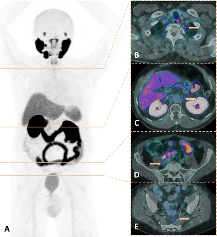
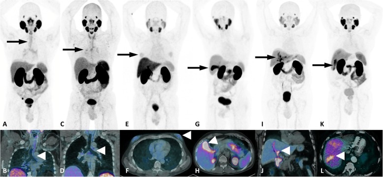
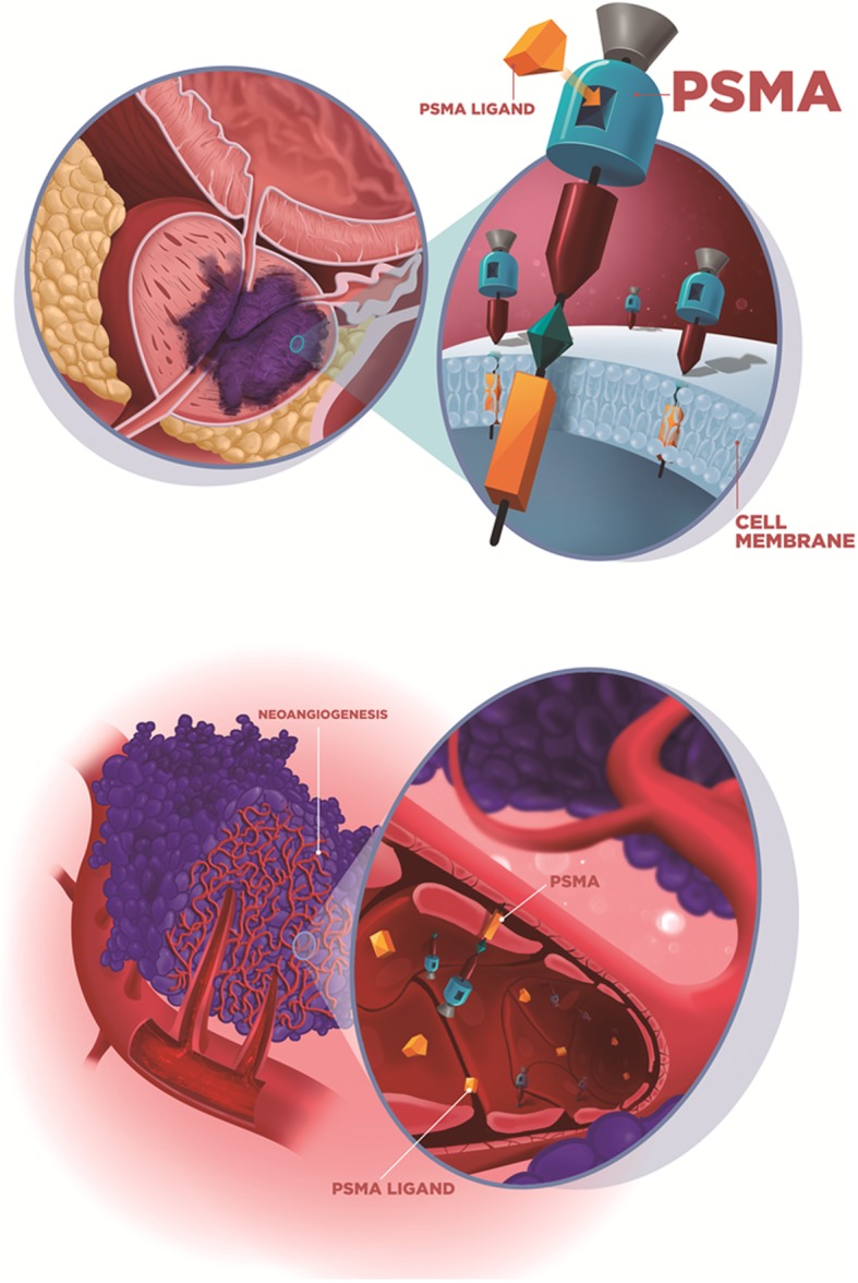
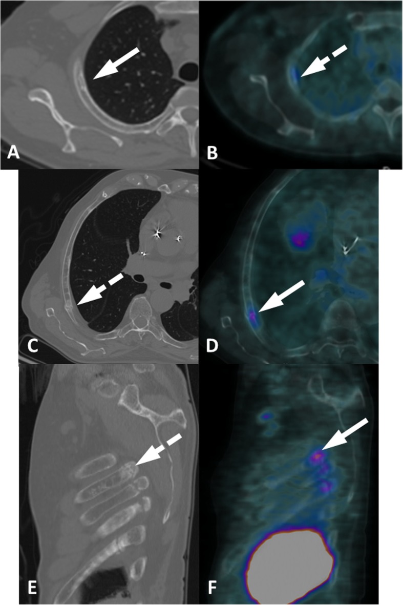
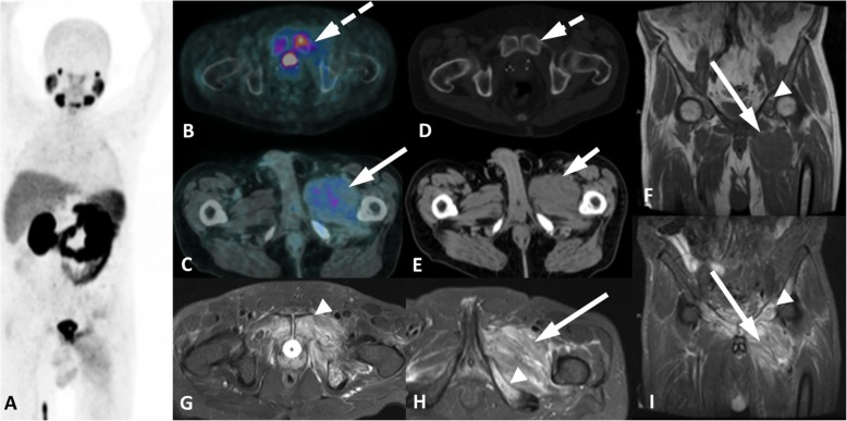
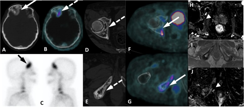
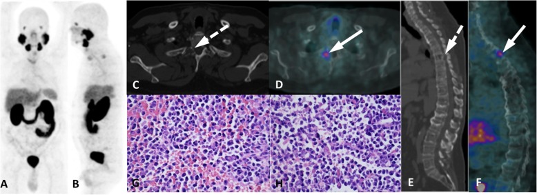
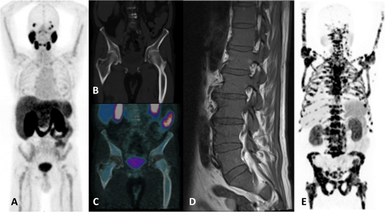
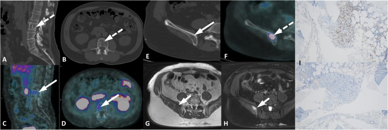
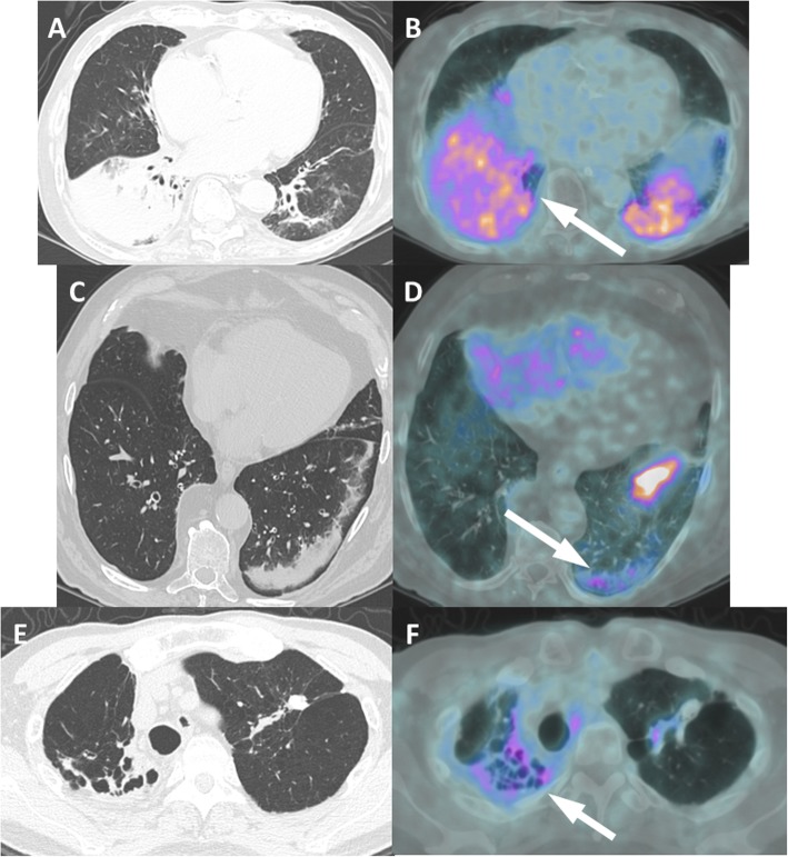
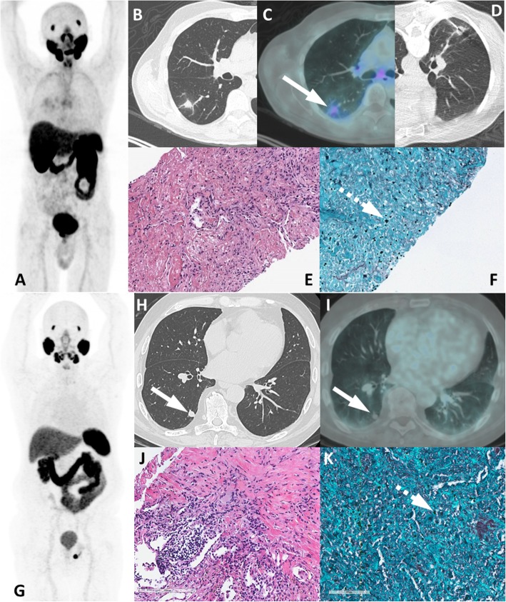
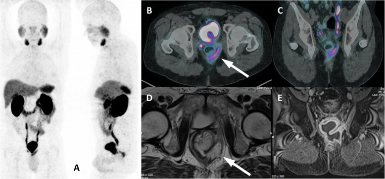
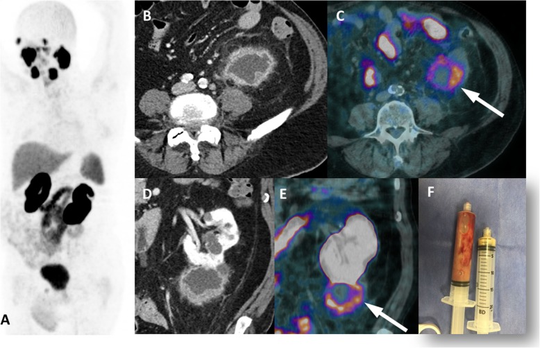
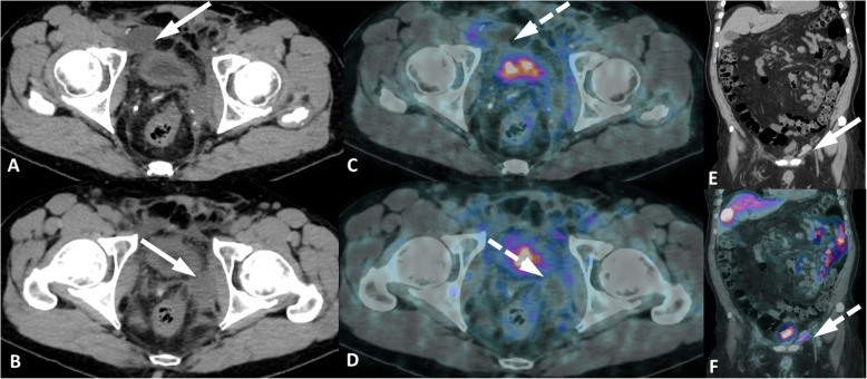
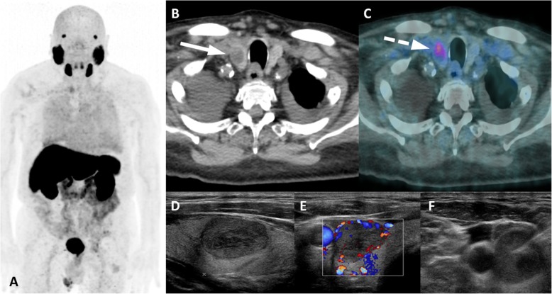
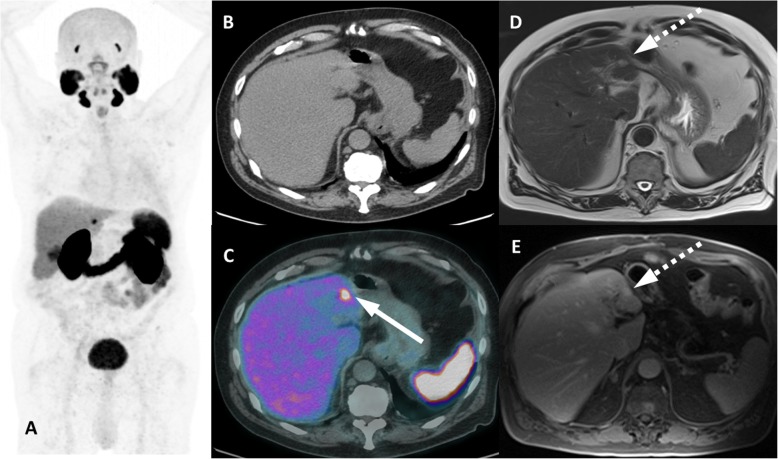
PSMA PET imaging was originally used to assess biochemical recurrence of prostate cancer (PCa), but its clinical use was promptly extended to detection, staging and therapy response assessment. The expanding use of PSMA PET worldwide has also revealed PSMA ligand uptake in diverse nonprostatic diseases, which raised questions about the specificity of this imaging modality. Although not very common initially, a growing number of pathologies presenting PSMA uptake on PET have been reported in the last few years, and a proper interpretation of PSMA PET imaging findings suddenly became challenging and, to some extent, confusing. Compared to cytoplasmic PSMA expression in nonprostatic cells, the molecular features of apical PSMA expression in PCa cells can help to distinguish these various conditions. Correlations of imaging findings to patient history, to the expected pattern of disease spread and mainly to computed tomography (CT) and/or magnetic resonance imaging (MRI) characteristics will reinforce the distinction of lesions that are more likely related to PCa from those that could lead to an incorrect diagnosis. The overall benefits of endothelial PSMA expression, which is associated with the neovasculature of malignant neoplasms, will be highlighted, stating the potential use of PSMA ligand uptake as a theranostic tool. This review aims to cover the collection of nonprostatic diseases, including benign and malignant tumors, in a didactic approach according to disease etiology, with discussion of bone-related conditions and inflammatory and infectious processes.
Overview
The claims and statistics of this paper haven’t been checked yet.
Run the check — free, no account needed. It reads the open-access text and checks the sources it cites.
Integrity signals
Registry
Cites
Statistics
Figures
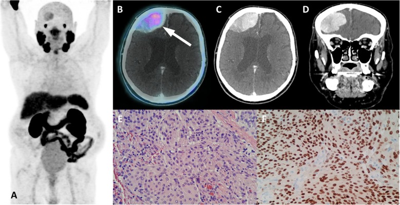
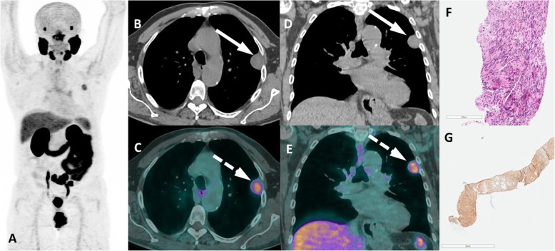
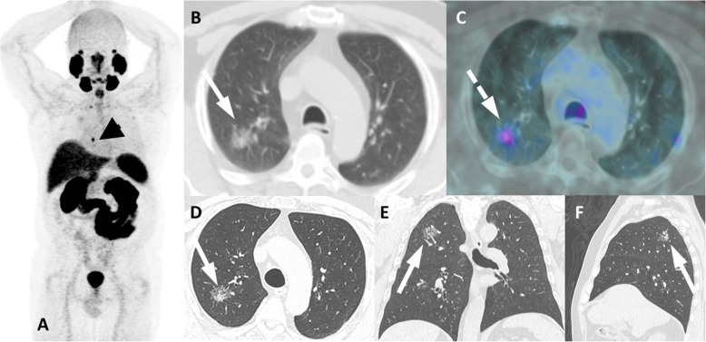
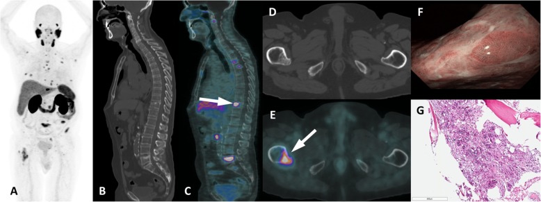
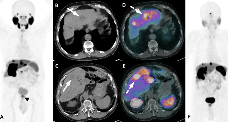
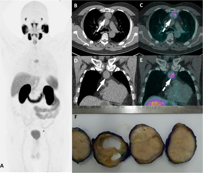
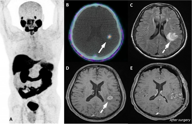
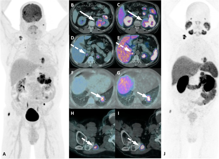
Verification
Claims the paper makes
The claims and statistics of this paper haven’t been checked yet.
Run the check — free, no account needed. It reads the open-access text and checks the sources it cites.
Claims other papers make about this one
Each is checked against this paper’s own text: does it say what the citing paper says it does?
The source backs part of the claim, not all of it — or backs it for a different population, condition or comparison.
In the citing paper
“However, sources of false positive or negative findings still remain with these tracers [131,132,133,134] and must be considered during image interpretation.”
From Influences on PET Quantification and Interpretation (2022) · cites this paper as [132]
Experimental — accuracy not yet measured
Self-check
Percentages, sums and reported differences re-computed from the paper’s own numbers.
The self-check needs the paper’s full text.
Citations
Cites
Perera, M., Papa, N., Roberts, M., Williams, M., Udovicich, C., Vela, I., Christidis, D., Bolton, D., Hofman, M. S., Lawrentschuk, N., et al. (2020) · European Urology · doi:10.1016/j.eururo.2019.01.049 · Cited by 958
Chang, S. S., Reuter, V. E., Heston, W. D. W., Bander, N. H., Grauer, L. S., & Gaudin, P. B. (1999) · PubMed · Cited by 807
Giesel, F. L., Hadaschik, B. A., Cardinale, J., Radtke, J. P., Vinsensia, M., Lehnert, W., Kesch, C. V., Tolstov, Y. L., Singer, S., Grabe, N., et al. (2016) · European Journal of Nuclear Medicine and Molecular Imaging · doi:10.1007/s00259-016-3573-4 · Cited by 595
Prostate-specific membrane antigen is produced in tumor-associated neovasculature.
Chang, S. S., O’Keefe, D. S., Bacich, D. J., Reuter, V. E., Heston, W. D. W., & Gaudin, P. B. (1999) · PubMed · Cited by 465
Constitutive and antibody-induced internalization of prostate-specific membrane antigen.
Liu, H., Rajasekaran, A. K., Moy, P., Xia, Y., Kim, S., Navarro, V., Rahmati, R., & Bander, N. H. (1998) · PubMed · Cited by 446
Hofman, M. S., Hicks, R. J., Maurer, T., & Eiber, M. J. (2018) · Radiographics · doi:10.1148/rg.2018170108 · Cited by 393
Horoszewicz, J. S., KAWINSKI, E., & Murphy, G. P. (1993) · Biotechnology Advances · doi:10.1016/0734-9750(93)90273-p · Cited by 379
Carter, R., Feldman, A., & Coyle, J. T. (1996) · Proceedings of the National Academy of Sciences · doi:10.1073/pnas.93.2.749 · Cited by 374
Ost, P., Bossi, A., Decaestecker, K., De Meerleer, G., Giannarini, G., Karnes, R. J., Roach, M., & Briganti, A. (2015) · European Urology · doi:10.1016/j.eururo.2014.09.004 · Cited by 349
Sheikhbahaei, S., Afshar‐Oromieh, A., Eiber, M. J., Sólnes, L. B., Javadi, M. S., Ross, A. E., Pienta, K. J., Allaf, M. E., Haberkorn, U., Pomper, M. G., et al. (2017) · European Journal of Nuclear Medicine and Molecular Imaging · doi:10.1007/s00259-017-3780-7 · Cited by 299
Mhawech-Fauceglia, P. Y., Zhang, S., Terracciano, L., Sauter, G., Chadhuri, A., Herrmann, F. R., & Penetrante, R. B. (2007) · Histopathology · doi:10.1111/j.1365-2559.2007.02635.x · Cited by 299
Haffner, M. C., Kronberger, I. E., Ross, J. S., Sheehan, C. E., Zitt, M., Mühlmann, G., Öfner, D., Zelger, B. G., Ensinger, C., Yang, X. J., et al. (2009) · Human Pathology · doi:10.1016/j.humpath.2009.06.003 · Cited by 239
Lowe, A. D., Bawazeer, S., Watson, D. G., McGill, S., Burchmore, R., Pomeroy, P. P., & Kennedy, M. W. (2017) · Scientific Reports · doi:10.1038/s41598-017-16187-7 · Cited by 231
Musculoskeletal Fibromatoses: Radiologic-Pathologic Correlation
Murphey, M. D., Ruble, C. M., Tyszko, S. M., Zbojniewicz, A. M., Potter, B. K., & Miettinen, M. (2009) · Radiographics · doi:10.1148/rg.297095138 · Cited by 231
Rajasekaran, S. A., Anilkumar, G., Oshima, E., Bowie, J. U., Liu, H., Heston, W., Bander, N. H., & Rajasekaran, A. K. (2003) · Molecular Biology of the Cell · doi:10.1091/mbc.e02-11-0731 · Cited by 226 · cited 1×
“In contrast with other clinically relevant prostate-related antigens, such as prostate-specific antigen or prostatic acid phosphatase, which are secretory proteins, the PSMA transmembrane conformational structure enables it to exhibit an internalization functionality by means of endosomal complexes, which is a highly…”
BackgroundAggressive Fibromatosis: MRI Features with Pathologic Correlation
Lee, J. C., Thomas, J. M., Phillips, S., Fisher, C., & Moskovic, E. C. (2005) · American Journal of Roentgenology · doi:10.2214/ajr.04.1674 · Cited by 217
Jeong, Y. J., Lee, K. S., Kim, J., Shim, Y. M., Han, J., & Kwon, O. (2004) · American Journal of Roentgenology · doi:10.2214/ajr.183.2.1830283 · Cited by 206
Computed tomography and magnetic resonance imaging of mediastinal tumors
Takahashi, K., & Al‐Janabi, N. J. (2010) · Journal of Magnetic Resonance Imaging · doi:10.1002/jmri.22377 · Cited by 203
O’Keefe, D. S., Bacich, D. J., Huang, S. S.-L., & Heston, W. D. W. (2018) · Journal of Nuclear Medicine · doi:10.2967/jnumed.117.203877 · Cited by 197
PSMA Theranostics: Current Status and Future Directions
Rahbar, K., Afshar‐Oromieh, A., Jadvar, H., & Ahmadzadehfar, H. (2018) · Molecular Imaging · doi:10.1177/1536012118776068 · Cited by 180
Normal distribution pattern and physiological variants of 68Ga-PSMA-11 PET/CT imaging
Demirci, E., Şahin, O. E., Ocak, M., Akovalı, B., Nematyazar, J., & Kabasakal, L. (2016) · Nuclear Medicine Communications · doi:10.1097/mnm.0000000000000566 · Cited by 167
Fragomeni, R. A. S., Amir, T., Sheikhbahaei, S., Harvey, S. C., Javadi, M. S., Sólnes, L. B., Kiess, A. P., Allaf, M. E., Pomper, M. G., Gorin, M. A., et al. (2018) · Journal of Nuclear Medicine · doi:10.2967/jnumed.117.203570 · Cited by 158
Targeting PSMA by radioligands in non-prostate disease—current status and future perspectives
Backhaus, P., Noto, B., Avramović, N., Grubert, L. S., Huss, S., Bögemann, M., Stegger, L., Weckesser, M., Schäfers, M. A., & Rahbar, K. (2018) · European Journal of Nuclear Medicine and Molecular Imaging · doi:10.1007/s00259-017-3922-y · Cited by 143
Rhee, H., Blazak, J., Tham, C. M., Ng, K. L., Shepherd, B., Lawson, M., Preston, J. M., Vela, I., Thomas, P. A., & Wood, S. (2016) · EJNMMI Research · doi:10.1186/s13550-016-0231-6 · Cited by 138
Pyka, T., Weirich, G., Einspieler, I., Maurer, T., Theisen, J., Hatzichristodoulou, G., Schwamborn, K., Schwaiger, M., & Eiber, M. J. (2015) · Journal of Nuclear Medicine · doi:10.2967/jnumed.115.164442 · Cited by 133
Metastatic renal cell carcinoma neovasculature expresses prostate-specific membrane antigen
Chang, S. S., Reuter, V. E., Heston, W. D. W., & Gaudin, P. B. (2001) · Urology · doi:10.1016/s0090-4295(00)01094-3 · Cited by 131
Rischpler, C., Beck, T. I., Okamoto, S., Schlitter, A. M., Knorr, K., Schwaiger, M., Gschwend, J. E., Maurer, T., Meyer, P. T., & Eiber, M. J. (2018) · Journal of Nuclear Medicine · doi:10.2967/jnumed.117.204677 · Cited by 128
68Ga-PSMA-HBED-CC PET imaging in breast carcinoma patients
Sathekge, M. M., Lengana, T., Modiselle, M., Vorster, M., Zeevart, J., Maes, A., Ebenhan, T., & de Wiele, C. V. (2016) · European Journal of Nuclear Medicine and Molecular Imaging · doi:10.1007/s00259-016-3563-6 · Cited by 120
Pitfalls in Gallium-68 PSMA PET/CT Interpretation—A Pictorial Review
Shetty, D., Patel, D., Le, K., Bui, C., & Mansberg, R. (2018) · Tomography · doi:10.18383/j.tom.2018.00021 · Cited by 116
Siva, S., Callahan, J., Pryor, D. I., Martin, J. M., Lawrentschuk, N., & Hofman, M. S. (2017) · Journal of Medical Imaging and Radiation Oncology · doi:10.1111/1754-9485.12590 · Cited by 103
Barbosa, F. D. G., Queiroz, M. A., Nunes, R. F., Viana, P. C. C., Marin, J. F. G., Cerri, G. G., & Buchpiguel, C. A. (2019) · Radiographics · doi:10.1148/rg.2019180079 · Cited by 100
Self-citationPrasad, V., Steffen, I. G., Diederichs, G., Makowski, M. R., Wust, P., & Brenner, W. (2016) · Molecular Imaging and Biology · doi:10.1007/s11307-016-0945-x · Cited by 97
PSMA expression by microvasculature of thyroid tumors – Potential implications for PSMA theranostics
Bychkov, A., Vutrapongwatana, U., Tepmongkol, S., & Keelawat, S. (2017) · Scientific Reports · doi:10.1038/s41598-017-05481-z · Cited by 97
Late-onset deep mesh infection after inguinal hernia repair
Delikoukos, S., Tzovaras, G., Liakou, P., Mantzos, F., & Hatzitheofilou, C. (2007) · Hernia · doi:10.1007/s10029-006-0131-1 · Cited by 89
Janssen, J.-C., Meißner, S., Woythal, N., Prasad, V., Brenner, W., Diederichs, G., Hamm, B., & Makowski, M. R. (2017) · European Radiology · doi:10.1007/s00330-017-4994-6 · Cited by 72
Rischpler, C., Maurer, T., Schwaiger, M., & Eiber, M. J. (2015) · European Journal of Nuclear Medicine and Molecular Imaging · doi:10.1007/s00259-015-3235-y · Cited by 69
Follicular Lymphoma Showing Avid Uptake on 68Ga PSMA-HBED-CC PET/CT
Kanthan, G. L., Coyle, L. A., Kneebone, A. B., Schembri, G. P., & Hsiao, E. (2016) · Clinical Nuclear Medicine · doi:10.1097/rlu.0000000000001169 · Cited by 65
Verma, P., Malhotra, G., Goel, A. H., Rakshit, S., Chandak, A., Chedda, R., Banerjee, S., & Asopa, R. V. (2019) · Clinical Nuclear Medicine · doi:10.1097/rlu.0000000000002520 · Cited by 60
Nulent, T. J. W. K., van Es, R. J. J., Krijger, G. C., de Bree, R., Willems, S. M., & de Keizer, B. (2017) · European Journal of Nuclear Medicine and Molecular Imaging · doi:10.1007/s00259-017-3737-x · Cited by 59
Vertebral Hemangioma Mimicking Bone Metastasis in 68Ga—PSMA Ligand PET/CT
Artigas, C., Otte, F.-X., Lemort, M., van Velthoven, R., & Flamen, P. (2017) · Clinical Nuclear Medicine · doi:10.1097/rlu.0000000000001631 · Cited by 56
Osman, M., Iravani, A., Hicks, R. J., & Hofman, M. S. (2017) · Journal of Nuclear Medicine · doi:10.2967/jnumed.117.190215 · Cited by 53
PSMA expression in Schwannoma: A potential clinical mimicker of metastatic prostate carcinoma
Wang, W., Tavora, F. R. F., Sharma, R. B., Eisenberger, M., & Netto, G. J. (2008) · Urologic Oncology Seminars and Original Investigations · doi:10.1016/j.urolonc.2008.04.007 · Cited by 52
Prostate-Specific Membrane Antigen PET/CT Findings for Hepatic Hemangioma
Bhardwaj, H., Stephens, M., Bhatt, M. K., & Thomas, P. A. (2016) · Clinical Nuclear Medicine · doi:10.1097/rlu.0000000000001384 · Cited by 51
Prostate-Specific Membrane Antigen PET/CT: False-Positive Results due to Sarcoidosis
Hermann, R. M., Djannatian, M., Czech, N., & Nitsche, M. (2016) · Case Reports in Oncology · doi:10.1159/000447688 · Cited by 49
Mesh migration following abdominal hernia repair: a comprehensive review
Cunningham, H. B., Weis, J. J., Taveras, L. R., & Huerta, S. (2019) · Hernia · doi:10.1007/s10029-019-01898-9 · Cited by 48
Metastases From Colorectal Cancer Avid on 68Ga-PSMA PET/CT
Hangaard, L., Jochumsen, M. R., Vendelbo, M. H., & Bouchelouche, K. (2017) · Clinical Nuclear Medicine · doi:10.1097/rlu.0000000000001700 · Cited by 46
68Ga-Labeled PSMA Uptake in Nonprostatic Malignancies
Malik, D., Kumar, R., Mittal, B. R., Singh, H., Bhattacharya, A., & Singh, S. K. (2018) · Clinical Nuclear Medicine · doi:10.1097/rlu.0000000000002103 · Cited by 46
68Ga-PSMA Uptake in Hepatocellular Carcinoma
Taneja, S., Taneja, R., Kashyap, V., Jha, A., & Jena, A. (2016) · Clinical Nuclear Medicine · doi:10.1097/rlu.0000000000001355 · Cited by 43
Schwannoma Showing Avid Uptake on 68Ga-PSMA-HBED-CC PET/CT
Kanthan, G. L., Izard, M. A., Emmett, L., Hsiao, E., & Schembri, G. P. (2016) · Clinical Nuclear Medicine · doi:10.1097/rlu.0000000000001281 · Cited by 36
Jochumsen, M. R., Dias, A. H., & Bouchelouche, K. (2017) · Clinical Nuclear Medicine · doi:10.1097/rlu.0000000000001871 · Cited by 32
Janssen, J.-C., Woythal, N., Meißner, S., Prasad, V., Brenner, W., Diederichs, G., Hamm, B., & Makowski, M. R. (2017) · Molecular Imaging and Biology · doi:10.1007/s11307-017-1101-y · Cited by 30
Subcutaneous Lobular Capillary Hemangioma on 68Ga-PSMA PET/CT
Jochumsen, M. R., Vendelbo, M. H., Høyer, S., & Bouchelouche, K. (2017) · Clinical Nuclear Medicine · doi:10.1097/rlu.0000000000001542 · Cited by 29
Strong PSMA Radioligand Uptake by Rectal Carcinoma
Stoykow, C., Huber-Schumacher, S., Almanasreh, N., Jilg, C. A., & Ruf, J. (2016) · Clinical Nuclear Medicine · doi:10.1097/rlu.0000000000001484 · Cited by 27
Parihar, A. S., Vadi, S. K., Mittal, B. R., Kumar, R., Bal, A., & Singh, S. K. (2018) · Clinical Nuclear Medicine · doi:10.1097/rlu.0000000000002172 · Cited by 25
Radiolabeled prostate-specific membrane antigen small-molecule inhibitors
Will, L., Sonni, I., Kopka, K., Kratochwil, C., Giesel, F. L., & Haberkorn, U. (2017) · The Quarterly Journal of Nuclear Medicine and Molecular Imaging · doi:10.23736/s1824-4785.17.02977-6 · Cited by 24
Malik, D., Mittal, B. R., Kumar, R., Singh, H., Bhattacharya, A., & Singh, S. K. (2018) · Clinical Nuclear Medicine · doi:10.1097/rlu.0000000000002189 · Cited by 21
Vadi, S. K., Kumar, R., Singh, H., Singh, S. K., & Mittal, B. R. (2018) · Clinical Nuclear Medicine · doi:10.1097/rlu.0000000000001990 · Cited by 20
Intense 68Ga-PSMA Uptake in Diverticulum of the Sigmoid Colon
Jochumsen, M. R., & Bouchelouche, K. (2017) · Clinical Nuclear Medicine · doi:10.1097/rlu.0000000000001927 · Cited by 19
Gastric GIST Incidentally Detected on 68Ga-PSMA-PET/CT
Vaz, S. C., Oliveira, C., Castanheira, J. C., Silva, Â. F., & Costa, D. C. (2018) · Clinical Nuclear Medicine · doi:10.1097/rlu.0000000000002347 · Cited by 19
68Ga-PSMA Uptake in Angiolipoma
Dekker, I., van der Leest, M., van Rijk, M. C., Gerritsen, W. R., & Arens, A. I. J. (2018) · Clinical Nuclear Medicine · doi:10.1097/rlu.0000000000002236 · Cited by 18
Ahuja, A., Taneja, S., Thorat, K., & Jena, A. (2017) · Clinical Nuclear Medicine · doi:10.1097/rlu.0000000000001874 · Cited by 15
68Ga-PSMA-Ligand PET/CT Uptake in Anthracosilicosis
McGuiness, M., & Sounness, B. (2017) · Clinical Nuclear Medicine · doi:10.1097/rlu.0000000000001782 · Cited by 15
Itabashi, T., Emori, M., Terashima, Y., Hasegawa, T., Shimizu, J., Nagoya, S., & Yamashita, T. (2017) · Acta Radiologica Open · doi:10.1177/2058460117728416 · Cited by 15
68Ga-PSMA Uptake in Anal Fistula on PET/CT Scan
Klingenberg, S., Jochumsen, M. R., Nielsen, T. F., & Bouchelouche, K. (2018) · Clinical Nuclear Medicine · doi:10.1097/rlu.0000000000002370 · Cited by 15
Pitfalls in Ga-68-PSMA-PET/CT: incidental finding of parathyroid adenoma
Pfob, C. H., Karimov, I., Jesinghaus, M., Novotny, A., Weber, W., Eiber, M., & Feuerecker, B. (2018) · European Journal of Nuclear Medicine and Molecular Imaging · doi:10.1007/s00259-018-4239-1 · Cited by 14
Angiolipoma: a rare benign soft tissue tumor with a malignant arteriographic appearance
Finberg, H. J., & Dc, L. (1977) · American Journal of Roentgenology · doi:10.2214/ajr.128.4.697 · Cited by 13
Splenic Hemangioma as a Potential Pitfall on PSMA-Targeted 18F-DCFPyL PET/CT
Chaussé, G., Laufer, J., Abikhzer, G., & Probst, S. (2019) · Clinical Nuclear Medicine · doi:10.1097/rlu.0000000000002428 · Cited by 12
Krishnaraju, V. S., Basher, R. K., Singh, H., Singh, S. K., Bal, A., & Mittal, B. R. (2018) · Clinical Nuclear Medicine · doi:10.1097/rlu.0000000000002000 · Cited by 9
Shenderov, E., Gorin, M. A., Kim, S., Johnson, P. T., Allaf, M. E., Partin, A. W., Pomper, M. G., Antonarakis, E. S., Pienta, K. J., & Rowe, S. P. (2017) · Urology Case Reports · doi:10.1016/j.eucr.2017.12.011 · Cited by 9
Patro, K. C., Palla, M., & Kashyap, R. K. (2018) · Clinical Nuclear Medicine · doi:10.1097/rlu.0000000000002203 · Cited by 9
Gorin, M. A., Marashdeh, W. M., Ross, A. E., Allaf, M. E., Pienta, K. J., Pomper, M. G., & Rowe, S. P. (2017) · Nuclear Medicine Communications · doi:10.1097/mnm.0000000000000716 · Cited by 8
68Ga-PSMA Uptake in Middle Lobe Syndrome
Pedersen, M. A., Bouchelouche, K., & Vendelbo, M. H. (2018) · Clinical Nuclear Medicine · doi:10.1097/rlu.0000000000002213 · Cited by 5
Orbital Hemangiopericytoma in 68Ga–Prostate-Specific Membrane Antigen–HBED–CC PET/CT
Hammes, J., Kobe, C., Hilgenberg, U., Lieb, W., & Drzezga, A. E. (2017) · Clinical Nuclear Medicine · doi:10.1097/rlu.0000000000001791 · Cited by 5
Prostate-Specific Membrane Antigen–Positive Manifestations of Chronic Beryllium Lung Disease
Huang, K., Baur, A. D., & Furth, C. (2018) · Clinical Nuclear Medicine · doi:10.1097/rlu.0000000000002323 · Cited by 5
Research works printed but not linked: 1
- Ruf J, Huber-Schumacher S, Stoykow C, Almanasreh N, Jilg C. Strong PSMA Radioligand uptake by rectal carcinoma. Clin Nucl Med. 2016;42:225–6.
Cited by
Narayan, V., Barber-Rotenberg, J. S., Jung, I.-Y., Lacey, S. F., Rech, A. J., Davis, M. M., Hwang, W.-T., Lal, P., Carpenter, E. L., Maude, S. L., et al. (2022) · Nature Medicine · doi:10.1038/s41591-022-01726-1 · Cited by 533
Chow, K. M., So, W. Z., Lee, H. J., Lee, A. Y. M., Yap, D. W. T., Takwoingi, Y., Tay, K. J., Tuan, J. K. L., Thang, S. P., Lam, W. W.-C., et al. (2023) · European Urology · doi:10.1016/j.eururo.2023.03.001 · Cited by 195
PSMA-Targeting Imaging and Theranostic Agents—Current Status and Future Perspective
Debnath, S., Zhou, N., McLaughlin, M. F., Rice, S. L., Pillai, A. K., Hao, G., & Sun, X. (2022) · International Journal of Molecular Sciences · doi:10.3390/ijms23031158 · Cited by 97
PSMA PET in Imaging Prostate Cancer
Tsechelidis, I., & Vrachimis, A. (2022) · Frontiers in Oncology · doi:10.3389/fonc.2022.831429 · Cited by 75
Best Approaches and Updates for Prostate Cancer Biochemical Recurrence
Simon, N. I., Parker, C. C., Hope, T. A., & Paller, C. J. (2022) · American Society of Clinical Oncology Educational Book · doi:10.1200/edbk_351033 · Cited by 70
Metadata
- Authors
- de Galiza Barbosa, F., Queiroz, M. A., Nunes, R. F., Costa, L. B., Zaniboni, E. C., Marin, J. F. G., Cerri, G. G., & Buchpiguel, C. A.
- Affiliations
- Hospital Sírio-Libanês (BR)
- Corresponding author
- Queiroz, Marcelo Araujo
- ORCID
- Venue
- Cancer Imaging
- Published
- 2020-03-14
- Volume
- 20
- Issue
- 1
- Pages
- 23-23
- ISSN
- 1470-7330 (ISSN-L)
- PMC
- PMC7071711
- OpenAlex
- W3011546611
- Cited by
- 288
- References
- 75
- Citations by year
- 2026: 27 · 2025: 48 · 2024: 67 · 2023: 53 · 2022: 51 · 2021: 27 · 2020: 15
- FWCI
- 5.38
- Type
- journal article
- Language
- en
- Open access
- gold
- doi.org
- Licence
- cc-by
- creativecommons.org/licenses/by/4.0/ (tdm)
- creativecommons.org/licenses/by/4.0/ (vor)
- Locations
- Cancer Imaging (publishedVersion, cc-by)
- PubMed Central (publishedVersion, cc-by)
- PubMed (publishedVersion)
- DOAJ (DOAJ: Directory of Open Access Journals) (submittedVersion, cc-by-sa)
- Topics
- Health Sciences › Medicine › Radiology, Nuclear Medicine and Imaging › Medical Imaging Techniques and Applications
- Health Sciences › Medicine › Radiology, Nuclear Medicine and Imaging › Radiopharmaceutical Chemistry and Applications
- Health Sciences › Medicine › Urology › Prostate Cancer Diagnosis and Treatment
- Keywords
- PSMA PET, prostate-specific membrane antigen, prostate cancer, theranostics, benign and malignant tumors
- MeSH terms
- Positron Emission Tomography Computed Tomography, Gallium Isotopes, Gallium Radioisotopes, Humans, Membrane Glycoproteins, Neoplasms, Organometallic Compounds, Sensitivity and Specificity, Radiopharmaceuticals
About these checks
Registry, retraction, Cites and statistics results are computed, not judged, with one exception: a printed reference that no index lists is read once by a model before it counts as a citation error. Claims are extracted by a model and kept only when their quote appears verbatim in the text; each cited source is then checked, with a model’s help, for whether it says what the claim relies on. Those verdicts can be wrong — read the claim’s quote and the cited source before relying on one.
Statistics and arithmetic are recomputed, not judged.
Confidence bands are shown only for fields calibrated on our annotated test set. None is calibrated yet, so no band is shown.
What counts as a claim ›How we measure confidence ›How we check this ›
Are you an author, or do you see a problem with this page? Request a review
Data sources
Bibliographic data: Semantic Scholar, OpenAlex, Crossref.
Contains information from the Semantic Scholar Academic Graph Datasets, made available under the ODC Attribution License (ODC-BY 1.0).
Semantic Scholar’s data is described in: Kinney, R., et al. (2023). The Semantic Scholar Open Data Platform. arXiv:2301.10140.
