

The DOI matches its Crossref record; 209 of the 209 references it cites were found in a registry or an index.
ApoE expression across the CNS: Who, What, Where, When, and How (much)?
Open accessAbstract
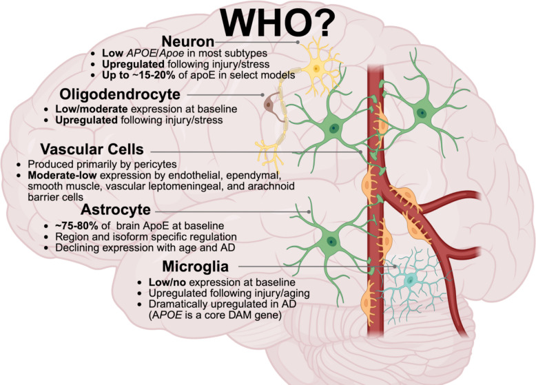
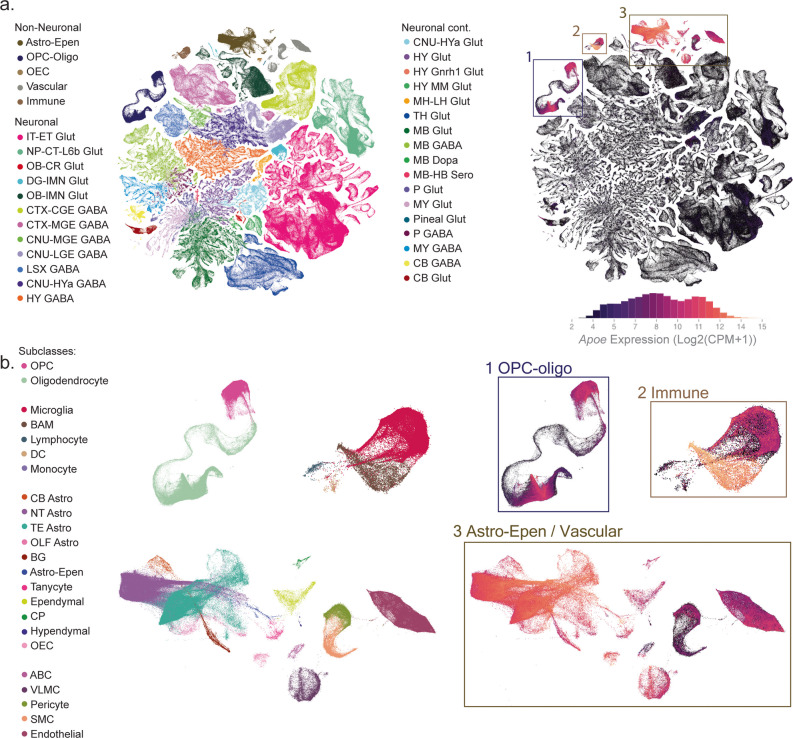
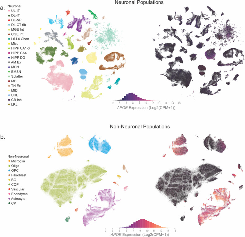
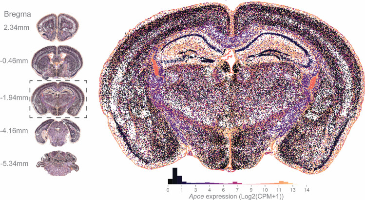
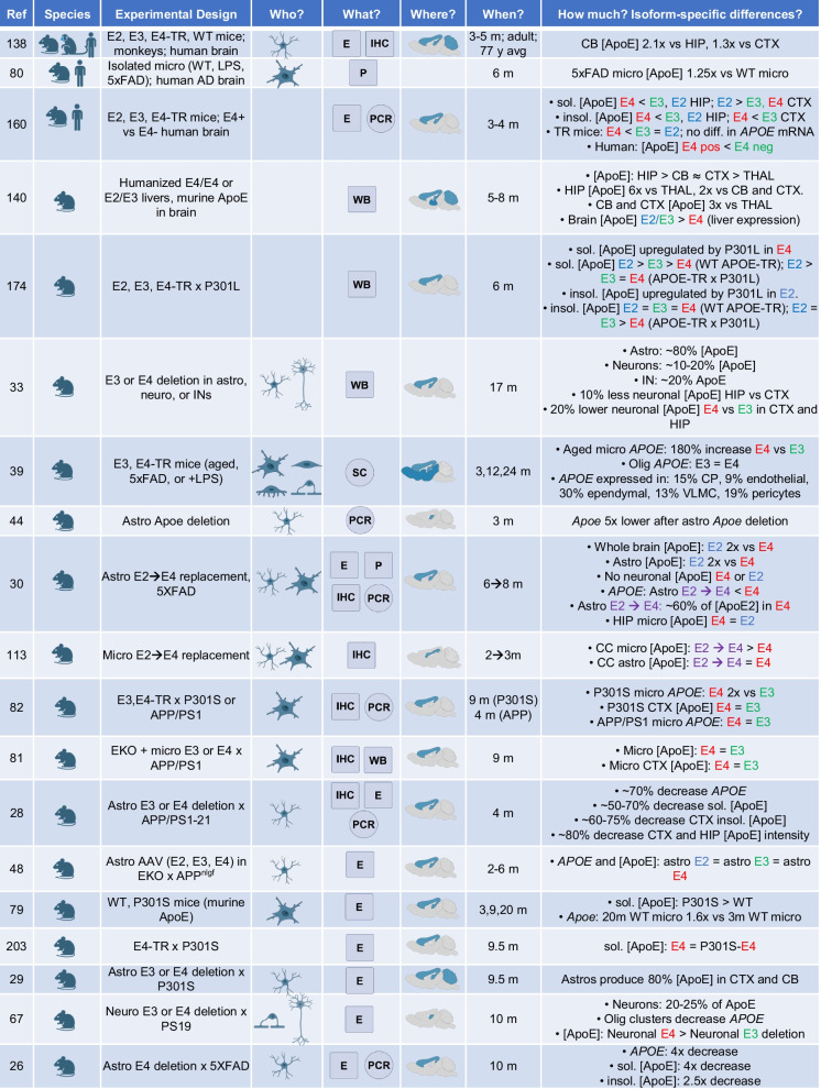
Apolipoprotein E ( APOE ) is the strongest genetic risk factor for late-onset Alzheimer’s disease (AD) and encodes a major lipid transporter in the central nervous system (CNS). Although apoE has been studied extensively, fundamental questions remain regarding its expression across the CNS. For example, which CNS cell types express APOE ? Where is APOE expression enriched across brain regions? When is APOE dynamically regulated – across development, aging, injury, and neurodegeneration? How much apoE is produced at the mRNA and protein level – particularly across the common E2, E3, and E4 isoforms? Here, we synthesize transcriptomic, spatial, biochemical, and genetic evidence to provide an organized framework for the “who, what, where, when, and how (much)” of CNS APOE /apoE. By assessing large-scale single-cell datasets along with in vivo and cell-type–specific genetic studies, we highlight that astrocytes are the predominant source of CNS apoE at baseline, while microglia, oligodendrocyte-lineage cells, vascular-associated populations, and border-interface macrophages exhibit context-dependent induction that is strongly influenced by aging, injury, and AD-related pathology. We also evaluate unresolved discrepancies, such as the extent of neuronal APOE expression and its contribution to total brain apoE, and describe how – across modalities – apoE abundance often follows an isoform-dependent hierarchy (typically E2 > E3 > E4 at the protein level) while transcript–protein discordance and model-specific effects underscore substantial post-transcriptional and contextual regulation. Finally, we discuss how clarifying spatiotemporal, cell-specific, and isoform-dependent apoE expression will enable better informed and more precise apoE-targeted therapeutic strategies.
Overview
The claims and statistics of this paper haven’t been checked yet.
Run the check — free, no account needed. It reads the open-access text and checks the sources it cites.
Integrity signals
Registry
Cites
Statistics
Figures
Verification
Claims the paper makes
The claims and statistics of this paper haven’t been checked yet.
Run the check — free, no account needed. It reads the open-access text and checks the sources it cites.
Self-check
Percentages, sums and reported differences re-computed from the paper’s own numbers.
The self-check needs the paper’s full text.
Citations
Cites
Items 201–209 of 209
APOE genotype-dependent differences in human astrocytic energy metabolism
Budny, V., Bodenmann, C., Zürcher, K. J., Krüger, M., de Leeuw, S. M., Weber, R. Z., Rust, R., Ravotto, L., Ruminot, I., Barros, L. F., et al. (2025) · Frontiers in Cellular Neuroscience · doi:10.3389/fncel.2025.1603657 · Cited by 6
Al‐Amin, M. M., Kim, B., Karahan, H., Tate, M. D., Walsh, S. P., Puntambekar, S. S., Bissel, S. J., Lamb, B. T., Wang, N., & Kim, J. (2025) · Alzheimer's & Dementia · doi:10.1002/alz.70791 · Cited by 6
Lipid droplets in astrocytes: Key organelles for CNS homeostasis and disease (Review)
Wang, Y., Wang, B., Hou, J., Huo, X., Liu, C., Guan, R., Chen, H., Zhou, Y., Zhang, J., Zhuang, C., et al. (2025) · International Journal of Molecular Medicine · doi:10.3892/ijmm.2025.5691 · Cited by 5
Microglia-derived APOE2 improves remyelination even in the presence of endogenous APOE4
Nolt, G. L., Golden, L. R., Thorpe, S. P., Funnell, J. L., Stephens, I. O., Hernandez, G., MacLean, S. M., Lucido, C. C., Brock, C. R., Pallerla, A. V., et al. (2025) · Journal of Neuroinflammation · doi:10.1186/s12974-025-03639-5 · Cited by 5
Self-citationden Hoedt, S., Dorst‐Lagerwerf, K. Y., de Vries, H. E., Rozemüller, A. J. M., Scheltens, P., Walter, J., Sijbrands, E. J. G., Martínez‐Martínez, P., Verhoeven, A. J. M., Teunissen, C. E., et al. (2023) · Journal of Alzheimer s Disease Reports · doi:10.3233/adr220072 · Cited by 4
Gim, J. (2025) · Journal of Alzheimer s Disease · doi:10.1177/13872877251366662 · Cited by 3
Akay, L. A., Bright, A., Boix, C. A., Louderback, K., Medrano, J. A., Sun, D., Lin, H., King, O., Welch, G., Nawaid, H., et al. (2025) · bioRxiv (Cold Spring Harbor Laboratory) · doi:10.1101/2025.10.16.682845 · Cited by 3
Kim, M. J., Blumenfeld, J. R., Li, Y., Yip, O., Yao, L., Ancheta, S., Leon, S. D., Platow, Z., Liang, Z., Shostak, D. M., et al. (2025) · bioRxiv (Cold Spring Harbor Laboratory) · doi:10.64898/2025.12.04.692390 · Cited by 1
Cuní-López, C., Root, J. T., Hao, Y., Blomberg, N., Koppes-den Hertog, S. J., Sacks, D., Trujillo, A. E., Ghirlando, R., Kowal, I., Epstein, J., et al. (2026) · Nature Communications · doi:10.1038/s41467-026-76565-6
Research works printed but not linked: 1
- Iadecola C, et al. Cell autonomous role of border associated macrophages in ApoE4 neurovascular dysfunction and susceptibility to white matter injury. Res Sq. 2023. · DOI
Cited by
Sorted by citations
Heart and Brain: Linking Obesity-Related Inflammation, Cardiovascular Disease and Cognitive Function
Reiss, A. B., Gulkarov, S., Syed, U., Ben-Dor, G. A., Srivastava, A., Singh, M., & Leon, J. D. (2026) · Journal of Cardiovascular Development and Disease · doi:10.3390/jcdd13100477
Gelperina, S. E., & Wacker, M. G. (2026) · Expert Opinion on Drug Delivery · doi:10.1080/17425247.2026.2755180
Metadata
- Authors
- Nolt, G. L., Lucido, C. C., Smith, L. M., & Johnson, L. A.
- Affiliations
- University of Kentucky (US)
- Corresponding author
- Johnson, Lance A.
- Venue
- Molecular Neurodegeneration Advances
- Published
- 2026-04-17
- Volume
- 2
- Issue
- 1
- Pages
- 24-24
- ISSN
- 3059-4944 (ISSN-L)
- PMC
- PMC13216210
- OpenAlex
- W7154709482
- Cited by
- 2
- References
- 193
- Citations by year
- 2026: 2
- FWCI
- 2.09
- Type
- journal article
- Language
- en
- Open access
- hybrid
- doi.org
- Licence
- Locations
- Molecular Neurodegeneration Advances (publishedVersion, cc-by-nc-nd)
- PubMed (publishedVersion)
- PubMed Central (publishedVersion, cc-by-nc-nd)
- Funders
- National Institutes of Health (R01AG081421)
- National Institute of General Medical Sciences (COBRE P20 GM148326)
- Alzheimer's Association (ABA-22-972169)
- Topics
- Life Sciences › Neuroscience › Cellular and Molecular Neuroscience › Alzheimer's disease research and treatments
- Life Sciences › Neuroscience › Neurology › Neuroinflammation and Neurodegeneration Mechanisms
- Life Sciences › Neuroscience › Neurology › Barrier Structure and Function Studies
- Keywords
- apolipoprotein E, APOE isoforms, Alzheimer's disease, central nervous system, astrocytes, microglia, oligodendrocyte lineage, spatiotemporal gene expression, post-transcriptional regulation, aging, neuroinflammation
About these checks
Registry, retraction, Cites and statistics results are computed, not judged, with one exception: a printed reference that no index lists is read once by a model before it counts as a citation error. Claims are extracted by a model and kept only when their quote appears verbatim in the text; each cited source is then checked, with a model’s help, for whether it says what the claim relies on. Those verdicts can be wrong — read the claim’s quote and the cited source before relying on one.
Statistics and arithmetic are recomputed, not judged.
Confidence bands are shown only for fields calibrated on our annotated test set. None is calibrated yet, so no band is shown.
What counts as a claim ›How we measure confidence ›How we check this ›
Are you an author, or do you see a problem with this page? Request a review
