

The DOI matches its Crossref record; 40 of the 40 references it cites were found in a registry or an index.
In vivo staging of regional amyloid deposition
Open accessAbstract
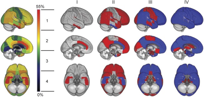
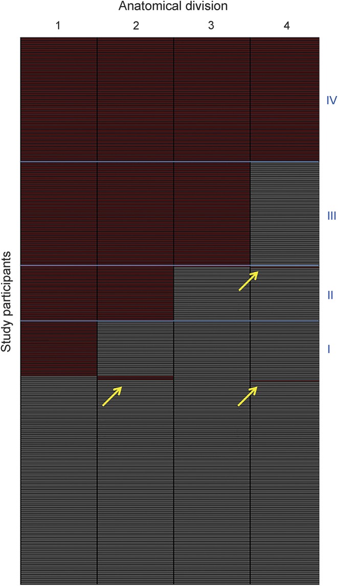
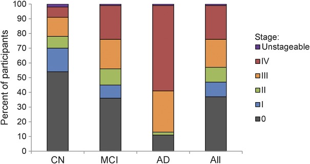
OBJECTIVES: To estimate a regional progression pattern of amyloid deposition from cross-sectional amyloid-sensitive PET data and evaluate its potential for in vivo staging of an individual's amyloid pathology. METHODS: F-AV45)-PET data was used to determine individual amyloid distribution profiles in a sample of 667 participants from the Alzheimer's Disease Neuroimaging Initiative cohort, including cognitively normal older individuals (CN) as well as patients with mild cognitive impairment and Alzheimer disease (AD) dementia. The frequency of regional amyloid positivity across CN individuals was used to construct a 4-stage model of progressing amyloid pathology, and individual distribution profiles were used to evaluate the consistency of this hierarchical stage model across the full cohort. RESULTS: According to a 4-stage model, amyloid deposition begins in temporobasal and frontomedial areas, and successively affects the remaining associative neocortex, primary sensory-motor areas and the medial temporal lobe, and finally the striatum. Amyloid deposition in these brain regions showed a highly consistent hierarchical nesting across participants, where only 2% exhibited distribution profiles that deviated from the staging scheme. The earliest in vivo amyloid stages were mostly missed by conventional dichotomous classification approaches based on global florbetapir-PET signal, but were associated with significantly reduced CSF Aβ42 levels. Advanced in vivo amyloid stages were most frequent in patients with AD and correlated with cognitive impairment in individuals without dementia. CONCLUSIONS: The highly consistent regional hierarchy of PET-evidenced amyloid deposition across participants resembles neuropathologic observations and suggests a predictable regional sequence that may be used to stage an individual's progress of amyloid pathology in vivo.
Overview
The claims and statistics of this paper haven’t been checked yet.
Run the check — free, no account needed. It reads the open-access text and checks the sources it cites.
Integrity signals
Registry
Cites
Statistics
Figures
Verification
Claims the paper makes
The claims and statistics of this paper haven’t been checked yet.
Run the check — free, no account needed. It reads the open-access text and checks the sources it cites.
Claims other papers make about this one
Each is checked against this paper’s own text: does it say what the citing paper says it does?
In the citing paper
“Aβ deposition occurs in a step-wise manner, starting in the temporobasal and frontomedial areas, then spreading to the remaining neocortex, the primary sensory-motor cortex and finally the striatum”
From The amyloid hypothesis in Alzheimer disease: new insights from new therapeutics (2022) · cites this paper as [15]
Self-check
Percentages, sums and reported differences re-computed from the paper’s own numbers.
The self-check needs the paper’s full text.
Citations
Cites
Neuropathological stageing of Alzheimer-related changes.
Braak, H., & Braak, E. (1991) · Acta Neuropathologica · doi:10.1007/bf00308809 · Cited by 16,123 · cited 2×
“While the first Thal phase encompasses the whole associative neocortex without further regional distinction, previous neuropathologic studies specifically implicated basal portions of the temporal and frontomedial cortex in the initial phase of amyloid accumulation (corresponding to the first stage of the Braak A-B-C…”
BackgroundResultDesikan, R. S., Ségonne, F., Fischl, B., Quinn, B. T., Dickerson, B. C., Blacker, D., Buckner, R. L., Dale, A. M., Maguire, R. P., Hyman, B. T., et al. (2006) · NeuroImage · doi:10.1016/j.neuroimage.2006.01.021 · Cited by 13,324
Self-citationStaging of brain pathology related to sporadic Parkinson’s disease
Braak, H., Tredici, K. D., Rüb, U., de Vos, R. A. I., Jansen Steur, E. N. H., & Braak, E. (2003) · Neurobiology of Aging · doi:10.1016/s0197-4580(02)00065-9 · Cited by 10,777
Hypothetical model of dynamic biomarkers of the Alzheimer's pathological cascade
Jack, C. R., Knopman, D. S., Jagust, W. J., Shaw, L. M., Aisen, P. S., Weiner, M. W., Petersen, R. C., & Trojanowski, J. Q. (2010) · The Lancet Neurology · doi:10.1016/s1474-4422(09)70299-6 · Cited by 4,806
Self-citationPhases of Aβ-deposition in the human brain and its relevance for the development of AD
Thal, D. R., Rüb, U., Orantes, M., & Braak, H. (2002) · Neurology · doi:10.1212/wnl.58.12.1791 · Cited by 3,490
Cerebrospinal fluid biomarker signature in Alzheimer's disease neuroimaging initiative subjects
Shaw, L. M., Vanderstichele, H., Knapik‐Czajka, M., Clark, C. M., Aisen, P. S., Petersen, R. C., Blennow, K., Soares, H., Simon, A., Lewczuk, P., et al. (2009) · Annals of Neurology · doi:10.1002/ana.21610 · Cited by 2,065
Self-citationUse of Florbetapir-PET for Imaging β-Amyloid Pathology
Clark, C. M. (2011) · JAMA · doi:10.1001/jama.2010.2008 · Cited by 1,117
Price, J. L., Davis, P. B., Morris, J. C., & White, D. L. (1991) · Neurobiology of Aging · doi:10.1016/0197-4580(91)90006-6 · Cited by 818
Self-citationMüller‐Gärtner, H. W., Links, J. M., Prince, J. L., Bryan, R. N., McVeigh, E. R., Leal, J. P., Davatzikos, C. A., & Frost, J. J. (1992) · Journal of Cerebral Blood Flow & Metabolism · doi:10.1038/jcbfm.1992.81 · Cited by 756
Florbetaben PET imaging to detect amyloid beta plaques in Alzheimer's disease: Phase 3 study
Sabri, O., Sabbagh, M. N., Seibyl, J., Barthel, H., Akatsu, H., Ouchi, Y., Senda, K., Murayama, S., Ishii, K., Takao, M., et al. (2015) · Alzheimer's & Dementia · doi:10.1016/j.jalz.2015.02.004 · Cited by 602
Self-citation
Cited by
Sorted by citations
Scheltens, P., De Strooper, B., Kivipelto, M., Holstege, H., Chételat, G., Teunissen, C. E., Cummings, J., & van der Flier, W. M. (2021) · The Lancet · doi:10.1016/s0140-6736(20)32205-4 · Cited by 5,465
Knopman, D. S., Amieva, H., Petersen, R. C., Chételat, G., Holtzman, D. M., Hyman, B. T., Nixon, R. A., & Jones, D. T. (2021) · Nature Reviews Disease Primers · doi:10.1038/s41572-021-00269-y · Cited by 2,606
Self-citationThe Amyloid-β Pathway in Alzheimer’s Disease
Hampel, H., Hardy, J., Blennow, K., Chen, C., Perry, G., Kim, S. H., Villemagne, V. L., Aisen, P., Vendruscolo, M., Iwatsubo, T., et al. (2021) · Molecular Psychiatry · doi:10.1038/s41380-021-01249-0 · Cited by 1,952
Self-citationThe amyloid hypothesis in Alzheimer disease: new insights from new therapeutics
Karran, E., & De Strooper, B. (2022) · Nature Reviews Drug Discovery · doi:10.1038/s41573-022-00391-w · Cited by 742
Amyloid-β-independent regulators of tau pathology in Alzheimer disease
van der Kant, R., Goldstein, L. S. B., & Ossenkoppele, R. (2020) · Nature reviews. Neuroscience · doi:10.1038/s41583-019-0240-3 · Cited by 580
Metadata
- Authors
- Grothe, M. J., Barthel, H., Sepulcre, J., Dyrba, M., Sabri, O., Teipel, S. J., Initiative, F. T. A. D. N., Initiative, A. D. N., Weiner, M., Aisen, P., Weiner, M., Aisen, P., Petersen, R., Jack, C. R., Jagust, W., Trojanowki, J. Q., Toga, A. W., Beckett, L., Green, R. C., Saykin, A. J., Morris, J., Liu, E., Green, R. C., Montine, T., Petersen, R., Aisen, P., Gamst, A., Thomas, R. G., Donohue, M., Walter, S., Gessert, D., Sather, T., Beckett, L., Harvey, D., Gamst, A., Donohue, M., Kornak, J., Jack, C. R., Dale, A., Bernstein, M., Felmlee, J., Fox, N., Thompson, P., Schuff, N., Alexander, G., DeCarli, C., Jagust, W., Bandy, D., Koeppe, R. A., Foster, N., Reiman, E. M., Chen, K., Mathis, C., Morris, J., Cairns, N. J., Taylor-Reinwald, L., Trojanowki, J. Q., Shaw, L., Lee, V. M. Y., Korecka, M., Toga, A. W., Crawford, K., Neu, S., Saykin, A. J., Foroud, T. M., Potkin, S., Shen, L., Kachaturian, Z., Frank, R., Snyder, P. J., Molchan, S., Kaye, J., Quinn, J., Lind, B., Dolen, S., Schneider, L. S., Pawluczyk, S., Spann, B. M., Brewer, J., Vanderswag, H., Heidebrink, J. L., Lord, J. L., Petersen, R., Johnson, K., Doody, R. S., Villanueva-Meyer, J., Chowdhury, M., Stern, Y., Honig, L. S., Bell, K. L., Morris, J. C., Ances, B., Carroll, M., Leon, S., Mintun, M. A., Schneider, S., Marson, D., Griffith, R., Clark, D., Grossman, H., Mitsis, E., Romirowsky, A., deToledo-Morrell, L., Shah, R. C., Duara, R., Varon, D., Roberts, P., Albert, M., Onyike, C., Kielb, S., Rusinek, H., de Leon, M. J., Glodzik, L., De Santi, S., Doraiswamy, P. M., Petrella, J. R., Coleman, R. E., Arnold, S. E., Karlawish, J. H., Wolk, D., Smith, C. D., Jicha, G., Hardy, P., Lopez, O. L., Oakley, M., Simpson, D. M., Porsteinsson, A. P., Goldstein, B. S., Martin, K., Makino, K. M., Ismail, M. S., Brand, C., Mulnard, R. A., Thai, G., Mc-Adams-Ortiz, C., Womack, K., Mathews, D., Quiceno, M., Diaz-Arrastia, R., King, R., Weiner, M., Martin-Cook, K., DeVous, M., Levey, A. I., Lah, J. J., Cellar, J. S., Burns, J. M., Anderson, H. S., Swerdlow, R. H., Apostolova, L., Lu, P. H., Bartzokis, G., Silverman, D. H. S., Parfitt, F., Johnson, H., Farlow, M. R., Hake, A. M., Matthews, B. R., Herring, S., van Dyck, C. H., Carson, R. E., MacAvoy, M. G., Chertkow, H., Bergman, H., Hosein, C., Black, S., Stefanovic, B., Caldwell, C., Robin Hsiung, G.-Y., Feldman, H., Mudge, B., Assaly, M., Kertesz, A., Rogers, J., Trost, D., Bernick, C., Munic, D., Kerwin, D., Mesulam, M.-M., Lipowski, K., Wu, C.-K., Johnson, N., Sadowsky, C., Martinez, W., Villena, T., Turner, R. S., Johnson, K., Reynolds, B., Sperling, R. A., Johnson, K. A., Marshall, G., Frey, M., Yesavage, J., Taylor, J. L., Lane, B., Rosen, A., Tinklenberg, J., Sabbagh, M., Belden, C., Jacobson, S., Kowall, N., Killiany, R., Budson, A. E., Norbash, A., Johnson, P. L., Obisesan, T. O., Wolday, S., Bwayo, S. K., Lerner, A., Hudson, L., Ogrocki, P., Fletcher, E., Carmichael, O., Olichney, J., DeCarli, C., Kittur, S., Borrie, M., Lee, T.-Y., Bartha, R., Johnson, S., Asthana, S., Carlsson, C. M., Potkin, S. G., Preda, A., Nguyen, D., Tariot, P., Fleisher, A., Reeder, S., Bates, V., Capote, H., Rainka, M., Scharre, D. W., Kataki, M., Zimmerman, E. A., Celmins, D., Brown, A. D., Pearlson, G. D., Blank, K., Anderson, K., Saykin, A. J., Santulli, R. B., Schwartz, E. S., Sink, K. M., Williamson, J. D., Garg, P., Watkins, F., Ott, B. R., Querfurth, H., Tremont, G., Salloway, S., Malloy, P., Correia, S., Rosen, H. J., Miller, B. L., Mintzer, J., Longmire, C. F., Spicer, K., Finger, E., Rachinsky, I., Rogers, J., Kertesz, A., Drost, D., Pomara, N., Hernando, R., Sarrael, A., Schultz, S. K., Boles Ponto, L. L., Shim, H., Smith, K. E., Relkin, N., Chaing, G., Raudin, L., Smith, A., Fargher, K., & Raj, B. A.
- Affiliations
- German Center for Neurodegenerative Diseases (DE)
- Corresponding author
- Grothe, Michel J.
- ORCID
- Sepulcre, Jorge
- Dale, Anders
- Fox, Nick
- Thompson, Paul
- Reiman, Eric M.
- Foroud, Tatiana M.
- Shen, Li
- Kaye, Jeffrey
- Stern, Yaakov
- Honig, Lawrence S.
- Clark, David
- Albert, Marilyn
- Arnold, Steven E.
- Karlawish, Jason H.
- Wolk, David
- Smith, Charles D.
- Hardy, Peter
- Weiner, Myron
- Levey, Allan I.
- Lah, James J.
- Burns, Jeffrey M.
- Swerdlow, Russell H.
- Apostolova, Liana
- Silverman, Daniel H.S.
- Johnson, Heather
- Farlow, Martin R.
- Hake, Ann Marie
- Chertkow, Howard
- Bergman, Howard
- Sperling, Reisa A.
- Johnson, Keith A.
- Marshall, Gad
- Frey, Meghan
- Yesavage, Jerome
- Taylor, Joy L.
- Lane, Barton
- Rosen, Allyson
- Jacobson, Sandra
- Kowall, Neil
- Killiany, Ronald
- Budson, Andrew E.
- Norbash, Alexander
- Salloway, Stephen
- Miller, Bruce L.
- Venue
- Neurology
- Published
- 2017-11-14
- Volume
- 89
- Issue
- 20
- Pages
- 2031-2038
- ISSN
- 0028-3878 (ISSN-L)
- 1526-632X
- PMC
- PMC5711511
- OpenAlex
- W2766782573
- Cited by
- 440
- References
- 40
- Citations by year
- 2026: 39 · 2025: 42 · 2024: 53 · 2023: 61 · 2022: 56 · 2021: 62 · 2020: 52 · 2019: 40 · 2018: 31 · 2017: 3
- FWCI
- 28.31
- Type
- journal article
- Language
- en
- Open access
- hybrid
- doi.org
- Licence
- cc-by-nc-nd
- Locations
- Neurology (publishedVersion, cc-by-nc-nd)
- Europe PMC (PubMed Central) (publishedVersion, cc-by-nc-nd)
- PubMed Central (publishedVersion, cc-by-nc-nd)
- PubMed (publishedVersion)
- Digital Access to Scholarship at Harvard (DASH) (Harvard University) (submittedVersion, cc-by)
- Funders
- U.S. Department of Defense (U01 AG024904, W81XWH, W81XWH-12-2-, W81XWH1220012, AG024904)
- Foundation for the National Institutes of Health (W81XWH-12-2-0012, U01AG024904, AG024904)
- Alzheimer's Association (U01 AG024904, W81XWH-12-2-0012)
- Bristol-Myers Squibb (U01 AG024904, AG024904, W81XWH-12-2-0012)
- Eli Lilly and Company (U01 AG024904, AG024904, W81XWH-12-2-0012)
- Pfizer (AG024904, W81XWH-12-2-0012, U01 AG024904)
- Biogen (W81XWH-12-2-0012, U01 AG024904, AG024904)
- University of Southern California (W81XWH-12-2-0012, U01 AG024904)
- F. Hoffmann-La Roche (W81XWH-12-2-0012, AG024904, U01 AG024904)
- Meso Scale Diagnostics (U01 AG024904, AG024904, W81XWH-12-2-0012)
- Alzheimer's Disease Neuroimaging Initiative (W81XWH-12-2–0012, U01AG024904, AG024904, W81XWH-12-2-, Department of Defense award number W81XWH-12-2-0012)
- BioClinica (U01 AG024904, W81XWH-12-2-0012, AG024904)
- Novartis Pharmaceuticals Corporation (W81XWH-12-2-0012, U01 AG024904, AG024904)
- Northern California Institute for Research and Education (U01 AG024904, W81XWH-12-2-0012)
- Eisai (W81XWH-12-2-0012, U01 AG024904, AG024904)
- Deutsches Zentrum für Neurodegenerative Erkrankungen
- Servier (AG024904, W81XWH-12-2-0012, U01 AG024904)
- H. Lundbeck A/S (U01 AG024904, AG024904, W81XWH-12-2-0012)
- IXICO (AG024904, W81XWH-12-2-0012, U01 AG024904)
- National Institutes of Health (3u01ag024904-10s2, W81XWH, W81XWH-12-2-0012, U01-AG024904, AG024904)
- Genentech (W81XWH-12-2-0012, AG024904, U01 AG024904)
- University of California, San Diego
- Canadian Institutes of Health Research (U01 AG024904, W81XWH-12-2-0012)
- National Institute on Aging (U01-AG-024904, P30AG010129, W81XWH-12-2–0012, AG 024904)
- National Institute of Biomedical Imaging and Bioengineering (W81XWH-12-2-0012, U01 AG024904, P41EB015922, AG024904)
- Topics
- Health Sciences › Medicine › Psychiatry and Mental health › Dementia and Cognitive Impairment Research
- Life Sciences › Neuroscience › Cellular and Molecular Neuroscience › Alzheimer's disease research and treatments
- Health Sciences › Medicine › Radiology, Nuclear Medicine and Imaging › Medical Imaging Techniques and Applications
- Keywords
- amyloid deposition, florbetapir PET, Alzheimer's disease, mild cognitive impairment, CSF amyloid-beta 42, medial temporal lobe, striatum, cognitive impairment without dementia, Alzheimer's Disease Neuroimaging Initiative
- MeSH terms
- Aged, Aged, 80 and over, Aging, Alzheimer Disease, Aniline Compounds, Cerebral Cortex, Cross-Sectional Studies, Ethylene Glycols, Female, Humans, Male, Peptide Fragments, Biomarkers, Amyloid beta-Peptides, Neostriatum, Disease Progression, Positron-Emission Tomography, Cognitive Dysfunction
About these checks
Registry, retraction, Cites and statistics results are computed, not judged, with one exception: a printed reference that no index lists is read once by a model before it counts as a citation error. Claims are extracted by a model and kept only when their quote appears verbatim in the text; each cited source is then checked, with a model’s help, for whether it says what the claim relies on. Those verdicts can be wrong — read the claim’s quote and the cited source before relying on one.
Statistics and arithmetic are recomputed, not judged.
Confidence bands are shown only for fields calibrated on our annotated test set. None is calibrated yet, so no band is shown.
What counts as a claim ›How we measure confidence ›How we check this ›
Are you an author, or do you see a problem with this page? Request a review
How to cite
Cite CoChat for the checks on this page, and the paper for its own findings.
- This page
- CoChat. (2026). Citation check: In vivo staging of regional amyloid deposition. https://app.cochat.ai/papers/10.1212/wnl.0000000000004643
- The paper
- Grothe, M. J., Barthel, H., Sepulcre, J., Dyrba, M., Sabri, O., Teipel, S. J., Initiative, F. T. A. D. N., Initiative, A. D. N., Weiner, M., Aisen, P., Weiner, M., Aisen, P., Petersen, R., Jack, C. R., Jagust, W., Trojanowki, J. Q., Toga, A. W., Beckett, L., Green, R. C., . . . Raj, B. A. (2017). In vivo staging of regional amyloid deposition. Neurology. https://doi.org/10.1212/wnl.0000000000004643
