Cited by 12

The DOI matches its Crossref record; 3 of the 3 references it cites were found in a registry or an index.

Construction of a human hTERT RPE-1 cell line with inducible Cre for editing of endogenous genes

Hindul, Naushin L., Jhita, Amarjot, Oprea, Daiana G., Hussain, Tasnim Alamgir, Gonchar, Oksana, Campillo, Miguel Angel Muro, O'Regan, Laura, Kanemaki, Masato T., Fry, Andrew M., Hirota, Kouji, Tanaka, Kayoko·2022·Biology Open

Abstract

The human retinal pigment epithelial RPE-1 cell line immortalized with hTERT retains a stable karyotype with a modal chromosome number of 46 and has been widely used to study physiological events in human cell culture systems. To facilitate inducible knock-out or knock-in experiments in this cell line, we have modified the AAVS1 locus to harbour a DNA fragment encoding ERT2-Cre-ERT2 fusion protein under regulation of a Tet-On expression system. In the generated cell line, active Cre recombinase was induced by simple addition of doxycycline and tamoxifen to the culture medium. As proof of concept, we successfully introduced an oncogenic point mutation to the endogenous KRAS gene locus of this cell line. The cell line will serve as a powerful tool to conduct functional analyses of human genes.

Overview

The claims and statistics of this paper haven’t been checked yet.

Run the check — free, no account needed. It reads the open-access text and checks the sources it cites.

A few free checks a day · papers already verified open instantly · results are public

Integrity signals

Registry

Crossref · 10.1242/bio.059056

Cites

3 of 3 references verified0 citation errors · 0 not checkable

Statistics

Figures

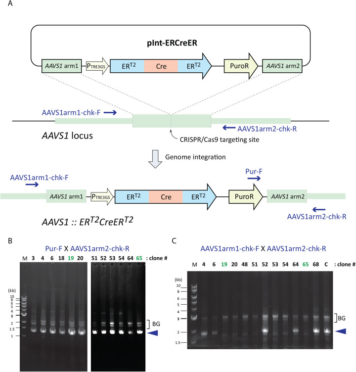
Fig. 1. Construction of hTERT RPE-1 ERT2-Cre-ERT2 cell line. (A) A schematic diagram of the integration donor plasmid pInt-ERCreER and the targeted AAVS1 locus. Locations of the primers used to genotype the integration event are indicated in blue. (B) Representative genome PCR results using primers Pur-F and AAVS1arm2-chk-R. Successful integration of the ERT2-Cre-ERT2 cassette into AAVS1 locus of at least one of the chromosomes yields a 1330 bp PCR product (indicated by a blue arrowhead). Numbers indicated for each lane represent clones isolated through the puromycin resistance screening. Clones 19 and 65 (shown in green) were further analysed in this study. BG indicates background bands. (C) Representative genome PCR results using primers AAVS1arm1-chk-F and AAVS1arm2-chk-R. This pair of primers amplifies an 1836 bp fragment (indicated by a blue arrowhead) if the wild-type unedited AAVS1 locus is present. The result indicates that clones 19, 20, 48, 51, 53, 54 and 65 do not retain the wild-type AAVS1 locus. Together with the result from panel B, we concluded that these clones carry the ERT2-Cre-ERT2 cassette as a homozygous insertion at the AAVS1 locus. Clones 19 and 65 were further analysed for Cre expression and activity in Figs 2 and 3. BG indicates background bands.
Fig. 1. Construction of hTERT RPE-1 ERT2-Cre-ERT2 cell line. (A) A schematic diagram of the integration donor plasmid pInt-ERCreER and the targeted AAVS1 locus. Locations of the primers used to genotype the integration event are indicated in blue. (B) Representative genome PCR results using primers Pur-F and AAVS1arm2-chk-R. Successful integration of the ERT2-Cre-ERT2 cassette into AAVS1 locus of at least one of the chromosomes yields a 1330 bp PCR product (indicated by a blue arrowhead). Numbers indicated for each lane represent clones isolated through the puromycin resistance screening. Clones 19 and 65 (shown in green) were further analysed in this study. BG indicates background bands. (C) Representative genome PCR results using primers AAVS1arm1-chk-F and AAVS1arm2-chk-R. This pair of primers amplifies an 1836 bp fragment (indicated by a blue arrowhead) if the wild-type unedited AAVS1 locus is present. The result indicates that clones 19, 20, 48, 51, 53, 54 and 65 do not retain the wild-type AAVS1 locus. Together with the result from panel B, we concluded that these clones carry the ERT2-Cre-ERT2 cassette as a homozygous insertion at the AAVS1 locus. Clones 19 and 65 were further analysed for Cre expression and activity in Figs 2 and 3. BG indicates background bands.CC BY · PubMed Central
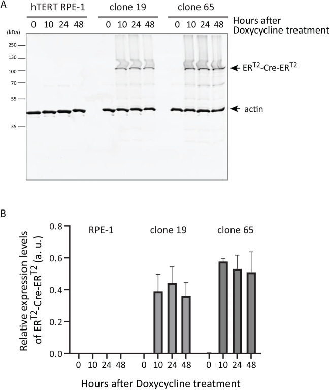
Fig. 2. Expression of ERT2-Cre-ERT2 is induced within 10 h after doxycycline treatment. hTERT RPE-1 ERT2-Cre-ERT2 clones 19 and 65 were treated with doxycycline (1 µg/ml) and the expression levels of ERT2-Cre-ERT2 at time 0, 10, 24 and 48 h after the treatment were analysed by western blotting. (A) A representative result of the parental hTERT RPE-1, clone 19 and clone 65. (B) Three biological replicates of the western blotting were quantitated using actin as an internal control. Mean and SD values are presented.
Fig. 2. Expression of ERT2-Cre-ERT2 is induced within 10 h after doxycycline treatment. hTERT RPE-1 ERT2-Cre-ERT2 clones 19 and 65 were treated with doxycycline (1 µg/ml) and the expression levels of ERT2-Cre-ERT2 at time 0, 10, 24 and 48 h after the treatment were analysed by western blotting. (A) A representative result of the parental hTERT RPE-1, clone 19 and clone 65. (B) Three biological replicates of the western blotting were quantitated using actin as an internal control. Mean and SD values are presented.CC BY · PubMed Central
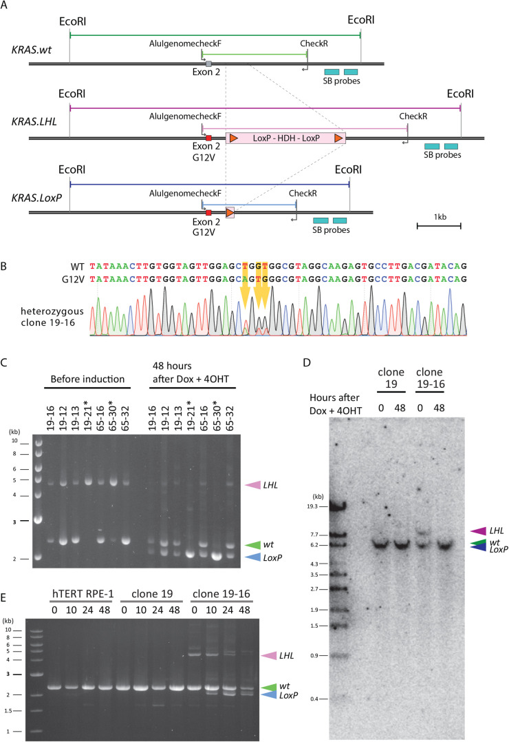
Fig. 3. Cre-LoxP excision of the targeted chromosome in 48 h after doxycycline and 4-OHT treatment. (A) A schematic diagram of a proof-of-principle test case, chromosome editing of the KRAS gene locus. A DNA fragment encoding histidinol dehydrogenase (HDH), flanked by LoxP sites (LoxP-HDH-LoxP, LHL) was inserted into intron 2 of KRAS gene to create KRAS.LHL locus. Active Cre is expected to excise the LHL cassette, leaving one copy of LoxP site, to generate KRAS.LoxP allele. A pair of primers, AlugenomecheckF and CheckR, is indicated in the diagram. Two probes used for the Southern blotting are indicated as ‘SB probes’ and EcoRI sites used for Southern blotting are indicated in the diagram. (B) Some of the clones with the LoxP cassette insertion also carried the KRAS.G12V mutation. The genome isolated from clone 19-16, which has a heterozygous integration of the ERT2-Cre-ERT2 cassette, was sequenced using the primer AlugenomecheckF. Three substitutions of the nucleic acids resulting in G12V mutation are indicated with orange arrows together with the sequencing chromatogram. (C) A typical example of successful excision of the LHL cassette 48 h after doxycycline (1 µg/ml) and 4-OHT (0.5 µM) exposure in seven independent clones. Clones 19-16, 19-12, 19-13 and 19-21 were generated by integrating the LHL cassette into the KRAS locus of clone 19 and clones 65-16, 65-30 and 65-32 were generated in the same way but using clone 65. Among these clones, clones 19-21 and 65-30 have KRAS.LHL as a homozygous insertion. PCR genotyping was conducted using primers AlugenomecheckF and CheckR. The PCR reaction produced the expected products of 4621 bp for KRAS.LHL, 2370 bp for KRAS.wt and 2115 bp for KRAS.LoxP. (D) Southern blotting to confirm the successful LHL excision 48 h after doxycycline and 4-OHT treatment. Genomes from clone 19 and clone 19-16 were digested by EcoRI and analysed by Southern blotting using a cocktail of two probes indicated in the diagram A (SB probes). Signals were detected at the expected size of 8797 bp for KRAS.LHL. For KRAS.wt and KRAS.LoxP, the expected sizes were 6546 bp and 6291 bp, respectively, and these two bands were not resolved in the agarose gel. Nonetheless, the KRAS.LHL signal became undetectable in clone 19-16 (time 48) sample, and therefore we concluded that LHL excision was successful 48 h after exposure to doxycycline and 4-OHT. (E) Time-course profile of the LHL excision. Parental hTERT RPE-1, clone 19 and clone 19-16 were exposed to doxycycline (1 µg/ml) and 4-OHT (0.5 µM), and samples collected at 0, 10, 24 and 48 h after treatment. Genome PCR was conducted using primers AlugenomecheckF and CheckR. A PCR product of KRAS.LoxP was detectable 10 h after the start of the treatment. By 48 h, most of the KRAS.LHL signal disappeared and the signal intensity of KRAS.wt and KRAS.LoxP became comparable.
Fig. 3. Cre-LoxP excision of the targeted chromosome in 48 h after doxycycline and 4-OHT treatment. (A) A schematic diagram of a proof-of-principle test case, chromosome editing of the KRAS gene locus. A DNA fragment encoding histidinol dehydrogenase (HDH), flanked by LoxP sites (LoxP-HDH-LoxP, LHL) was inserted into intron 2 of KRAS gene to create KRAS.LHL locus. Active Cre is expected to excise the LHL cassette, leaving one copy of LoxP site, to generate KRAS.LoxP allele. A pair of primers, AlugenomecheckF and CheckR, is indicated in the diagram. Two probes used for the Southern blotting are indicated as ‘SB probes’ and EcoRI sites used for Southern blotting are indicated in the diagram. (B) Some of the clones with the LoxP cassette insertion also carried the KRAS.G12V mutation. The genome isolated from clone 19-16, which has a heterozygous integration of the ERT2-Cre-ERT2 cassette, was sequenced using the primer AlugenomecheckF. Three substitutions of the nucleic acids resulting in G12V mutation are indicated with orange arrows together with the sequencing chromatogram. (C) A typical example of successful excision of the LHL cassette 48 h after doxycycline (1 µg/ml) and 4-OHT (0.5 µM) exposure in seven independent clones. Clones 19-16, 19-12, 19-13 and 19-21 were generated by integrating the LHL cassette into the KRAS locus of clone 19 and clones 65-16, 65-30 and 65-32 were generated in the same way but using clone 65. Among these clones, clones 19-21 and 65-30 have KRAS.LHL as a homozygous insertion. PCR genotyping was conducted using primers AlugenomecheckF and CheckR. The PCR reaction produced the expected products of 4621 bp for KRAS.LHL, 2370 bp for KRAS.wt and 2115 bp for KRAS.LoxP. (D) Southern blotting to confirm the successful LHL excision 48 h after doxycycline and 4-OHT treatment. Genomes from clone 19 and clone 19-16 were digested by EcoRI and analysed by Southern blotting using a cocktail of two probes indicated in the diagram A (SB probes). Signals were detected at the expected size of 8797 bp for KRAS.LHL. For KRAS.wt and KRAS.LoxP, the expected sizes were 6546 bp and 6291 bp, respectively, and these two bands were not resolved in the agarose gel. Nonetheless, the KRAS.LHL signal became undetectable in clone 19-16 (time 48) sample, and therefore we concluded that LHL excision was successful 48 h after exposure to doxycycline and 4-OHT. (E) Time-course profile of the LHL excision. Parental hTERT RPE-1, clone 19 and clone 19-16 were exposed to doxycycline (1 µg/ml) and 4-OHT (0.5 µM), and samples collected at 0, 10, 24 and 48 h after treatment. Genome PCR was conducted using primers AlugenomecheckF and CheckR. A PCR product of KRAS.LoxP was detectable 10 h after the start of the treatment. By 48 h, most of the KRAS.LHL signal disappeared and the signal intensity of KRAS.wt and KRAS.LoxP became comparable.CC BY · PubMed Central
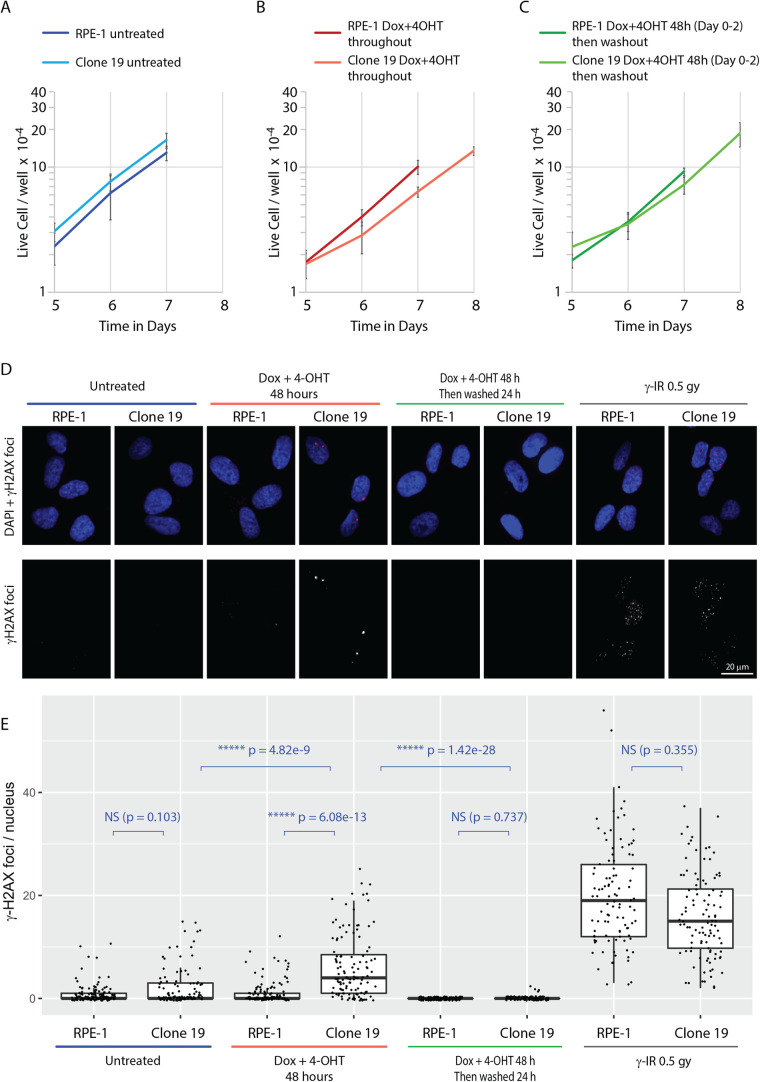
Fig. 4. The physiological impact of expression of ERT2-Cre-ERT2 in the clone 19. (A–C) Proliferation rates of RPE1 and clone 19 cells with or without doxycycline and 4-OHT treatment. A relatively small number of cells (2×10³ cells) were seeded per well in a six-well plate to allow observation of proliferation for up to 8 days. The panels show the proliferation rates from day 5. The cell count data for the whole course of the experiment is presented in the Fig. S2. All graphs have a logarithmic y-axis. (A) Proliferation rates of RPE1 and clone 19 cells without doxycycline and 4-OHT treatment. (B) Proliferation rates of RPE1 and clone 19 cells cultured in the presence of doxycycline and 4-OHT throughout the experiment. (C) Proliferation rates of RPE1 and clone 19 cells treated for 48 h with doxycycline and 4-OHT before replacement into fresh media. For the clone 19 samples treated with doxycycline and 4-OHT, cell counts were made for an additional day to reveal growth recovery of these samples. All experiments, n=3; error bars represent SD. (D) Formation of γ-H2AX foci upon expression of ERT2-Cre-ERT2. RPE-1 and Clone 19 cells were subject to the following treatments: incubation for 48 h with doxycycline and 4-OHT, incubation for 48 h with doxycycline and 4-OHT, which were then removed for 24 h before fixation, and γ-irradiation (0.5 Gy) 30 min before fixation. Untreated cells are included as a reference. γ-H2AX foci formation was detected by immunofluorescence microscopy as described in the Materials and Methods. Upper panels show merged images of γ-H2AX foci and DNA. Scale bar: 20 µm. (E) Summary of γ-H2AX foci formation results presented in (D). 100–150 nuclei were counted for each sample and the data presented using the boxplot format (RStudio) where median, and first and third quartiles are indicated using a box. Individual data points are also indicated. The data were analysed by Kruskal–Wallis test and pairwise comparisons conducted by Dunn’s test. NS, not significant; **P<0.01, ***P<0.001, ****P<0.0001, *****P<0.00001.
Fig. 4. The physiological impact of expression of ERT2-Cre-ERT2 in the clone 19. (A–C) Proliferation rates of RPE1 and clone 19 cells with or without doxycycline and 4-OHT treatment. A relatively small number of cells (2×10³ cells) were seeded per well in a six-well plate to allow observation of proliferation for up to 8 days. The panels show the proliferation rates from day 5. The cell count data for the whole course of the experiment is presented in the Fig. S2. All graphs have a logarithmic y-axis. (A) Proliferation rates of RPE1 and clone 19 cells without doxycycline and 4-OHT treatment. (B) Proliferation rates of RPE1 and clone 19 cells cultured in the presence of doxycycline and 4-OHT throughout the experiment. (C) Proliferation rates of RPE1 and clone 19 cells treated for 48 h with doxycycline and 4-OHT before replacement into fresh media. For the clone 19 samples treated with doxycycline and 4-OHT, cell counts were made for an additional day to reveal growth recovery of these samples. All experiments, n=3; error bars represent SD. (D) Formation of γ-H2AX foci upon expression of ERT2-Cre-ERT2. RPE-1 and Clone 19 cells were subject to the following treatments: incubation for 48 h with doxycycline and 4-OHT, incubation for 48 h with doxycycline and 4-OHT, which were then removed for 24 h before fixation, and γ-irradiation (0.5 Gy) 30 min before fixation. Untreated cells are included as a reference. γ-H2AX foci formation was detected by immunofluorescence microscopy as described in the Materials and Methods. Upper panels show merged images of γ-H2AX foci and DNA. Scale bar: 20 µm. (E) Summary of γ-H2AX foci formation results presented in (D). 100–150 nuclei were counted for each sample and the data presented using the boxplot format (RStudio) where median, and first and third quartiles are indicated using a box. Individual data points are also indicated. The data were analysed by Kruskal–Wallis test and pairwise comparisons conducted by Dunn’s test. NS, not significant; **P<0.01, ***P<0.001, ****P<0.0001, *****P<0.00001.CC BY · PubMed Central

Verification

Claims the paper makes

The claims and statistics of this paper haven’t been checked yet.

Run the check — free, no account needed. It reads the open-access text and checks the sources it cites.

A few free checks a day · papers already verified open instantly · results are public

Claims other papers make about this one

Each is checked against this paper’s own text: does it say what the citing paper says it does?

Self-check

Percentages, sums and reported differences re-computed from the paper’s own numbers.

The self-check needs the paper’s full text.

Citations

Cites

Show all 3 references

Research works printed but not linked: 32

  1. Abremski, 1984, Bacteriophage P1 site-specific recombination. Purification and properties of the Cre recombinase protein, J. Biol. Chem., 259, 1509, 10.1016/S0021-9258(17)43437-5 · DOI
  2. Bodnar, 1998, Extension of life-span by introduction of telomerase into normal human cells, Science, 279, 349, 10.1126/science.279.5349.349 · DOI · Paper page
  3. Casanova, 2002, ER-based double iCre fusion protein allows partial recombination in forebrain, Genesis, 34, 208, 10.1002/gene.10153 · DOI
  4. Cong, 2013, Multiplex genome engineering using CRISPR/Cas systems, Science, 339, 819, 10.1126/science.1231143 · DOI · Paper page
  5. Das, 2016, Tet-on systems for doxycycline-inducible gene expression, Curr. Gene Ther., 16, 156, 10.2174/1566523216666160524144041 · DOI · Paper page
  6. Feil, 1997, Regulation of Cre recombinase activity by mutated estrogen receptor ligand-binding domains, Biochem. Biophys. Res. Commun., 237, 752, 10.1006/bbrc.1997.7124 · DOI
  7. Hoess, 1982, P1 site-specific recombination: nucleotide sequence of the recombining sites, Proc. Natl. Acad. Sci. USA, 79, 3398, 10.1073/pnas.79.11.3398 · DOI
  8. Jiang, 1999, Telomerase expression in human somatic cells does not induce changes associated with a transformed phenotype, Nat. Genet., 21, 111, 10.1038/5056 · DOI
  9. Katoh, 2017, Practical method for targeted disruption of cilia-related genes by using CRISPR/Cas9-mediated, homology-independent knock-in system, Mol. Biol. Cell, 28, 898, 10.1091/mbc.e17-01-0051 · DOI
  10. Kotin, 1992, Characterization of a preferred site on human chromosome 19q for integration of adeno-associated virus DNA by non-homologous recombination, EMBO J., 11, 5071, 10.1002/j.1460-2075.1992.tb05614.x · DOI
  11. Laemmli, 1970, Cleavage of structural proteins during the assembly of the head of bacteriophage T4, Nature, 227, 680, 10.1038/227680a0 · DOI · Paper page
  12. Lakso, 1992, Targeted oncogene activation by site-specific recombination in transgenic mice, Proc. Natl. Acad. Sci. USA, 89, 6232, 10.1073/pnas.89.14.6232 · DOI
  13. Mali, 2013, RNA-guided human genome engineering via Cas9, Science, 339, 823, 10.1126/science.1232033 · DOI · Paper page
  14. Miyamoto, 2015, The microtubule-depolymerizing activity of a mitotic kinesin protein KIF2A drives primary cilia disassembly coupled with cell proliferation, Cell Rep, 10, 664, 10.1016/j.celrep.2015.01.003 · DOI
  15. Natsume, 2016, Rapid protein depletion in human cells by auxin-inducible degron tagging with short homology donors, Cell Rep, 15, 210, 10.1016/j.celrep.2016.03.001 · DOI · Paper page
  16. Ogata, 2003, Identification of an insulator in AAVS1, a preferred region for integration of adeno-associated virus DNA, J. Virol., 77, 9000, 10.1128/JVI.77.16.9000-9007.2003 · DOI
  17. Orban, 1992, Tissue- and site-specific DNA recombination in transgenic mice, Proc. Natl. Acad. Sci. USA, 89, 6861, 10.1073/pnas.89.15.6861 · DOI
  18. Rogakou, 1998, DNA double-stranded breaks induce histone H2AX phosphorylation on serine 139, J. Biol. Chem., 273, 5858, 10.1074/jbc.273.10.5858 · DOI · Paper page
  19. Sauer, 1988, Site-specific DNA recombination in mammalian cells by the Cre recombinase of bacteriophage P1, Proc. Natl. Acad. Sci. USA, 85, 5166, 10.1073/pnas.85.14.5166 · DOI · Paper page
  20. Schindelin, 2012, Fiji: an open-source platform for biological-image analysis, Nat. Methods, 9, 676, 10.1038/nmeth.2019 · DOI · Paper page
  21. Schmidt, 2000, Illegitimate Cre-dependent chromosome rearrangements in transgenic mouse spermatids, Proc. Natl. Acad. Sci. USA, 97, 13702, 10.1073/pnas.240471297 · DOI
  22. Sengupta, 2017, Viral Cre-LoxP tools aid genome engineering in mammalian cells, J Biol Eng, 11, 45, 10.1186/s13036-017-0087-y · DOI
  23. Senmatsu, 2021, lncRNA transcription induces meiotic recombination through chromatin remodelling in fission yeast, Commun. Biol., 4, 295, 10.1038/s42003-021-01798-8 · DOI
  24. Silver, 2001, Self-excising retroviral vectors encoding the Cre recombinase overcome Cre-mediated cellular toxicity, Mol. Cell, 8, 233, 10.1016/S1097-2765(01)00295-7 · DOI
  25. Smith, 2008, Robust, persistent transgene expression in human embryonic stem cells is achieved with AAVS1-targeted integration, Stem Cells, 26, 496, 10.1634/stemcells.2007-0039 · DOI
  26. Sternberg, 1981, Bacteriophage P1 site-specific recombination. I. Recombination between loxP sites, J. Mol. Biol., 150, 467, 10.1016/0022-2836(81)90375-2 · DOI · Paper page
  27. Uhl, 2015, Mouse models as predictors of human responses: evolutionary medicine, Curr Pathobiol Rep, 3, 219, 10.1007/s40139-015-0086-y · DOI
  28. Van Duyne, 2001, A structural view of cre-loxp site-specific recombination, Annu. Rev. Biophys. Biomol. Struct., 30, 87, 10.1146/annurev.biophys.30.1.87 · DOI
  29. Vara, 1986, Expression in mammalian cells of a gene from Streptomyces alboniger conferring puromycin resistance, Nucleic Acids Res., 14, 4617, 10.1093/nar/14.11.4617 · DOI
  30. Wang, 2009, Cre transgenic mouse lines, Methods Mol. Biol., 561, 265, 10.1007/978-1-60327-019-9_17 · DOI
  31. Weinrich, 1997, Reconstitution of human telomerase with the template RNA component hTR and the catalytic protein subunit hTRT, Nat. Genet., 17, 498, 10.1038/ng1297-498 · DOI · Paper page
  32. Zhang, 1996, Inducible site-directed recombination in mouse embryonic stem cells, Nucleic Acids Res., 24, 543, 10.1093/nar/24.4.543 · DOI

Cited by

See all 12

Metadata

Authors
Hindul, N. L., Jhita, A., Oprea, D. G., Hussain, T. A., Gonchar, O., Campillo, M. A. M., O'Regan, L., Kanemaki, M. T., Fry, A. M., Hirota, K., & Tanaka, K.
Venue
Biology Open
Published
2022-02-15
Volume
11
Issue
2
ISSN
2046-6390
Cited by
12
References
3
Language
en
Funders
  • JSPS (JP20H04337, JP16H01314, JP19KK0210)
  • University of Leicester

About these checks

Registry, retraction, Cites and statistics results are computed, not judged, with one exception: a printed reference that no index lists is read once by a model before it counts as a citation error. Claims are extracted by a model and kept only when their quote appears verbatim in the text; each cited source is then checked, with a model’s help, for whether it says what the claim relies on. Those verdicts can be wrong — read the claim’s quote and the cited source before relying on one.

Statistics and arithmetic are recomputed, not judged.

Confidence bands are shown only for fields calibrated on our annotated test set. None is calibrated yet, so no band is shown.

What counts as a claim ›How we measure confidence ›How we check this ›

Are you an author, or do you see a problem with this page? Request a review

How to cite

Cite CoChat for the checks on this page, and the paper for its own findings.

This page
CoChat. (2026). Citation check: Construction of a human hTERT RPE-1 cell line with inducible Cre for editing of endogenous genes. https://app.cochat.ai/papers/10.1242/bio.059056
The paper
Hindul, N. L., Jhita, A., Oprea, D. G., Hussain, T. A., Gonchar, O., Campillo, M. A. M., O'Regan, L., Kanemaki, M. T., Fry, A. M., Hirota, K., & Tanaka, K. (2022). Construction of a human hTERT RPE-1 cell line with inducible Cre for editing of endogenous genes. Biology Open. https://doi.org/10.1242/bio.059056