

The DOI matches its Crossref record; 129 of the 129 references it cites were found in a registry or an index.
The biological significance of brain barrier mechanisms: help or hindrance in drug delivery to the central nervous system?
Open accessAbstract
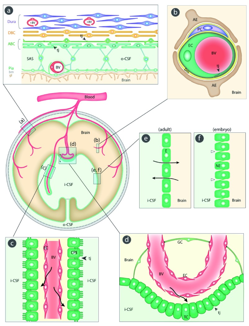
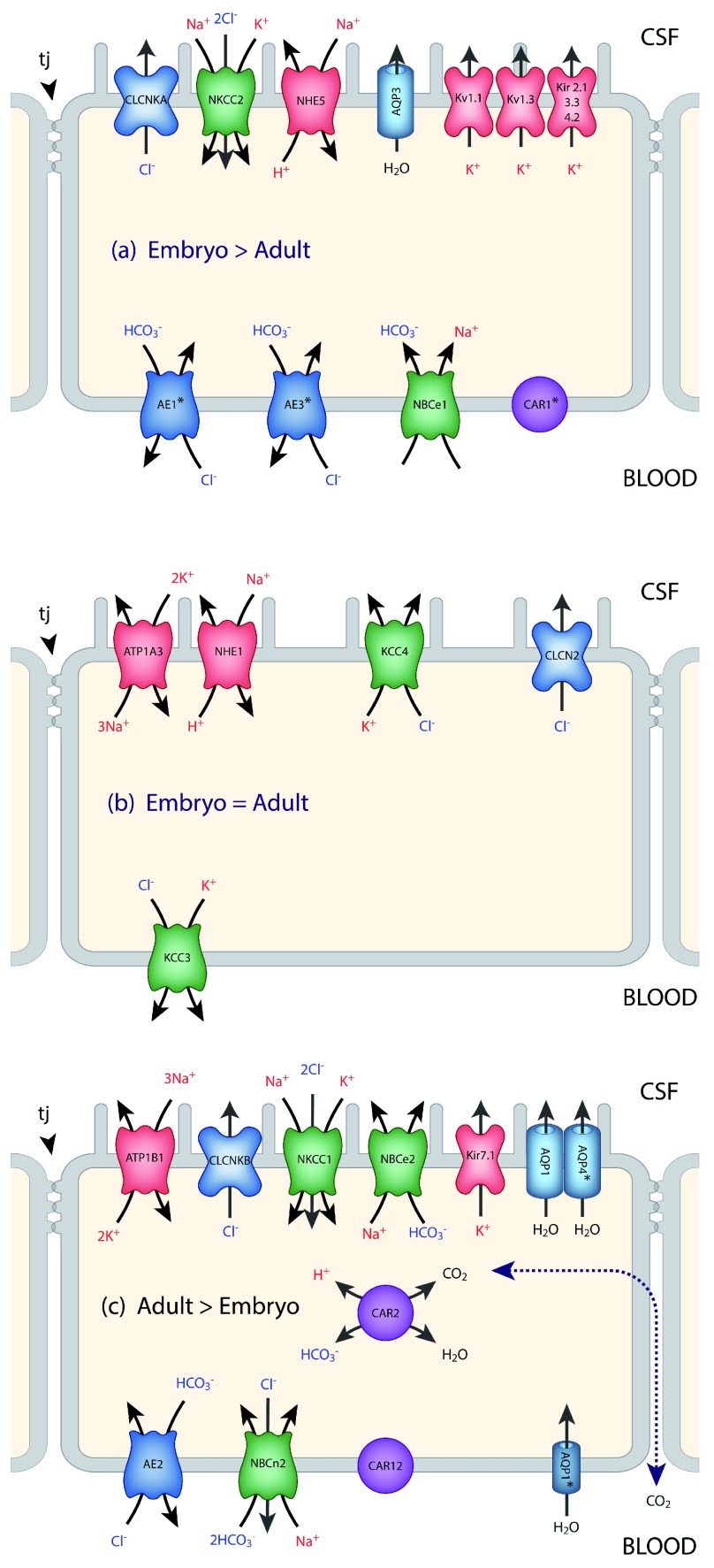
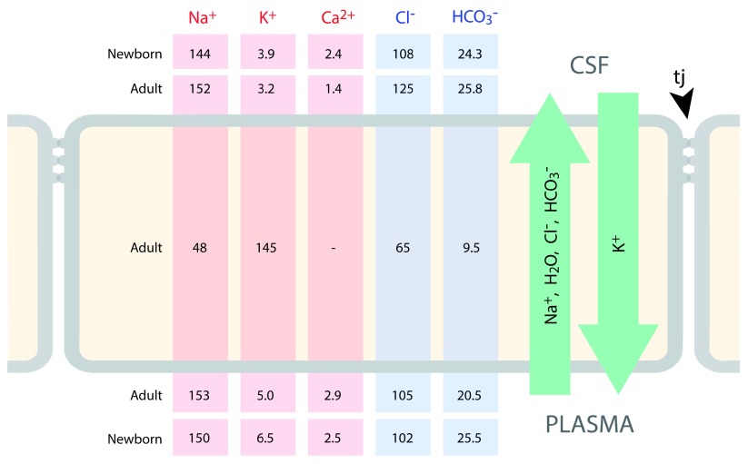
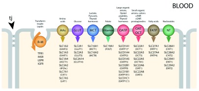
Barrier mechanisms in the brain are important for its normal functioning and development. Stability of the brain’s internal environment, particularly with respect to its ionic composition, is a prerequisite for the fundamental basis of its function, namely transmission of nerve impulses. In addition, the appropriate and controlled supply of a wide range of nutrients such as glucose, amino acids, monocarboxylates, and vitamins is also essential for normal development and function. These are all cellular functions across the interfaces that separate the brain from the rest of the internal environment of the body. An essential morphological component of all but one of the barriers is the presence of specialized intercellular tight junctions between the cells comprising the interface: endothelial cells in the blood-brain barrier itself, cells of the arachnoid membrane, choroid plexus epithelial cells, and tanycytes (specialized glial cells) in the circumventricular organs. In the ependyma lining the cerebral ventricles in the adult brain, the cells are joined by gap junctions, which are not restrictive for intercellular movement of molecules. But in the developing brain, the forerunners of these cells form the neuroepithelium, which restricts exchange of all but the smallest molecules between cerebrospinal fluid and brain interstitial fluid because of the presence of strap junctions between the cells. The intercellular junctions in all these interfaces are the physical basis for their barrier properties. In the blood-brain barrier proper, this is combined with a paucity of vesicular transport that is a characteristic of other vascular beds. Without such a diffusional restrain, the cellular transport mechanisms in the barrier interfaces would be ineffective. Superimposed on these physical structures are physiological mechanisms as the cells of the interfaces contain various metabolic transporters and efflux pumps, often ATP-binding cassette (ABC) transporters, that provide an important component of the barrier functions by either preventing entry of or expelling numerous molecules including toxins, drugs, and other xenobiotics. In this review, we summarize these influx and efflux mechanisms in normal developing and adult brain, as well as indicating their likely involvement in a wide range of neuropathologies. There have been extensive attempts to overcome the barrier mechanisms that prevent the entry of many drugs of therapeutic potential into the brain. We outline those that have been tried and discuss why they may so far have been largely unsuccessful. Currently, a promising approach appears to be focal, reversible disruption of the blood-brain barrier using focused ultrasound, but more work is required to evaluate the method before it can be tried in patients. Overall, our view is that much more fundamental knowledge of barrier mechanisms and development of new experimental methods will be required before drug targeting to the brain is likely to be a successful endeavor. In addition, such studies, if applied to brain pathologies such as stroke, trauma, or multiple sclerosis, will aid in defining the contribution of brain barrier pathology to these conditions, either causative or secondary.
Overview
The claims and statistics of this paper haven’t been checked yet.
Run the check — free, no account needed. It reads the open-access text and checks the sources it cites.
Integrity signals
Registry
Cites
Statistics
Figures
Verification
Claims the paper makes
The claims and statistics of this paper haven’t been checked yet.
Run the check — free, no account needed. It reads the open-access text and checks the sources it cites.
Self-check
Percentages, sums and reported differences re-computed from the paper’s own numbers.
The self-check needs the paper’s full text.
Citations
Cites
Structural and functional features of central nervous system lymphatic vessels
Louveau, A., Smirnov, I., Keyes, T. J., Eccles, J. D., Rouhani, S. J., Peske, J. D., Derecki, N. C., Castle, D., Mandell, J. W., Lee, K. S., et al. (2015) · Nature · doi:10.1038/nature14432 · Cited by 4,413
JUNCTIONS BETWEEN INTIMATELY APPOSED CELL MEMBRANES IN THE VERTEBRATE BRAIN
Brightman, M., & Reese, T. S. (1969) · The Journal of Cell Biology · doi:10.1083/jcb.40.3.648 · Cited by 2,457
Pericytes are required for blood–brain barrier integrity during embryogenesis
Daneman, R., Zhou, L., Kebede, A. A., & Barres, B. A. (2010) · Nature · doi:10.1038/nature09513 · Cited by 2,128
Drug Transport across the Blood–Brain Barrier
Pardridge, W. M. (2012) · Journal of Cerebral Blood Flow & Metabolism · doi:10.1038/jcbfm.2012.126 · Cited by 1,994
Developmental neurotoxicity of industrial chemicals
Grandjean, P., & Landrigan, P. (2006) · The Lancet · doi:10.1016/s0140-6736(06)69665-7 · Cited by 1,984
The Human ATP-Binding Cassette (ABC) Transporter SuperfamilyPreprint
Dean, M. C., Rzhetsky, A., & Allikmets, R. L. (2001) · Genome Research · doi:10.1101/gr.184901 · Cited by 1,820
Brain uptake of radiolabeled amino acids, amines, and hexoses after arterial injection
Oldendorf, W. (1971) · American Journal of Physiology-Legacy Content · doi:10.1152/ajplegacy.1971.221.6.1629 · Cited by 1,691
Noninvasive MR Imaging–guided Focal Opening of the Blood-Brain Barrier in Rabbits
Hynynen, K., McDannold, N. J., Vykhodtseva, N. I., & Jolesz, F. A. (2001) · Radiology · doi:10.1148/radiol.2202001804 · Cited by 1,463
The human ATP-binding cassette (ABC) transporter superfamily
Dean, M., Hamon, Y., & Chimini, G. (2001) · Journal of Lipid Research · doi:10.1016/s0022-2275(20)31588-1 · Cited by 1,302
Maliepaard, M., Scheffer, G. L., Faneyte, I. F., van Gastelen, M. A., Pijnenborg, A. C. L. M., Schinkel, A. H., van de Vijver, M. J., Scheper, R. J., & Schellens, J. H. M. (2001) · PubMed · Cited by 1,130
Research works printed but not linked: 2
- M Lewandowsky, 1900, Zur Lehre von der Cerebrospinalflüssgkeit., Z Clin Med., 40, 480-494
- G Cotzias, 1968, Experimental treatment of parkinsonism with L-Dopa., Neurology., 18, 276-277
Cited by
Sorted by citations
Löscher, W., Potschka, H., Sisodiya, S. M., & Vezzani, A. (2020) · Pharmacological Reviews · doi:10.1124/pr.120.019539 · Cited by 967
The blood-brain barrier: Physiology and strategies for drug delivery
Pandit, R., Chen, L., & Götz, J. (2020) · Advanced Drug Delivery Reviews · doi:10.1016/j.addr.2019.11.009 · Cited by 651
Unlocking the Power of Exosomes for Crossing Biological Barriers in Drug Delivery
Elliott, R. O., & He, M. (2021) · Pharmaceutics · doi:10.3390/pharmaceutics13010122 · Cited by 268
Weller, R. O., Sharp, M. M., Christodoulides, M., Carare, R. O., & Møllgård, K. (2018) · Acta Neuropathologica · doi:10.1007/s00401-018-1809-z · Cited by 222
“An additional sixth interface, (f) the interface between the CSF and the embryonic brain is present only in the foetus [62, 83].”
Self-citationHow Does the Immune System Enter the Brain?
Mapunda, J. A., Tibar, H., Regragui, W., & Engelhardt, B. (2022) · Frontiers in Immunology · doi:10.3389/fimmu.2022.805657 · Cited by 172
Metadata
- Authors
- Saunders, N. R., Habgood, M. D., Møllgård, K., & Dziegielewska, K. M.
- Affiliations
- The University of Melbourne (AU)
- University of Copenhagen (DK)
- Corresponding author
- Saunders, Norman R.
- Venue
- F1000Research
- Publisher
- F1000 Research Ltd
- Published
- 2016-03-10
- Volume
- 5
- Pages
- 313
- ISSN
- 2046-1402 (ISSN-L)
- PMC
- PMC4786902
- OpenAlex
- W2337076198
- Cited by
- 143
- References
- 129
- Citations by year
- 2026: 9 · 2025: 10 · 2024: 12 · 2023: 13 · 2022: 15 · 2021: 12 · 2020: 17 · 2019: 19 · 2018: 12 · 2017: 13 · 2016: 10
- FWCI
- 1.58
- Type
- journal article
- Language
- en
- Open access
- gold
- doi.org
- Locations
- F1000Research (publishedVersion, cc-by)
- Europe PMC (PubMed Central) (publishedVersion, cc-by)
- PubMed Central (publishedVersion, cc-by)
- PubMed (publishedVersion)
- Research at the University of Copenhagen (University of Copenhagen) (publishedVersion, cc-by)
- Minerva Access (University of Melbourne) (submittedVersion, cc-by)
- DOAJ (DOAJ: Directory of Open Access Journals) (submittedVersion)
- Topics
- Life Sciences › Neuroscience › Neurology › Barrier Structure and Function Studies
- Health Sciences › Medicine › Oncology › Drug Transport and Resistance Mechanisms
- Health Sciences › Medicine › Clinical Neurology › Cerebrospinal fluid and hydrocephalus
- Keywords
- blood-brain barrier, brain drug delivery, tight junctions, ABC transporters, efflux pumps, circumventricular organs, choroid plexus epithelium, arachnoid membrane, tanycytes, neuroepithelium, gap junctions, strap junction, focused ultrasound, blood-brain barrier disruption
About these checks
Registry, retraction, Cites and statistics results are computed, not judged, with one exception: a printed reference that no index lists is read once by a model before it counts as a citation error. Claims are extracted by a model and kept only when their quote appears verbatim in the text; each cited source is then checked, with a model’s help, for whether it says what the claim relies on. Those verdicts can be wrong — read the claim’s quote and the cited source before relying on one.
Statistics and arithmetic are recomputed, not judged.
Confidence bands are shown only for fields calibrated on our annotated test set. None is calibrated yet, so no band is shown.
What counts as a claim ›How we measure confidence ›How we check this ›
Are you an author, or do you see a problem with this page? Request a review
