

The DOI matches its Crossref record; 129 of the 129 references it cites were found in a registry or an index.
The biological significance of brain barrier mechanisms: help or hindrance in drug delivery to the central nervous system?
Open accessAbstract
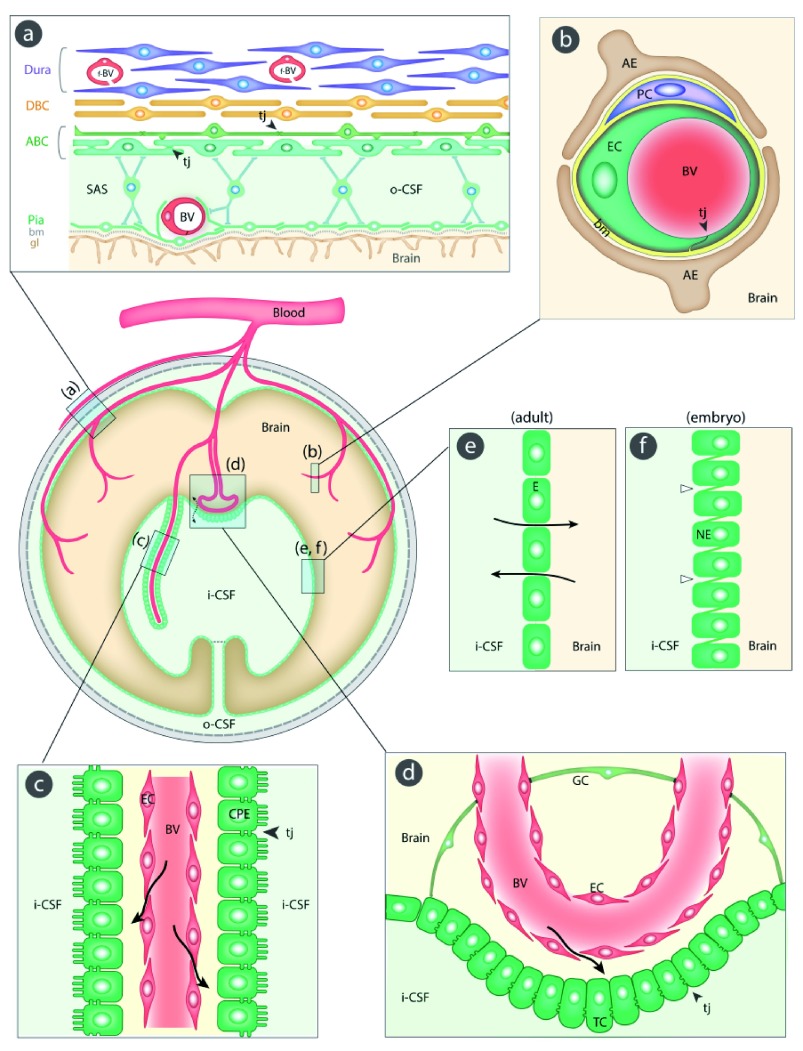
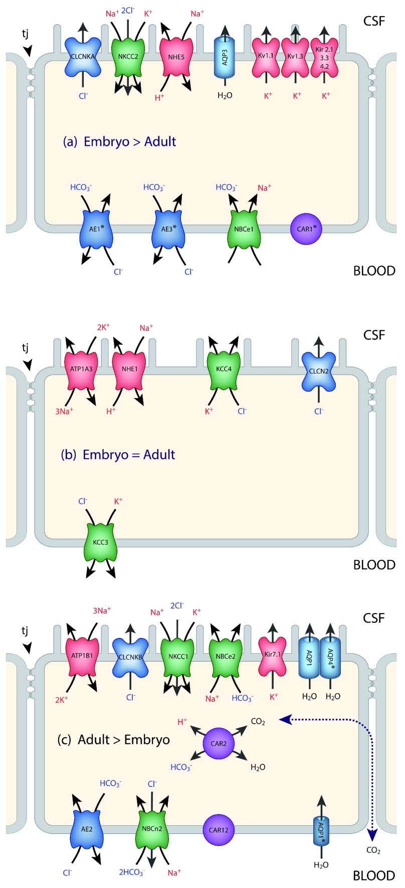
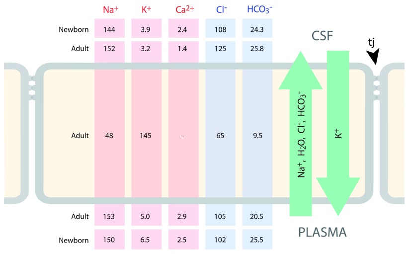
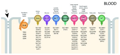
Barrier mechanisms in the brain are important for its normal functioning and development. Stability of the brain’s internal environment, particularly with respect to its ionic composition, is a prerequisite for the fundamental basis of its function, namely transmission of nerve impulses. In addition, the appropriate and controlled supply of a wide range of nutrients such as glucose, amino acids, monocarboxylates, and vitamins is also essential for normal development and function. These are all cellular functions across the interfaces that separate the brain from the rest of the internal environment of the body. An essential morphological component of all but one of the barriers is the presence of specialized intercellular tight junctions between the cells comprising the interface: endothelial cells in the blood-brain barrier itself, cells of the arachnoid membrane, choroid plexus epithelial cells, and tanycytes (specialized glial cells) in the circumventricular organs. In the ependyma lining the cerebral ventricles in the adult brain, the cells are joined by gap junctions, which are not restrictive for intercellular movement of molecules. But in the developing brain, the forerunners of these cells form the neuroepithelium, which restricts exchange of all but the smallest molecules between cerebrospinal fluid and brain interstitial fluid because of the presence of strap junctions between the cells. The intercellular junctions in all these interfaces are the physical basis for their barrier properties. In the blood-brain barrier proper, this is combined with a paucity of vesicular transport that is a characteristic of other vascular beds. Without such a diffusional restrain, the cellular transport mechanisms in the barrier interfaces would be ineffective. Superimposed on these physical structures are physiological mechanisms as the cells of the interfaces contain various metabolic transporters and efflux pumps, often ATP-binding cassette (ABC) transporters, that provide an important component of the barrier functions by either preventing entry of or expelling numerous molecules including toxins, drugs, and other xenobiotics. In this review, we summarize these influx and efflux mechanisms in normal developing and adult brain, as well as indicating their likely involvement in a wide range of neuropathologies. There have been extensive attempts to overcome the barrier mechanisms that prevent the entry of many drugs of therapeutic potential into the brain. We outline those that have been tried and discuss why they may so far have been largely unsuccessful. Currently, a promising approach appears to be focal, reversible disruption of the blood-brain barrier using focused ultrasound, but more work is required to evaluate the method before it can be tried in patients. Overall, our view is that much more fundamental knowledge of barrier mechanisms and development of new experimental methods will be required before drug targeting to the brain is likely to be a successful endeavor. In addition, such studies, if applied to brain pathologies such as stroke, trauma, or multiple sclerosis, will aid in defining the contribution of brain barrier pathology to these conditions, either causative or secondary.
Overview
The claims and statistics of this paper haven’t been checked yet.
Run the check — free, no account needed. It reads the open-access text and checks the sources it cites.
Integrity signals
Registry
Cites
Statistics
Figures
Verification
Claims the paper makes
The claims and statistics of this paper haven’t been checked yet.
Run the check — free, no account needed. It reads the open-access text and checks the sources it cites.
Self-check
Percentages, sums and reported differences re-computed from the paper’s own numbers.
The self-check needs the paper’s full text.
Citations
Cites
Structural and functional features of central nervous system lymphatic vessels
Louveau, A., Smirnov, I., Keyes, T. J., Eccles, J. D., Rouhani, S. J., Peske, J. D., Derecki, N. C., Castle, D., Mandell, J. W., Lee, K. S., et al. (2015) · Nature · doi:10.1038/nature14432 · Cited by 4,413
JUNCTIONS BETWEEN INTIMATELY APPOSED CELL MEMBRANES IN THE VERTEBRATE BRAIN
Brightman, M., & Reese, T. S. (1969) · The Journal of Cell Biology · doi:10.1083/jcb.40.3.648 · Cited by 2,457
Pericytes are required for blood–brain barrier integrity during embryogenesis
Daneman, R., Zhou, L., Kebede, A. A., & Barres, B. A. (2010) · Nature · doi:10.1038/nature09513 · Cited by 2,128
Drug Transport across the Blood–Brain Barrier
Pardridge, W. M. (2012) · Journal of Cerebral Blood Flow & Metabolism · doi:10.1038/jcbfm.2012.126 · Cited by 1,994
Developmental neurotoxicity of industrial chemicals
Grandjean, P., & Landrigan, P. (2006) · The Lancet · doi:10.1016/s0140-6736(06)69665-7 · Cited by 1,984
The Human ATP-Binding Cassette (ABC) Transporter SuperfamilyPreprint
Dean, M. C., Rzhetsky, A., & Allikmets, R. L. (2001) · Genome Research · doi:10.1101/gr.184901 · Cited by 1,820
Brain uptake of radiolabeled amino acids, amines, and hexoses after arterial injection
Oldendorf, W. (1971) · American Journal of Physiology-Legacy Content · doi:10.1152/ajplegacy.1971.221.6.1629 · Cited by 1,691
Noninvasive MR Imaging–guided Focal Opening of the Blood-Brain Barrier in Rabbits
Hynynen, K., McDannold, N. J., Vykhodtseva, N. I., & Jolesz, F. A. (2001) · Radiology · doi:10.1148/radiol.2202001804 · Cited by 1,463
The human ATP-binding cassette (ABC) transporter superfamily
Dean, M., Hamon, Y., & Chimini, G. (2001) · Journal of Lipid Research · doi:10.1016/s0022-2275(20)31588-1 · Cited by 1,302
Maliepaard, M., Scheffer, G. L., Faneyte, I. F., van Gastelen, M. A., Pijnenborg, A. C. L. M., Schinkel, A. H., van de Vijver, M. J., Scheper, R. J., & Schellens, J. H. M. (2001) · PubMed · Cited by 1,130
Research works printed but not linked: 2
- M Lewandowsky, 1900, Zur Lehre von der Cerebrospinalflüssgkeit., Z Clin Med., 40, 480-494
- G Cotzias, 1968, Experimental treatment of parkinsonism with L-Dopa., Neurology., 18, 276-277
Cited by
Sorted by citations
Löscher, W., Potschka, H., Sisodiya, S. M., & Vezzani, A. (2020) · Pharmacological Reviews · doi:10.1124/pr.120.019539 · Cited by 967
The blood-brain barrier: Physiology and strategies for drug delivery
Pandit, R., Chen, L., & Götz, J. (2020) · Advanced Drug Delivery Reviews · doi:10.1016/j.addr.2019.11.009 · Cited by 651
Unlocking the Power of Exosomes for Crossing Biological Barriers in Drug Delivery
Elliott, R. O., & He, M. (2021) · Pharmaceutics · doi:10.3390/pharmaceutics13010122 · Cited by 268
Weller, R. O., Sharp, M. M., Christodoulides, M., Carare, R. O., & Møllgård, K. (2018) · Acta Neuropathologica · doi:10.1007/s00401-018-1809-z · Cited by 222
“An additional sixth interface, (f) the interface between the CSF and the embryonic brain is present only in the foetus [62, 83].”
Self-citationHow Does the Immune System Enter the Brain?
Mapunda, J. A., Tibar, H., Regragui, W., & Engelhardt, B. (2022) · Frontiers in Immunology · doi:10.3389/fimmu.2022.805657 · Cited by 172
Structure and Junctional Complexes of Endothelial, Epithelial and Glial Brain Barriers
Dias, M. C., Mapunda, J. A., Vladymyrov, M., & Engelhardt, B. (2019) · International Journal of Molecular Sciences · doi:10.3390/ijms20215372 · Cited by 163
Review: The blood-brain barrier; protecting the developing fetal brain
Goasdoué, K., Miller, S. M., Colditz, P. B., & Bjorkman, S. T. (2016) · Placenta · doi:10.1016/j.placenta.2016.12.005 · Cited by 161
Physiology and molecular biology of barrier mechanisms in the fetal and neonatal brain
Saunders, N. R., Dzięgielewska, K. M., Møllgård, K., & Habgood, M. D. (2018) · The Journal of Physiology · doi:10.1113/jp275376 · Cited by 159
Self-citationPDGF-B Is Required for Development of the Glymphatic System
Munk, A. S. F., Wang, W., Bèchet, N. B., Eltanahy, A. M., Cheng, A. X., Sigurðsson, B., Benraiss, A., Mäe, M. A., Kress, B. T., Kelley, D. H., et al. (2019) · Cell Reports · doi:10.1016/j.celrep.2019.02.050 · Cited by 147
Self-citationTransport across the choroid plexus epithelium
Prætorius, J., & Damkier, H. H. (2017) · American Journal of Physiology-Cell Physiology · doi:10.1152/ajpcell.00041.2017 · Cited by 127
Gomez‐Zepeda, D., Taghi, M., Scherrmann, J., Declèves, X., & Menet, M. (2019) · Pharmaceutics · doi:10.3390/pharmaceutics12010020 · Cited by 131
Targeted Theranostic Nanoparticles for Brain Tumor Treatment
Mendes, M., Sousa, J., Pais, A. A. C. C., & Vitorino, C. (2018) · Pharmaceutics · doi:10.3390/pharmaceutics10040181 · Cited by 117
ABC Transporters Are Key Players in Alzheimer’s Disease
Pereira, C. D., Martins, F., Wiltfang, J., Silva, O. A. B. D. C. E., & Rebelo, S. (2017) · Journal of Alzheimer s Disease · doi:10.3233/jad-170639 · Cited by 94
Бажанова, Е. Д., Kozlov, А. А., & Litovchenko, A. V. (2021) · Brain Sciences · doi:10.3390/brainsci11050663 · Cited by 98
Nanoparticle-Based Technology Approaches to the Management of Neurological Disorders
Sim, T. M., Dinesh, T., Dheen, S. T., Bay, B., & Srinivasan, D. K. (2020) · International Journal of Molecular Sciences · doi:10.3390/ijms21176070 · Cited by 84
Roy, B., Lee, E. J., Li, T., & Rampersaud, M. (2022) · Genes · doi:10.3390/genes13030425 · Cited by 83
Deligne, C., Hachani, J., Duban‐Deweer, S., Meignan, S., Leblond, P., Carcaboso, A. M., Sano, Y., Shimizu, F., Kanda, T., Gosselet, F., et al. (2020) · Fluids and Barriers of the CNS · doi:10.1186/s12987-020-00198-0 · Cited by 77
Behl, T., Kaur, I., Sehgal, A., Kumar, A., Uddin, M. S., & BUNGAU, S. G. (2020) · Molecular Neurobiology · doi:10.1007/s12035-020-02211-x · Cited by 78
Jackson, S., WEINGART, J. D., Nduom, E. K.-E., Harfi, T. T., George, R. T., McAreavey, D., Ye, X., Anders, N. M., Peer, C. J., Figg, W. D., et al. (2018) · Fluids and Barriers of the CNS · doi:10.1186/s12987-017-0088-8 · Cited by 74
Challenges of gene delivery to the central nervous system and the growing use of biomaterial vectors
Puhl, D. L., D’Amato, A. R., & Gilbert, R. J. (2019) · Brain Research Bulletin · doi:10.1016/j.brainresbull.2019.05.024 · Cited by 74
Multiscale Mechanobiology in Brain Physiology and Diseases
Procès, A., Luciano, M., Kalukula, Y., Ris, L., & Gabriele, S. (2022) · Frontiers in Cell and Developmental Biology · doi:10.3389/fcell.2022.823857 · Cited by 73
The restorative role of annexin A1 at the blood–brain barrier
McArthur, S., Loiola, R. A., Maggioli, E., Errede, M., Virgintino, D., & Solito, E. (2016) · Fluids and Barriers of the CNS · doi:10.1186/s12987-016-0043-0 · Cited by 73
Mechanisms of DHA transport to the brain and potential therapy to neurodegenerative diseases
Van, A. L., Sakayori, N., Hachem, M., Belkouch, M., Picq, M., Lagarde, M., Osumi, N., & Bernoud‐Hubac, N. (2016) · Biochimie · doi:10.1016/j.biochi.2016.07.011 · Cited by 73
Møllgård, K., Dzięgielewska, K. M., Holst, C. B., Habgood, M. D., & Saunders, N. R. (2017) · Scientific Reports · doi:10.1038/s41598-017-11596-0 · Cited by 66
Self-citationRhaman, M. M., Islam, M. R., Akash, S., Mim, M., Noor alam, M., Nepovimova, E., Valis, M., Kuca, K., & Sharma, R. (2022) · Frontiers in Cell and Developmental Biology · doi:10.3389/fcell.2022.989471 · Cited by 63
Rönnbäck, C., & Hansson, E. (2019) · Frontiers in Neurology · doi:10.3389/fneur.2019.00533 · Cited by 63
Deng, F.-L., Pan, J.-X., Zheng, P., Xia, J.-J., Yin, B.-M., Liang, W.-W., Li, Y.-F., Wu, J., Xu, F., Wu, Q.-Y., et al. (2019) · Neuropsychiatric Disease and Treatment · doi:10.2147/ndt.s186071 · Cited by 58
Saunders, N. R., Dzięgielewska, K. M., Møllgård, K., & Habgood, M. D. (2018) · The Annual Review of Pharmacology and Toxicology · doi:10.1146/annurev-pharmtox-010818-021430 · Cited by 56
Self-citationAmino Acid Transporters as Disease Modifiers and Drug Targets
Bröer, S. (2018) · SLAS DISCOVERY · doi:10.1177/2472555218755629 · Cited by 53
Blood Brain Barrier Permeability Prediction Using Machine Learning Techniques: An Update
Saxena, D., Sharma, A., Siddiqui, M. H., & Kumar, R. (2019) · Current Pharmaceutical Biotechnology · doi:10.2174/1389201020666190821145346 · Cited by 57
A Reconfigurable In Vitro Model for Studying the Blood–Brain Barrier
Moya, M. L., Triplett, M., Simon, M. G., Alvarado, J. A., Booth, R., Osburn, J. J., Soscia, D. A., Qian, F., Fischer, N. O., Kulp, K. S., et al. (2019) · Annals of Biomedical Engineering · doi:10.1007/s10439-019-02405-y · Cited by 51
Sex-Dependent Macromolecule and Nanoparticle Delivery in Experimental Brain Injury
Bharadwaj, V. N., Copeland, C., Mathew, E., Newbern, J. M., Anderson, T. R., Lifshitz, J., Kodibagkar, V. D., & Stabenfeldt, S. E. (2020) · Tissue Engineering Part A · doi:10.1089/ten.tea.2020.0040 · Cited by 52
Herold, R., Schroten, H., & Schwerk, C. (2019) · International Journal of Molecular Sciences · doi:10.3390/ijms20215393 · Cited by 48
The role of circadian rhythm in choroid plexus functions
Quintela, T., Furtado, A., Duarte, A. C., Gonçalves, I., Myung, J., & Santos, C. R. A. (2021) · Progress in Neurobiology · doi:10.1016/j.pneurobio.2021.102129 · Cited by 45
Nanomedicine for glioblastoma: Progress and future prospects
Khan, I., Baig, M. H., Mahfooz, S., Imran, M. A., Khan, M. I., Dong, J.-J., Choi, Y., & Hatiboğlu, M. A. (2022) · Seminars in Cancer Biology · doi:10.1016/j.semcancer.2022.06.007 · Cited by 43
The effect of regadenoson on the integrity of the human blood–brain barrier, a pilot study
Jackson, S., George, R. T., Lodge, M. A., Piotrowski, A. F., Wahl, R. L., Gujar, S. K., & Grossman, S. A. (2017) · Journal of Neuro-Oncology · doi:10.1007/s11060-017-2404-1 · Cited by 42
Gutti, G., Ramakrishna, K., Kumar, D., Beohar, M., Ganeshpurkar, A., Kumar, A., Krishnamurthy, S., & Singh, S. K. (2019) · European Journal of Medicinal Chemistry · doi:10.1016/j.ejmech.2019.111613 · Cited by 42
Ystgaard, M. B., Sejersted, Y., Løberg, E. M., Lien, E., Yndestad, A., & Saugstad, O. D. (2015) · Neonatology · doi:10.1159/000437247 · Cited by 41
Delivery of synthetic mRNAs for tissue regeneration
Steinle, H., Weber, M., Stoppelkamp, S., Große‐Berkenbusch, K., Golombek, S., Weber, M., Çanak-Ipek, T., Trenz, S.-M., Schlensak, C., & Avci‐Adali, M. (2021) · Advanced Drug Delivery Reviews · doi:10.1016/j.addr.2021.114007 · Cited by 42
Age-Related 1H NMR Characterization of Cerebrospinal Fluid in Newborn and Young Healthy Piglets
Ventrella, D., Laghi, L., Barone, F., Elmi, A., Romagnoli, N., & Bacci, M. L. (2016) · PLoS ONE · doi:10.1371/journal.pone.0157623 · Cited by 40
Forcione, M., Chiarelli, A. M., Davies, D. J., Perpetuini, D., Sawosz, P. L., Merla, A., & Belli, A. (2020) · Journal of Cerebral Blood Flow & Metabolism · doi:10.1177/0271678x20921973 · Cited by 39
Benmimoun, B., Papastefanaki, F., Périchon, B., Segklia, K., Roby, N., Miriagou, V., Schmitt, C., Dramsi, S., Matsas, R., & Spéder, P. (2020) · Nature Communications · doi:10.1038/s41467-020-19826-2 · Cited by 41
Recent advances in understanding the human host immune response in tuberculous meningitis
Barnacle, J. R., Davis, A., & Wilkinson, R. J. (2024) · Frontiers in Immunology · doi:10.3389/fimmu.2023.1326651 · Cited by 45
Microphysiological Blood‐Brain Barrier Systems for Disease Modeling and Drug Development
Mulay, A. R., Hwang, J., & Kim, D. (2024) · Advanced Healthcare Materials · doi:10.1002/adhm.202303180 · Cited by 37
Current progress on microRNAs ‐based therapeutics in neurodegenerative diseases
Pereira, P., Queiroz, J. A., Figueiras, A., & de Sousa, F. P. (2016) · Wiley Interdisciplinary Reviews - RNA · doi:10.1002/wrna.1409 · Cited by 37
Electroresponsive Hydrogels for Therapeutic Applications in the Brain
Khan, Z. M., Wilts, E. M., Vlaisavljevich, E., Long, T. E., & Verbridge, S. S. (2021) · Macromolecular Bioscience · doi:10.1002/mabi.202100355 · Cited by 37
Bera, S., Kar, R. K., Mondal, S., Pahan, K., & Bhunia, A. (2016) · Biochemistry · doi:10.1021/acs.biochem.6b00518 · Cited by 34
Paracetamol and morphine for infant and neonatal pain; still a long way to go?
Baarslag, M. A., Allegaert, K., van den Anker, J. N., Knibbe, C. A. J., van Dijk, M., Simons, S. H. P., & Tibboel, D. (2016) · Expert Review of Clinical Pharmacology · doi:10.1080/17512433.2017.1254040 · Cited by 30
Practical approaches to evaluating and optimizing brain exposure in early drug discovery
Freeman, B. B., Yang, L., & Ranković, Z. (2019) · European Journal of Medicinal Chemistry · doi:10.1016/j.ejmech.2019.111643 · Cited by 31
Prodrug strategy for enhanced therapy of central nervous system disease
Xia, X., Zhou, Y., & Gao, H. (2021) · Chemical Communications · doi:10.1039/d1cc02940a · Cited by 28
The senses of the choroid plexus
Santos, C. R. A., Duarte, A. C., Costa, A. R., Tomás, J., Quintela, T., & Gonçalves, I. (2019) · Progress in Neurobiology · doi:10.1016/j.pneurobio.2019.101680 · Cited by 30
Varnamkhasti, B. S., Jafari, S., Taghavi, F., Alaei, L., Izadi, Z., Lotfabadi, A., Dehghanian, M., Jaymand, M., Derakhshankhah, H., & Saboury, A. A. (2020) · Current Drug Delivery · doi:10.2174/1567201817666200415111755 · Cited by 29
Fahoum, F., & Eyal, S. (2023) · Epilepsia · doi:10.1111/epi.17625 · Cited by 27
Koehn, L. M., Dzięgielewska, K. M., Møllgård, K., Saudrais, É., Strazielle, N., Ghersi‐Egea, J., Saunders, N. R., & Habgood, M. D. (2019) · Scientific Reports · doi:10.1038/s41598-019-42402-8 · Cited by 23
Self-citationCharacterization, isolation, and in vitro culture of leptomeningeal fibroblasts
Remšík, J., Saadeh, F. S., Tong, X., Li, M., Snyder, J., Bale, T., Wu, J. G., Derderian, C., Guber, D., Chi, Y., et al. (2021) · Journal of Neuroimmunology · doi:10.1016/j.jneuroim.2021.577727 · Cited by 21
Current Principles in the Management of Drug-Resistant Epilepsy
Shlobin, N. A., & Sander, J. W. A. S. (2022) · CNS Drugs · doi:10.1007/s40263-022-00922-4 · Cited by 22
Wang, D., Wu, F., Wen, M., Ding, L., Du, L., Xue, C., Xu, J., & Wang, Y. (2018) · Journal of Food Science · doi:10.1111/1750-3841.14017 · Cited by 19
Koehn, L. M. (2020) · Neural Regeneration Research · doi:10.4103/1673-5374.272568 · Cited by 19
Meister, S. W., Hjelm, L. C., Dannemeyer, M., Tegel, H., Lindberg, H., Ståhl, S., & Löfblom, J. (2020) · International Journal of Molecular Sciences · doi:10.3390/ijms21082999 · Cited by 21
Qiu, F., Habgood, M. D., & Schneider‐Futschik, E. K. (2020) · ACS Pharmacology & Translational Science · doi:10.1021/acsptsci.0c00098 · Cited by 18
Self-citationOrganic small-molecule NIR-II fluorophores for tumor phototheranostics
Xiang, D., Wang, Z., Zheng, H., Tang, Y., & Li, Q. (2026) · Light: Science & Applications · doi:10.1038/s41377-026-02212-w · Cited by 22
Chang, J.-H., Greene, C., Frudd, K., dos Santos, L. A., Futter, C. E., Nichols, B. J., Campbell, M., & Turowski, P. (2022) · Cell Reports Medicine · doi:10.1016/j.xcrm.2021.100497 · Cited by 16
Beyond the Blood–Brain Barrier
Kumar, M., Sharma, P. A., Maheshwari, R., Tekade, M., Shrivastava, S. K., & Tekade, R. K. (2018) · Elsevier eBooks · doi:10.1016/b978-0-12-812218-1.00015-4 · Cited by 16
Ueno, M. (2017) · Rinsho Shinkeigaku · doi:10.5692/clinicalneurol.cn-001004 · Cited by 15
Evolutionary concept of inflammatory response and stroke
Duris, K., & Jurajda, M. (2019) · Journal of Neuroscience Research · doi:10.1002/jnr.24392 · Cited by 15
Yu, S., Fu, L., Lu, J., Wang, Z., & Fu, W. (2019) · Journal of Ethnopharmacology · doi:10.1016/j.jep.2019.112165 · Cited by 15
Mentor, S., Makhathini, K. B., & Fisher, D. (2022) · International Journal of Molecular Sciences · doi:10.3390/ijms23020742 · Cited by 15
Ibrutinib disrupts blood-tumor barrier integrity and prolongs survival in rodent glioma model
Lim, S., Kwak, M., Kang, J. H., Cesaire, M., Tang, K., Robey, R. W., Frye, W. J. E., Karim, B. O., Butcher, D. O., Lizak, M. J., et al. (2024) · Acta Neuropathologica Communications · doi:10.1186/s40478-024-01763-6 · Cited by 14
Brain-Targeted Drug Delivery with Surface-Modified Nanoparticles
Lahkar, S., & Das, M. K. (2019) · Springer eBooks · doi:10.1007/978-3-030-06115-9_15 · Cited by 14
Brain Distribution of Drugs: Brain Morphology, Delivery Routes, and Species Differences
Bourasset, F., Auvity, S., Thorne, R. G., & Scherrmann, J. (2020) · Handbook of experimental pharmacology · doi:10.1007/164_2020_402 · Cited by 13
Botryllamide G is an ABCG2 inhibitor that improves lapatinib delivery in mouse brain
Strope, J. D., Peer, C. J., Sissung, T. M., Hall, O. M., Huang, P. A., Harris, E. M., Gustafson, K. R., Henrich, C. J., Sigano, D. M., Pauly, G. T., et al. (2019) · Cancer Biology & Therapy · doi:10.1080/15384047.2019.1683324 · Cited by 13
Nduom, E. K.-E., Glod, J. W., Brown, D. A., Fagan, M., Dalmage, M., Heiss, J. D., Steinberg, S. M., Peer, C. J., Figg, W. D., & Jackson, S. (2023) · PLoS ONE · doi:10.1371/journal.pone.0291068 · Cited by 12
The Function and Expression of ATP-Binding Cassette Transporters Proteins in the Alzheimer's Disease
Aykaç, A., & Sehirli, A. Ö. (2021) · Global Medical Genetics · doi:10.1055/s-0041-1735541 · Cited by 12
Regulation of Physiological Barrier Function by the Commensal Microbiota
McArthur, S. (2023) · Life · doi:10.3390/life13020396 · Cited by 12
Modvig, S., Jeyakumar, J., Marquart, H. V., & Christensen, C. (2023) · Cancers · doi:10.3390/cancers15092504 · Cited by 14
Min, W., Dang, Q., Li, Z., Zhao, X., Shen, Y., Li, F., & Liu, C. (2024) · Food Research International · doi:10.1016/j.foodres.2024.115302 · Cited by 11
The Meninges as CNS Interfaces and the Roles of Meningeal Macrophages
Hiraki, C., & Tsuruta, F. (2025) · Biomolecules · doi:10.3390/biom15040497 · Cited by 12
Improving drug delivery to the brain: the prodrug approach
Huttunen, K. M. (2024) · Expert Opinion on Drug Delivery · doi:10.1080/17425247.2024.2355180 · Cited by 10
Response to Selpercatinib in a Patient With Recurrent Glioblastoma and RET Amplification
Czech, C., Chen, A., Morgan, K. P., Zamora, C. A., El-Refai, S. M., Poynter, N., & Khagi, S. (2022) · Journal of the National Comprehensive Cancer Network · doi:10.6004/jnccn.2022.7030 · Cited by 10
Harnessing the circadian nature of the choroid plexus and cerebrospinal fluid
Fame, R. M. (2025) · npj Biological Timing and Sleep · doi:10.1038/s44323-025-00033-5 · Cited by 11
Developmental changes in the extent of drug binding to rat plasma proteins
Qiu, F., Dzięgielewska, K. M., Huang, Y., Habgood, M. D., Fitzpatrick, G., & Saunders, N. R. (2023) · Scientific Reports · doi:10.1038/s41598-023-28434-1 · Cited by 9
Self-citationBlood-brain barriers and drug pharmacokinetics: mechanisms and models
Tregub, P. P., Bystrov, D. A., Kushnir, I. A., Korsakova, S. A., Yurchenko, S. O., & Salmina, A. B. (2025) · European Journal of Pharmacology · doi:10.1016/j.ejphar.2025.177872 · Cited by 9
Nanoparticles in Cancer Theranostics: Focus on Gliomas
Azadani, M. N., Abed, A., Mirzaei, S. A., Mahjoubin‐Tehran, M., Hamblin, M. R., Rahimian, N., & Mirzaei, H. (2024) · BioNanoScience · doi:10.1007/s12668-024-01752-y · Cited by 9
Biology of the Blood-Brain and Blood-Brain Tumor Barriers
Engelhard, H. H., Arnone, G. D., Mehta, A. I., & Nicholas, M. K. (2018) · Elsevier eBooks · doi:10.1016/b978-0-12-812100-9.00008-5 · Cited by 7
Using miniature brain implants in rodents for novel drug discovery
Waldau, B. (2019) · Expert Opinion on Drug Discovery · doi:10.1080/17460441.2019.1577816 · Cited by 8
Harby, S. A., Khalil, N. A., El-Sayed, N. S., Thabet, E. H., Saleh, S. R., & Fathelbab, M. H. (2023) · Naunyn-Schmiedeberg s Archives of Pharmacology · doi:10.1007/s00210-023-02485-7 · Cited by 7
Effect of Efflux Transporter Inhibition on the Distribution of Fluconazole in the Rat Brain
Wang, W., Zheng, N., Zhang, J., Huang, X., & Yu, S. (2017) · Neurochemical Research · doi:10.1007/s11064-017-2240-x · Cited by 7
Páez, P., Sebastian, J. L., Ceron-Funez, R., Jimenez, A. J., & Rodríguez‐Pérez, L. (2023) · Frontiers in Neuroscience · doi:10.3389/fnins.2023.1204197 · Cited by 6
Intracranial Gene Delivery Mediated by Albumin-Based Nanobubbles and Low-Frequency Ultrasound
Koga, T., Kida, H., Yamasaki, Y., Feril, L. B., Endo, H., Itaka, K., Abe, H., & Tachibana, K. (2024) · Nanomaterials · doi:10.3390/nano14030285 · Cited by 6
Devnarain, N., Ramharack, P., & Soliman, M. E. S. (2017) · RSC Advances · doi:10.1039/c7ra05918c · Cited by 7
Medications for pregnant women: A balancing act between the interests of the mother and of the fetus
Saunders, N. R., & Dzięgielewska, K. M. (2020) · Prenatal Diagnosis · doi:10.1002/pd.5720 · Cited by 6
Self-citationWhat the Body Does to A Drug: Pharmacokinetics
Bereda, G. (2022) · Clinical Endocrinology and Metabolism · doi:10.31579/2834-8761/006 · Cited by 5
Affibody Molecules Intended for Receptor-Mediated Transcytosis via the Transferrin Receptor
Hjelm, L. C., Lindberg, H., Ståhl, S., & Löfblom, J. (2023) · Pharmaceuticals · doi:10.3390/ph16070956 · Cited by 7
Gap Junction Coupled Cells, Barriers and Systemic Inflammation
Rönnbäck, C. (2017) · International Journal of open Access Ophthalmology · doi:10.15226/2474-9249/2/1/00117 · Cited by 5
Alcohol withdrawal and pain: Peripheral mechanisms join central circuits
Neugebauer, V., & Ponomarev, I. (2024) · Neuron · doi:10.1016/j.neuron.2023.12.006 · Cited by 5
Biologics for the management of dementia
Chaurasiya, A., Katke, S., Panchal, K., & Nirmal, J. (2022) · Elsevier eBooks · doi:10.1016/b978-0-12-824331-2.00001-7 · Cited by 4
Qiu, F., Huang, Y., Dzięgielewska, K. M., Habgood, M. D., & Saunders, N. R. (2024) · European Journal of Neuroscience · doi:10.1111/ejn.16452 · Cited by 5
Self-citationBypassing the barrier: new routes for delivery of macromolecules to the central nervous system
Liddelow, S. A. (2017) · The Journal of Physiology · doi:10.1113/jp275499 · Cited by 4
Petronilho, F. C., Goldman, J. L., & Barichello, T. (2018) · Neuromethods · doi:10.1007/978-1-4939-8946-1_12 · Cited by 4
Biomarkers of Disease Progression
Petzold, A. (2017) · Springer eBooks · doi:10.1007/978-3-319-65921-3_6 · Cited by 4
Metadata
- Authors
- Saunders, N. R., Habgood, M. D., Møllgård, K., & Dziegielewska, K. M.
- Affiliations
- The University of Melbourne (AU)
- University of Copenhagen (DK)
- Corresponding author
- Saunders, Norman R.
- Venue
- F1000Research
- Publisher
- F1000 Research Ltd
- Published
- 2016-03-10
- Volume
- 5
- Pages
- 313
- ISSN
- 2046-1402 (ISSN-L)
- PMC
- PMC4786902
- OpenAlex
- W2337076198
- Cited by
- 143
- References
- 129
- Citations by year
- 2026: 9 · 2025: 10 · 2024: 12 · 2023: 13 · 2022: 15 · 2021: 12 · 2020: 17 · 2019: 19 · 2018: 12 · 2017: 13 · 2016: 10
- FWCI
- 1.58
- Type
- journal article
- Language
- en
- Open access
- gold
- doi.org
- Locations
- F1000Research (publishedVersion, cc-by)
- Europe PMC (PubMed Central) (publishedVersion, cc-by)
- PubMed Central (publishedVersion, cc-by)
- PubMed (publishedVersion)
- Research at the University of Copenhagen (University of Copenhagen) (publishedVersion, cc-by)
- Minerva Access (University of Melbourne) (submittedVersion, cc-by)
- DOAJ (DOAJ: Directory of Open Access Journals) (submittedVersion)
- Topics
- Life Sciences › Neuroscience › Neurology › Barrier Structure and Function Studies
- Health Sciences › Medicine › Oncology › Drug Transport and Resistance Mechanisms
- Health Sciences › Medicine › Clinical Neurology › Cerebrospinal fluid and hydrocephalus
- Keywords
- blood-brain barrier, brain drug delivery, tight junctions, ABC transporters, efflux pumps, circumventricular organs, choroid plexus epithelium, arachnoid membrane, tanycytes, neuroepithelium, gap junctions, strap junction, focused ultrasound, blood-brain barrier disruption
About these checks
Registry, retraction, Cites and statistics results are computed, not judged, with one exception: a printed reference that no index lists is read once by a model before it counts as a citation error. Claims are extracted by a model and kept only when their quote appears verbatim in the text; each cited source is then checked, with a model’s help, for whether it says what the claim relies on. Those verdicts can be wrong — read the claim’s quote and the cited source before relying on one.
Statistics and arithmetic are recomputed, not judged.
Confidence bands are shown only for fields calibrated on our annotated test set. None is calibrated yet, so no band is shown.
What counts as a claim ›How we measure confidence ›How we check this ›
Are you an author, or do you see a problem with this page? Request a review
